cluster of epitopes defined by the MAb, termed AE5, AC8, and. AE12, remains accessible in the hGH-R complex whereas over- lapping epitopes 3C11 and HG3 ...
0013-7227/90/1273-1009$02.00/0 Endocrinology Copyright © 1990 by The Endocrine Society
Vol. 127, No. 3 Printed in U.S.A.
Monoclonal Antibodies as Probes to Study the Human Growth Hormone-Binding Domain to Lactogenic Rat Liver Receptors* L. P. ROGUIN, C. E. COHEN, AND L. A. RETEGUI Institute de Quimica y Fisicoquimica Biologicas (UBA-CONICET), Facultad de Farmacia y Bioquimica, 1113 Buenos Aires, Argentina
HG3, and the closely related MAb 10C1 and NA71) failed to inhibit binding of the preformed [125I]MAb AE5-hGH complex to the receptors, suggesting a hormone modification after MAb AE5 binding. Accordingly competition experiments indicated an increase in the affinity of hGH for its receptors induced by this MAb. A higher hGH concentration was required to obtain 50% [125I]hGH binding to liver microsomes in the presence of MAb AE5 than in its absence. As the MAb used define epitopes that were previously correlated with the hGH structure, we concluded that a high flexible region (sequences 134-150) is exposed in the hGH-R complex. Furthermore, some MAb directed to this region enhance the hormone affinity for its rat liver receptors, probably through an induced conformational change. (Endocrinology 127: 10091015,1990)
ABSTRACT. A set of monoclonal antibodies (MAb) to human GH (hGH) was used to study the hormone binding orientation to its receptors (R) from female rat liver. The hGH antigenic region left exposed after its binding to liver microsomes was detected by measuring the ability of various [125I]MAb to bind to the preformed hGH-R complexes. Results indicated that a cluster of epitopes defined by the MAb, termed AE5, AC8, and AE12, remains accessible in the hGH-R complex whereas overlapping epitopes 3C11 and HG3 would define a hGH region involved in the binding site. Supporting these findings, solubilization and HPLC gel filtration of [125I]MAb-hGH-R complexes showed a radioactive peak of about 450,000 mol wt for MAb AE5 or AC8, but not for MAb 3C11 or HG3. [125I]MAb AE12 behaved differently, suggesting that epitope AE12 may be masked or altered in hGH-R-solubilized complexes. MAb directed to the putative hGH-binding site (MAb 3C11,
G
H, PRL, and placental lactogens (PL) are phylogenetically related hormones that share structural, functional, and antigenic properties (1). Human GH (hGH) displays a great variety of biological activities, including linear growth (somatogenesis), lactation, activation of macrophages, and insulin-like and diabetogenic effects (1-3). Although it has been shown that growth stimulation of cartilage and bone is mainly due to the hGH-dependent release of insulin-like growth factor-I from the liver (4), direct effects of hGH have also been described (3, 5, 6). Studies of the [125I]hGH interaction with receptors from different sources allowed them to be classified according to their respective specificities as somatogenic or lactogenic. The binding of [125I]hGH to somatogenic receptor is inhibited by GH of primates and nonprimates, whereas only PRL and human PL (hPL) effectively Received February 13, 1990. Address all correspondence and requests for reprints to: Dr. L. A. Retegui, Instituto de Quimica y Fisicoquimica Biologicas, Facultad de Farmacia y Bioquimica, Junin 956, 1113 Buenos Aires, Argentina. * This work was supported in part by grants from the Consejo Nacional de Investigaciones Cientificas y Tecnicas (CONICET), Fundacion Antorchas, and the Organization of American States.
compete with [125I]hGH for the binding to lactogenic receptors (1). Receptors that bind exclusively primate GH with high affinity have also been described in human IM-9 lymphocytes (7) and human liver (8). A few years ago, we explored whether hGH contained one or more binding sites to receptors from IM-9 cells and rabbit liver membranes (9). The different inhibitory activities of five monoclonal antibodies (MAb) anti-hGH on hormone binding led us to suggest that the same region of hGH was involved. More recently, we established the antigenic topography of hGH and proposed a tentative three-dimensional model of the relative distribution of 20 antigenic determinants (or epitopes) on the hormone surface (10). As the epitopes studied covered the whole hGH molecule, it was possible to allocate some epitopes on the primary structure of the protein (11). The knowledge of the hGH antigenic structure allowed us to study the orientation of the hGH molecule in its binding to specific receptors. The MAb, characterized previously (10), were used as probes to identify the hGH region left exposed after its binding to lactogenic receptors from female rat liver. Experiments were carried out with both particulate receptors and solubilized hGH-
1009
1010
hGH LACTOGENIC BINDING DOMAIN STUDIED WITH MAb
receptor (hGH-R) complexes. The results obtained indicated that at least a cluster formed by epitopes AE5, AE12, AC8, and AC3 remained exposed in the hGH-R complex, whereas epitopes 3C11 and HG3 were close to or included in the hGH-binding site. The data also showed that two MAb (AE5 and AC3) enhanced hGH binding to the receptors.
Endo•1990 Vol 127 • No 3
affinity chromatography on a protein-A-Sepharose 4B column (19). The pH 6.0 fraction was dialyzed against 50 mM Tris-HCl buffer, pH 7.4, concentrated, and stored at -20 C. [125I]MAb were prepared by the method of McConahey and Dixon (20). Specific radioactivities for most MAb ranged from 11-17 fiCi/ ng, except for the MAb HG3 which consistently displayed a lower value (6 nCi/fig). RIA
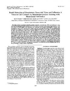
Materials and Methods GH hGH was prepared by the method of Mills et al. (12). Its purity and biological activity were established as indicated previously (13). Labeling of the hormone with 125I was performed following the method of Roth (14). Specific radioactivities of 90-120 ^Ci/Vg were usually achieved. Bovine GH (bGH) was obtained in our laboratory according to the procedures of Dellacha and Sonenberg (15). MAb MAb anti-hGH QA68, NA39, NA71, NA27, and anti-hPL, termed MAb EBl, EB2, and EB3, were kindly provided by Dr. J. Ivanyi (16, 17). MAb anti-hGH Fll, AE5, AE12, AC3, 3C11, HG3, 10C1, 10D6, AC8, 04C11, AE6, and AC6 have been prepared and characterized by the authors (10, 13, 18). As shown in Fig. 1, these MAb recognize epitopes covering the whole hGH surface and display a wide spectrum of affinities toward the hormone. Epitopes AE6 and AC6 define a hGH region only expressed when the protein is adsorbed on plastic surfaces (10). Purification of MAb from ascitic fluid was carried out by
MAb affinities were measured by a solid phase RIA, as previously described (18). The reactivity of each purified MAb was tested as follows. Polyvinyl microplates (Dynatech Laboratories, Inc., Alexandria, VA) were coated with the hormone (5 Mg/ml diluted in 5 mM glycine buffer, pH 9.2) and incubated with serial dilutions of ascitic fluid in PBS containing 5% (vol/ vol) fetal calf serum. After incubating for 16 h at 25 C the plates were washed with PBS containing 0.125 ml Tween-20/ liter and the reacting MAb revealed with [125I]Ab to mouse immunoglobulin (Ig; 100,000 cpm). A similar assay was used to test the reactivity of [125I]MAb, except that [125I]Ab to mouse Ig was omitted. Crude liver microsomal preparation Livers were obtained from Wistar female rats (210-250 g) during estrus. Microsomal preparation was performed as described by Bonifacino et al. (21). Briefly, livers were homogenized in 10 vol (vol/wt) chilled 0.3 M sucrose, 5 mM Tris-HCl buffer, pH 7.4., and centrifuged at 10,000 x g for 20 min and then at 100,000 x g for 1 h. The final 100,000 x g pellet was resuspended in 25 mM Tris-HCl buffer, pH 7.4. A sample of the suspension was solubilized by heating for 30 min at 100 C in 1 M NaOH, and the protein concentration was determined by the method of Lowry et al. (22). The crude microsomal suspension (protein concentration, 50 mg/ml) was frozen at -20 C until use. Binding of P25I]hGH to rat liver microsomes
FlG. 1. Schematic drawing showing the relative distribution of various hGH epitopes. The rectangle represents the hGH molecular area (7238 A2), and the squares correspond to the area of the epitope recognized by each MAb (690 A2). The two-dimensional design retains the essential features of the spatial distribution of the hGH epitopes established previously (10), with the restrictions imposed when transferring a three-dimensional model to a plane. Encircled numbers indicate the Ka (X108 M"1) of the corresponding MAb measured in a solid phase RIA as indicated in Materials and Methods. ND, Not determined.
[125I]hGH (0.5-1 ng) was incubated for 16 h at 25 C with 100 ng microsomal proteins and serial dilutions of competitors (unlabeled GH or MAb) in a total volume of 0.5 ml. The incubation medium (IM) consisted of 25 mM Tris-HCl and 10 mM CaCl2 (pH 7.4) containing 0.1% (wt/vol) BSA. The reaction was stopped by the addition of 4 ml ice-cold IM, the membranes were sedimented by centrifugation at 800 X g for 25 min at 4 C, and the bound radioactivity was measured in a 7-counter. [125I]hGH nonspecific binding was determined in the presence of 5 ng unlabeled hGH. When MAb were used as competitors, the microsomes were previously incubated for 2 h at room temperature with normal rat serum (final dilution, 1:100 in IM). Receptor specificity As has been reported by many researchers, liver microsomes from female rats are mainly lactogenic (for a review, see Ref. 1). Under our experimental conditions, 50% of [125I]hGH binding inhibition was obtained with 6 X 10~10 and 1 X 10~7 M hGH and bGH, respectively (data not shown).
hGH LACTOGENIC BINDING DOMAIN STUDIED WITH MAb
1011
Binding of P25IJMAb to hGH-R complexes
Microsomes (100 tig protein) were incubated in IM for 16 h at 25 C in the absence or presence (0.1-10 ^g/ml) of unlabeled hGH in a final 0.5-ml volume. Unbound hGH was removed by washing with 4 ml ice-cold IM and centrifuging at 800 X g for 25 min at 4 C. The pellets were resuspended in 0.5 ml IM containing 100,000 cpm [125I]MAb (4-12 ng) and incubated 2 h at 37 C. After washing, the bound radioactivity was counted. Results were expressed as the signal/noise ratio (S/N), defined as the ratio between radioactivity bound to the hGH-R complex and the radioactivity bound to the microsomes in the absence of hormone. Molecular size determination complexes
of solubilized
4
P25IJMAb-hGH-R
5
LOG ( 1/antibody
4
dilution)
125
Rat liver microsomes (10 mg protein) suspended in 0.5 ml IM were shaken for 2 h in the presence of 5 ng hGH and then incubated for 16 h at 25 C. After washing, the pellet was resuspended in 1 ml of the same buffer containing about 8 x 10⁶ cpm [125I]MAb and agitated for 3 h at 25 C. After overnight incubation at 4 C, the unbound radioactivity was removed by washing with ice-cold IM, and 2 ml 1% (wt/vol) Triton X-100 and 25 mM Tris-HCl buffer, pH 7.4, were added to the pellet. The suspension was shaken for 30 min and then centrifuged at 100,000 x g for 2 h. The supernatants containing the solubilized [125I]MAb-hGH-R complexes were submitted to HPLC as follows. One hundred microliters of supernatant were injected into a Superose 6HR 10/30 column (Pharmacia, Uppsala, Sweden). Elution was carried out at a flow rate of 0.4 ml/min with 50 mM Tris-HCl, 10 mM CaCl2, 0.1% (wt/vol) Triton X-100, and 0.02% (wt/vol) sodium azide buffer, pH 7.4. The radioactivity present in the effluent fractions (240 nl) was measured in a ycounter.
FIG. 2. Effects of various MAb on [ I]hGH binding to female rat liver receptors. The tracer (60,000-80,000 cpm) was incubated with microsomal proteins (100 ^g) in the presence of serial dilutions of ascitic fluid containing the indicated MAb. Microsomes were treated with normal rat serum before the assay, as indicated in Materials and Methods. Results are expressed as a percentage of hGH specific binding (100% = 6,500 cpm). Ascitic fluid devoid of immunological activity toward hGH was used as a control.
Binding of P25I]MAb-hGH complexes to rat liver microsomes
FIG. 3. Binding of [125I]MAb to hGH-R complex. Microsomes from female rat liver were preincubated with hGH at the indicated concentrations, and after eliminating the unbound hormone [125I]MAb (100,000 cpm) was added. Results are expressed as the S/N, which is defined as bound radioactivity to the hGH-R complex divided by the radioactivity bound in the absence of hormone. The values shown are the mean ± SE of three determinations for each hGH concentration.
[125I]MAb (-100,000 cpm) were incubated for 1 h at 37 C with 50 ng hGH in a final volume of 0.5 ml IM. Rat liver microsomes (100 ^g protein) treated with rat normal serum as previously indicated were added, and the mixture was incubated for 2 h at 37 C. After washing with ice-cold IM, the bound radioactivity was counted. MAb competitive effects were determined by adding each ascitic fluid (final dilution, 1:5000) to the mixture of [125I]MAb and hGH.
Results Effect of MAb on the binding of ^IJhGH liver microsomes
to female rat
The MAb tested differently affected the binding of the hormone to rat liver receptors (Fig. 2). Most of them were inhibitors, whereas MAb AE12 and 04C11 originated competition curves similar to that of the control. MAb AE5 and AC3 significantly enhanced [125I]hGH binding; their effects were proportional to the amount of antibody added. Similar results were obtained when pu-
AE12
hGH
0.1 0.2
1
2
10
0.2
1
2 10
2 10
2
10
2 10
rified MAb AE5, AE12, 04C11, 3C11, and AC8 were used, discarding any artifact due to some component of the ascitic fluid other than the antibody. Furthermore, all experiments described above were carried out with microsomes preincubated with normal rat serum to avoid nonspecific binding of the MAb to the membranes. Otherwise, MAb activities were masked, probably through interaction with Fc receptors (data not shown). Binding of P^IJMAb to the hGH-R complex To detect the antigenic sites of hGH that remained exposed after its binding to receptors we measured the ability of various [125I]MAb to bind to the preformed hGH-R complex. The results presented in Fig. 3 show that 125I-labeled MAb AE5, AC8, and AE12 originated
1012
hGH LACTOGENIC BINDING DOMAIN STUDIED WITH MAb
much higher S/N values than 125Habeled MAb 3C11 or HG3, suggesting that epitopes AE5, AC8, and AE12 are accessible in the hGH-R complex, whereas epitopes 3C11 and HG3 are near or covered by the receptor. MAb AE5 was the most effective in recognizing the exposed site of hGH. Values of bound radioactivity were already significant at 0.1 /ug/ml hGH, although the highest S/N values were obtained with 2-10 jug/ml hormone. [125I]MAb AE12 originated S/N values only at the maximal hormone concentration while [125I]MAb AC8 bound to the hGH-R complex preformed in the presence of either 2 or 10 Mg/ml hGH. Molecular size determination of solubilized P2bI]MAbhGH-R complexes To assess the specificity of the binding reactions described above, preformed hGH-R complexes incubated with [125I]MAb were solubilized in the presence of Triton X-100 and fractionated by HPLC (Fig. 4). Soluble complexes of about 450,000 mol wt were found with either [125I]MAb AE5 or MAb AC8, while a predominant component of about 300,000 mol wt was detected when 125Ilabeled MAb 3C11 or HG3 was used. A radioactive peak of about 150,000 mol wt, similar to the ones found in the controls without hGH, was obtained with [125I]MAb AE12. [125I]hGH-R solubilized and submitted to HPLC under the same experimental conditions had a mol wt of about 300,000 (data not shown). Therefore, the occurrence of soluble complexes of about 450,000 mol wt clearly indicated that MAb AE5 and AC8 recognized the hormone already bound to the receptor. Radioactive material with a mol wt of about 300,000 was not restricted to experiments carried out with 125Ilabeled MAb 3C11 or HG3. A shoulder in the main peak obtained upon incubation with 125I-labeled MAb AE5 or AC8 also indicated its presence (Fig. 4). To investigate the nature of such a component, we performed similar experiments but in the absence of receptor. When hGH (from 0.1-10 Mg/ml) was incubated with [125I]MAb 3C11 and the mixture submitted to HPLC, radioactive material eluted at a mol wt of about 300,000 (data not shown). Although MAb AE5 behaved similarly, the peak disappeared when the hGH concentration was below 1 ng/m\. As this behavior was never found when an excess of MAb was incubated with trace amounts of hGH (0.5 ng) we concluded that the aggregation of the hormone at high concentrations may play a role in the formation of complexes of about 300,000 mol wt. Binding of ^IJMAb-hGH complexes to rat liver microsomes: competition experiments When labeled MAb 3C11 or AE5 was preincubated with free hGH before the addition of microsomes, the
Endo • 1990 Vol 127 • No 3
450.000 150000 I 300.000 I
450.000 150000 I 300000
I
1 i I
•-AE12
ACB
o x
E Q. O >>
< o Q CC
5
3
„..,_...•• 12
13
V.
15
16
17
12
13
U
15
16
17
ELUTION VOLUME (ml) FIG. 4. Detection of solubilized [125I]MAb-hGH-R complexes. Rat liver microsomes (10 mg protein) were preincubated with 5 ng hGH, and after removing unbound hormone [125I]MAb (8 X 10⁶ cpm) was added. The free radioactivity was eliminated by washing, and the [128I]MAbhGH-R was solubilized in the presence of Triton X-100 before being submitted to HPLC in a Superose 6HR column, as indicated in Materials and Methods. Controls were performed by submitting the microsomes to the same experimental procedure but in the absence of hormone.
[125I]MAb 3Cll-hGH complex did not bind to the receptors, while the [125I]MAb AE5-hGH complex bound effectively (Table 1). Furthermore, the S/N obtained with 0.1 fig/m\ hGH was even higher (~20) than that obtained when [125I]MAb AE5 was added to the hGH-R complex preformed with the same amount of hormone (Table 1 and Fig. 3). Provided that epitope 3C11 would be closely related to the hGH-binding site, one can expect a strong inhibition produced by MAb 3C11 on the binding of [125I]MAb AE5-hGH complex to rat liver receptors. However, competition experiments (Fig. 5) showed that MAb 3C11 was totally unable to yield such an effect. On the other hand,
hGH LACTOGENIC BINDING DOMAIN STUDIED WITH MAb TABLE 1. Binding of [125I]MAb-hGH complexes to rat liver microsomes Bound radioactivity (cpm) hGH kg/ml) 0 0.1
126
A—a — a — a — a
100
5 80
1013
\ •
\
V
AE5
125
[ I]MAb AE5
[ I]MAb 3C11
580 ± 100 12,230 ± 300
560 ± 50 550 ± 50
Each [126I]MAb (100,000 cpm) was preincubated with or without hGH before the addition of microsomes from female rat liver (100 ng protein). After incubation, free and bound [125I]MAb-hGH complexes were separated by washing and centrifugation. Values are the mean ± SE of three determinations.
O
\
60
3C11
\
5 Z
40
•
m
\ \
20 -
\
\
CONTROL ^o—o
0.1
1
10
i 100
^ a
hGH(nM) FIG. 6. Effects of MAb AE5 and MAb 3C11 on the hGH affinity toward rat liver receptors. Microsomes (100 fig protein) were incubated with hGH (85,000 cpm; -0.5 ng), MAb AE5 (0.6 ng), or MAb 3C11 (ascitic fluid diluted 1:10,000; ~0.4 ng) or in the absence of any MAb (control), with different hGH concentrations as competitors. Results are expressed as percentage of bound radioactivity (100% = 13,500 cpm in the presence of MAb AE5; 7,360 cpm in the presence of MAb 3C11; 10,930 cpm in control).
m
LOG (1/antibody dilution)
FIG. 5. Effects of MAb AE5 and MAb 3C11 on the binding of [125I] MAb AE5-hGH complex to rat liver microsomes. The experimental design was the same as that described in Table 1, except that serial dilutions of ascitic fluid containing the indicated MAb were added simultaneously to the mixture of [125I]MAb AE5 and hGH. Results are expressed as a percentage of the maximal binding (100% = 12,200 cpm).
the addition of increasing concentrations of MAb AE5 decreased the bound radioactivity, as the unlabeled MAb competed with [125I]MAb AE5 for hGH binding. The experimental approach described in Fig. 5 was used to examine the behavior of a set of 18 MAb (Fig. 1). The lack of inhibition of [125I]MAb AE5-hGH binding to microsomes shown for MAb 3C11 was also found with MAb 10C1, HG3, and NA71, whose epitopes overlapped or were very close to epitope 3C11 (Fig. 1), whereas the rest of the MAb competed to different extents (data not shown). Taken together, these results suggested that MAb AE5 binding to hGH would modify the 3C11 antigenic domain, leading to an increase in the affinity of the hormone for its receptors. Ability of hGH to compete with P^IJhGH for binding to rat liver receptors in the presence of MAb AE5 and 3C11 To test whether MAb AE5 affected hGH binding affinity we measured the inhibitory effect of hGH on [125I] hGH binding to the receptors in both the presence and absence of MAb. Figure 6 shows an important shift of the competition curve when MAb AE5 was added to the reaction mixture. Fifty percent of [125I]hGH binding was
obtained with 0.5 nM hGH in the absence of any MAb, whereas a 30-nM hormone concentration was needed to elicit the same effect in the presence of MAb AE5. On the contrary, simultaneous incubation with MAb 3C11 did not greatly affect the inhibitory capacity of hGH (Fig. 6). Results similar to those obtained with MAb AE5 were found with MAb AC3 (data not shown), suggesting that both Ab induce an increase in hGH affinity for the lactogenic receptors. Discussion The availability of a set of MAb recognizing epitopes that cover the entire hGH surface allowed us to examine the orientation of hormone binding to its receptors, focusing the study on lactogenic receptors from female rat liver. Experiments designed to detect the hGH antigenic region left exposed after binding showed that [125I]MAb AE5, AC8, and AE12 were able to recognize bound hGH; hence, the cluster formed by the respective epitopes remains accessible in the hGH-R complex. On the contrary, overlapped epitopes 3C11 and HG3 would define a hGH region involved in the binding site. MAb AE12 failed to give high mol wt soluble complexes, although it displayed a significant S/N value at the same hGH concentration. Masking and/or alteration of AE12 epitope in solubilized hGH-R complex may explain these findings. S/N values for MAb AE5 were strikingly higher when the MAb-hGH complex was presented to the receptor than when the MAb was bound to preformed hGH-R complexes, suggesting that hGH binding was improved by MAb AE5. Failure of MAb 3C11, HG3, 10C1, and
1014
hGH LACTOGENIC BINDING DOMAIN STUDIED WITH MAb
NA71 to inhibit the binding of [125I]MAb AE5-hGH supported this hypothesis. These observations prompted us to carry out experiments to study whether binding of MAb AE5 to hormone affected its affinity for rat liver receptors. Competition curves showed that a higher hGH concentration was required to obtain 50% [125I]hGH binding inhibition in the presence of MAb AE5 than in its absence. On the other hand, the addition of MAb 3C11 only slightly affected the hGH inhibitory capacity. Results similar to those described for MAb AE5 were obtained with MAb AC3; hence, we inferred that both Ab are able to increase the affinity of hGH for its receptors, probably through an allosteric conformational change induced on the hormone. MAb AE5, AE12, and AC3 recognize a highly flexible hGH region, probably located in the 134-150 fragment (11). Previous reports have shown that proteolytic cleavage as well as spontaneous deamidation reactions occur easily in that hGH sequence (23). Coincidentally, this region does not have a defined structure, as shown in the hGH three-dimensional model proposed by Borisova et al. (24). Lewis (23) reported enhanced growth-promoting activity of hGH natural derivatives lacking some residues from the 134-150 sequence, although an increase in affinity toward the specific receptors has not been described. These findings together with the ones presented in this work would suggest that alterations in that flexible hGH region, produced by either enzymatic proteolysis or Ab binding, affect the biological properties of the hormone. Furthermore, we have recently found that MAb 3C11, 10C1, and NA71 are able to induce an allosteric conformational modification in the hGH molecule, which is recognized by a MAb termed 4D11. The new epitope 4D11 is located in the AE5/AC8 domain (25). Thus, our present view of hGH is that of a protein containing at least two regions susceptible to induced conformational changes. As described in the present work, epitopes 3C11 and HG3, both overlapped by epitope 10C1, would be involved in the hGH interaction with its lactogenic receptors. Results from our laboratory (11) suggest that the antigenic domain defined by these MAb is located in the central portion of sequence 1-32 (helix 1), partially interacting with helix 3 (residues 106-128) and helix 4 (residues 155-191), according to the hGH three-dimensional model proposed by Borisova et al. (24). Cunningham et al (26) studied the hGH-binding site to the extracellular portion of the cloned somatogenic liver receptor. They reported it to be the loop between residues 54-74, the central portion of helix 4 to the carboxyl-terminus, and, to a lesser extent, the N-terminal region of helix 1. Differences between the hGHbinding sites proposed by Cunningham et al. (26) and ourselves are not surprising, since they studied somato-
E n d o • 1990 Vol 127 • No 3
genic receptors and not lactogenic ones as we did. Due to the different specificities of both receptors, one cannot discard the existence of more than one binding domain in the hGH molecule. Furthermore, Cunningham et al. (26) supported their hypothesis of the three-dimensional structure of porcine GH (27), whereas our data fit better with the hGH structure proposed by Borisova et al. (24).
Acknowledgments We are particularly indebted to Prof. Alejandro C. Paladini for helpful discussions and critical revision of the manuscript. The technical assistance of Miss M. Ramirez in the preparation of the manuscript is gratefully acknowledged.
References 1. Paladini AC, Pefia C, Poskus E 1983 Molecular biology of growth hormone. In: Hurrell JGR (ed) CRC Critical Review of Biochemistry. CRC Press, Cleveland, vol 215:25 2. Chawla RK, Parks JS, Rudman D 1983 Structural variants of human growth hormone: biochemical, genetic, and clinical aspects. Annu Rev Med 34:519 3. Edwards CK, Ghiasuddin SM, Schepper JM, Yunger LM, Kelly KW 1988 A newly defined property of somatotropin: priming of macrophages for production of superoxide anion. Science 239:769 4. Daughaday WH, Italk K, Raben MS, Salmon WD, Van den Brande JL, Van Wyk JJ 1972 Somatomedin: proposed designation for sulphation factor. Nature 236:107 5. Madsen K, Friberg U, Roos P, Eden S, Isaksson O 1983 Growth hormone stimulates the proliferation of cultured chondrocytes from rabbit ear and rat rib growth cartilage. Nature 304:545 6. Tanaka T, Shiu RPC, Gout PW, Beer CT, Noble RL, Friesen HG 1980 A new sensitive and specific bioassay for lactogenic hormones: measurement of prolactin and growth hormone in human serum. J Clin Endocrinol Metab 51:1058 7. Lesniak MA, Gorden P, Roth J, Gavin JR 1974 Binding of 12SIhuman growth hormone to specific receptors in human cultured lymphocytes. J Biol Chem 249:1661 8. Carr D, Friesen HG 1976 Growth hormone and insulin binding to human liver. J Clin Endocrinol Metab 42:484 9. Retegui LA, De Meyts P, Pefia C, Masson PL 1982 The same region of human growth hormone is involved in its binding to various receptors. Endocrinology 111:668 10. Mazza MM, Retegui LA 1989 The antigenic topography of human growth hormone. Mol Immunol 26:231 11. Mazza MM, Gobet MG, Biscoglio MJ, Mihajlovich V, Guillemot JC, Vita N, Ferrara P, Retegui LA 1990 Relationship between the antigenic topography and the structure of human growth hormone. Endocrinology 127:1002 12. Mills JB, Ashworth RB, Wilhelmi AE, Hartree AS 1969 Improved method for the extraction and purification of human growth hormone. J Clin Endocrinol Metab 29:1456 13. Retegui LA, Masson PL, Paladini AC 1985 Specificities of antibodies to hGH in patients treated with hGH. Longitudinal study and comparison with the specificities of animal antisera. J Clin Endocrinol Metab 60:184 14. Roth J 1975 Methods for assessing immunologic and biological properties of iodinated peptide hormones. In: Colowick SO, Kaplan NO (eds) Methods in Enzymology, part B. Academic Press, New York, vol 37:223 15. Dellacha JM, Sonenberg M 1964 Purification of bovine growth hormone. J Biol Chem 239:1515 16. Ivanyi J 1982 Analysis of monoclonal antibodies to human growth hormone and related proteins. In: Hurrell JGR (ed) Monoclonal Hybridoma Antibodies: Techniques and Applications. CRC Press, Boca Raton, pp 59-79 17. Aston R, Ivanyi J 1985 Monoclonal antibodies to growth hormone
hGH LACTOGENIC BINDING DOMAIN STUDIED WITH MAb and prolactin. Pharmacol Ther 27:403 18. Retegui LA, Milne RW, Cambiaso CL, Masson PL 1982 The recognition by monoclonal antibodies of various portions of a major antigenic site of human growth hormone. Mol Immunol 19:865 19. Ey PL, Prowse SJ, Jenkin CR 1978 Isolation of pure IgG2a and IgG2b immunoglobulins from mouse serum using protein A-Sepharose. Immunochemistry 15:429 20. McConahey PJ, Dixon FJ 1966 A method of trace iodination of proteins for immunologic studies. Int Arch Allergy 29:185 21. Bonifacino JS, Sanchez SH, Paladini AC 1981 Characterization of human somatotropin binding to detergent-solubilized lactogenic receptors from rat liver. Biochem J 194:385 22. Lowry OH, Rosebrough NJ, Farr AL, Randall RJ 1951 Protein measurement with the Folin phenol reagent. J Biol Chem 193:265 23. Lewis UJ 1984 Variants of growth hormone and prolactins and
their posttranslational modifications. Annu Rev Physiol 46:33 24. Borisova SN, Pavlovsky AG, Naktinis VI, Yanulaitis E-AA, Rubtsov PM, Skryabin KG, Bayev AA, Vainshtein BK 1988 Crystallization and preliminar x-ray studies of genetically engineered human somatotropin. DAN USSR 301:474 25. Mazza MM, Retegui LA 1989 Monoclonal antibodies to human growth hormone induce an allosteric conformational change in the antigen. Immunology 67:148 26. Cunningham BC, Jhurani P, Ng P, Wells JA 1989 Receptor and antibody epitopes in human growth hormone identified by homolog-scanning mutagenesis. Science 243:1330 27. Abdel-Meguid SS, Shieh H-S, Smith WW, Dayringer HE, Violand BN, Bentle LA 1987 Three-dimensional structure of a genetically engineered variant of porcine growth hormone. Proc Natl Acad Sci USA 84:6434
Methods in Basic and Clinical Bone Biology An instructional course in theory and practice, sponsored by ICCRH Inc., with the cooperation of the Japanese Society for Bone Metabolism Research. Subjects include methods of bone organ culture, culture of osteoblasts and osteoclasts, application of approaches of molecular and cell biology, and methods of studying clinical bone disease. Date: January 30-February 2, 1991 Location: Oiso Prince Hotel, Japan (one and a half hours from Tokyo) Faculty: C. Christiansen P. Delmas H. Fleisch T. J. Martin
H. Orimo J. W. Pike J. G. Raisz G. A. Rodan
Enquiries and Registration: Prof. Hajime Orimo Department of Geriatrics University of Tokyo 7-3-1 Hongo, Bunkyo-ku Tokyo, Japan
1015
R. G. G. Russell R. Schenk T. Suda A. H. Tashjian, Jr.