0 1991 by The American Society for Biochemistry and Molecular Biology, Inc. Vol. 266, No. ..... 358-365, American Society of Microbiology, Washington,. D. C..
Vol. 266, No. 33, Issue of November 25, pp. 22129-22135,1991 Printed in U.S.A .
THEJOURNALOF BIOLOGICAL CHEMISTRY 0 1991by The American Society for Biochemistry and Molecular Biology, Inc.
Monoclonal Antibodiesto Escherichia coliRibosomal Proteins L9 and L10 EFFECTS ON RIBOSOME FUNCTION ANDLOCALIZATION RIBOSOMAL SUBUNIT*
OF L9 ON THE SURFACE OF THE 50 S (Received for publication, July 18, 1991)
Bishwajit Nag$, Sesha S . Akellaj, PaulineA. Cannll, Dinesh S . Tewarill , Dohn G. Glitzl, and Robert R. Traut** From the Departmentof Biological Chemistry, School of Medicine, University of California, Davis, California 95616 and the TDepartment of Biological Chemistry, UCLASchool of Medicine, University of California, Los Angeles, California 90024
Monoclonal antibodies against Escherichia coli ri- seems to be universal in the ribosome, not only in its amino bosomal proteins L9 and L10 were obtained and their acid sequence (Matheson et al., 1990), but also in its particispecificity confirmed by Western blot analysis of total pation in a unique quaternary structure (Casiano et al., 1990). ribosomal protein. This was particularly important for Less is known concerning possible functional involvement the L9antibody, since theimmunizing antigen mixture of L9. It has been mapped by immune electron microscopy contained predominantly L l l . Each antibody recog- with a polyclonal antibody (Stoffler-Meilicke et al., 1983) to nized both 70 S ribosomes and 60 S subunits. Affinity- a site near protein L1, not far from the peptidyltransferase purified antibodies were tested for their effect on in domain. It has been cross-linked not only to L1, but also to vitro assays of ribosome function. Anti-L10 and anti- L2 (Traut et al., 1986), a major candidate for involvement in L9 inhibited poly(U)-directed polyphenylalanine syn- the peptidyltransferase reaction (Romero et al., 1990). L9 is thesis almost completely. The antibodies hadno effect not among the proteins that have been observed to be lacking on subunit association or dissociation and neither antibody inhibited peptidyltransferase activity.Both an- in viable E. coli strains (Dabbs, 1986). Like L1, L9 is one of to tibodies inhibited the binding of the ternary complex the primary RNA-binding proteins, and both proteins bind the 12 S 3‘ end fragment of 23 S RNA (Rohl and Nierhaus, that consisted of aminoacyl-tRNA, guanylyl &y-methylenediphosphonate, and elongation factor Tu, and the 1982) that contains domain V and sites associated with pepbinding of elongation factor G to the ribosome. The tidyltransferase activity (Barta et al., 1990; Egebjerg et al., intact antibodies were more potent inhibitors than the 1990). We havebeen interested in L7/L12, L10, and other proteins Fab fragments. In contrast to the previously established location of L10 at the base of the L7/L12 stalk of the factor-binding domain (Traut et al., 1986). We atnear thefactor-binding site,the siteof anti-19 binding tempted to prepare monoclonal antibodies against both L10 to 50 S subunits was shown by immune electron mi- and L11, a near neighbor of L10. We obtained a hybridoma croscopy to be on the L1 lateralprotuberance opposite cell line that makes antibodies to L10; however, hybridomas the L7/L12 stalk as viewed in the quasisymmetric pro- made with mice injected with an L11 preparation that conjection. The inhibition of factor binding by both anti- tained a minor contamination of L9 did not recognize L11, bodies, although consistent with established properties but instead, reacted strongly with L9. We continued studies of L10 in the ribosome, suggests a long range effect on with the purified anti-19 and anti-Ll0 monoclonal antibodies. subunit structure that is triggered by the binding of anti-19. EXPERIMENTAL PROCEDURES AND RESULTS’ Production and Characterization of Monoclonal Antibodies-Hybridoma cell lines producing monoclonal antibodies Ribosomal protein L10 of Escherichia coli is the protein against E. coli ribosomal proteins L9 and L10 were obtained that binds both L7/L12 dimers to form a pentamericcomplex by immunizing 8-week-old BALB/C female mice either with that is stable in solution. Protein L10 anchors both L7/L12 an HPLC’ fraction that contained predominantly L11 with a dimers to the large ribosomal subunit through its interactions minor amount ((2%) of L9 or pure L10. The cell supernatant with both ribosomal RNA and other proteins. Its location is fractions were screened by ELISA using intact 50 S subunits at the base of the L7/L12 stalk, a siteconsidered to comprise and subcloned. The antibodies obtained from mouse ascites fluid were purified by affinity chromatography using Affi-Gel the factor-binding domain (Liljas, 1990). Like L7/L12, L10 coupled with total proteins from the 50 S subunit. The ho* This work was supported by Grant GM 17924 from the United mogeneity of the purified antibodies was analyzed by SDSStates Public Health Service and Grant PCM 09853 from the National Science Foundation. The costs of publication of this article were defrayed in part by the payment of page charges. This article must therefore be hereby marked “advertisement” in accordance with 18 U.S.C. Section 1734 solelyto indicate this fact. $Present address: Biospan Corp.,301 Penobscot Dr.,Redwood City, CA 94063. Present address: 465 Lynbrook Dr., Pacifica, CA 94044. 11 Present address: Dept. of Human Genetics, School of Medicine, University of Pennsylvania, Philadelphia, PA 19174. ** To whom proofs should be sent. Tel.: 916-752-3354.
~~
~
Portions of this paper (including “Experimental Procedures,” part of “Results,” and Figs. 1and 2) are presented in miniprint at theend of this paper. Miniprint is easily read with the aid of a standard magnifying glass. Full size photocopies are included in the microfilm edition of the Journal thatis available from Waverly Press. * The abbreviations used are: HPLC, high pressure liquid chromatography; ELISA, enzyme-linked immunoabsorbent assay; SDS, sodium dodecyl sulfate; PAGE, polyacrylamide gel electrophoresis; EF, elongation factor; GMP-PCP, guanylyl j3,y-methylenediphosphonate.
22129
22130
L9 a n d LlO
Monoclonal Antibodies Ribosomal to Proteins
PAGE. The specificity of anti-L9 and anti-Ll0 antibodies was confirmed by one- and two-dimensional gel electrophoresis followed by Western blot analysis (Fig. 1, Miniprint). Anti-L9 andanti-Ll0 react strongly with L9 and L10, respectively, without any detectable cross-reactivity with any other 50 or 30 S ribosomal proteins. Both antibodies recognize 50 S ribosomal subunits and intact 70 S ribosomes (Fig. 2). The binding of anti-19 to 70 S ribosomes appeared greater than to 50 S subunits. Anti-L9 and anti-Ll0belonged to subclasses IgG 1 and 2B, respectively, both with K-light chains. Effect of Anti-L9 and Anti-LlO Monoclonal Antibodies on Polyphenylalanine Synthesis-Antibodies and their Fabfragments were incubated with 70 S ribosomes before the initiation of the assay for protein synthesis by addition of the supernatant fraction. As shown in Fig. 3, both antibodies strongly inhibited polyphenylalanine synthesis. Preincubation of ribosomes with a 4-fold molar excess of antibody resulted in 70% inhibition of polyphenylalanine synthesis for anti-19 and 95% for anti-Ll0 (Fig. 1B). Additional excess antibody did not increase the inhibition. The Fab fragments gave similar inhibition. Nonimmune mouse IgG had no significant effect on polyphenylalanine synthesis. Following incubation of antibodies with 70 S ribosomes, the mixtures were analyzed by sucrose density centrifugation to determine if the antibodies caused subunit dissociation. There was no dissociation of 70 S ribosomes. In similar experiments, 50 S subunits were preincubated with antibody and tested for reassociation with 30 S subunits. The 50 S subunits treated with antibody reassociated normally (results not shown). The location of L9 near L2 (see below) suggested that antiL9 might inhibit peptidyltransferase activity. Table I gives the results for anti-19, anti-Ll0, and as a positive control, anti-12. Anti-L2 gave 85% inhibition of peptidyltransferase as found previously (Nag et al., 1986),but neither anti-19 nor anti-Ll0 gave any significant inhibition. Effect of Anti-L9 and Anti-LlO on the Binding of Elongation Factors and Factor-associated GTPase Actiuity-Both antibodies were tested for their effect on the binding of elongation factors EF-Tu and EF-G, aswell as factor-ribosome-dependent GTPase activities. Ribosomes (70 S) were preincubated with antibody before addition of factors. The effect of the antibodies on the binding of the EF-Tu. Phe tRNA. GMP3
m
E
Y
.
"'m 8
E
10 10
0
2
4
6
8
1
0
0
2
4
6
8
1
0
Ab or Fob : 50 S (molar ratio)
FIG. 3. Inhibition of poly(U)-directed polyphenylalanine synthesis by anti-L9 and anti-Ll0 monoclonal antibodies and Fab fragments. Antibodies and or Fab fragments were incubated with 70 S ribosomes (25 pmol) at the molar ratios indicated for 1 h at room temperature, after which the remaining components of the protein synthesis assay were added. A , anti-Ll0 antibody; B , anti-19 antibody. 0,intact antibody; 0,Fab fragments, 0, nonimmune mouse IgG.
TABLEI Effect of monoclonal antibodies on peptidyltransferase activity Seventy-eight pmol of70 S ribosomes were incubated with the molar excess of antibody indicated and the formation of N-acetyl['4C]phenylalanyl puromycin was assayed as indicated in the Miniprint. 100% activity corresponded to 200 fmolof N-a~etyl-['~C]phenylalanyl puromycin formed per pmol of 70 S ribosomes. Molar excess activity Percent Antibody Control 100 Mab-L10 4 86 8 79 2 93 Mab-L9 4 97 8 92 Mab-L2 4 17 8 12
E
"5 . . . . . 0
6
2 . 4
0
8 1 0
2
4
8
1
6
8
-
0 4 . . 8 ' ' ~ ' ' ~ " ~ ' . l 0
2
4
6
8
10 02
4
6
0
Ab or Fab :50 S (molar rotlo)
FIG. 4. Inhibition of binding of EF-Tu ternary complex to 70 S ribosomes and of EF-Tu.ribosome-coupled GTPase activity. Ribosomes (50 pmol, 70 S) were preincubated with different molar ratios of antibodies or Fab fragments, mixed with freshly prepared EF-Tu ternary complex, and thebinding of ternary complex was measured by filtration through nitrocellulose membrane, as indicated in the Miniprint. GTPase activity was assayed by the release of [32P]GTP (Naget al., 1987b). 100% activity refers to the value obtained from control ribosomes in the absence of antibodies and corresponded to the release of 2.5 nmol of 32Pby 10 pmol of70 S ribosomes in 15 min. A and C, effect of anti-Ll0 on EF-Tu binding ( A ) and EF-Tu-dependent GTPase activity (C); B and D, effect of anti-19 on EF-Tu-dependent GTPase activity ( B ) and EF-Tu-dependent GTPase activity (D). 0, intact antibody; 0, Fab fragments; 0 nonimmune mouse IgG.
PCP ternary complex and associated GTPase activity is shown in Fig.4. The binding of the ternary complex and GTPase activity were inhibited by approximately 40% at 2fold molar excess of anti-19 or its Fab fragments. Increased antibody concentration did not increase the inhibition. AntiL10 gave more complete inhibition than anti-19, up to 95% at 8-fold antibody excess. The effect of the antibodies on the binding of EF-G and this factor-ribosome GTPase activity is shown in Fig. 5. Againthe anti-Ll0gave nearly complete and anti-19 partialinhibition of both factor binding and GTPase activity. Nonimmune mouse IgG had no significant effect on
Monoclonal Antibodiesto Ribosomal ProteinsL9 and L10
22131
- 0 0
2
4
6
0
2
4
6
8
8
1
1
0 0
2
4
6
8
1
0
0
Ab or Fob :70 S (malar ratio)
FIG. 5. Inhibition of the bindingof EF-G to 70 S ribosomes and ribosome. EF-G-dependentGTPase activity.Ribosomes (50 pmol, 70 S) were preincubated with different molar concentrationsof anti-19 and anti-Ll0 antibodies, or with Fab fragments or nonimmune mouse serum, as indicated above. IgG and the EF-G binding, as well as EF-G-dependent GTPase activity were measured as indicated in the Miniprint. 100% activity refers to the value obtained from control ribosomes in the absenceof antibody, and corresponded to the release of 3 nmol of "P by pmol of ribosomes in 15 min a t 0 "C. A and C, effect of anti-Ll0 on EF-G binding ( A ) and EF-Gdependent GTPase activity( C ) ;B and D , effect of anti-19 on EF-Gdependent GTPase activity( B )and EF-G-dependent GTPase activity ( D ) . 0, intact antibody; 0, Fab fragments; 0 nonimmune mouse IgG.
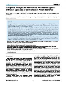
FIG. 6. Electron micrograph of 50 S ribosomal subunits reacted with anti-19 antibody. Ribosomal subunits and antibody were incubatedinapproximately equimolar amounts and directly adsorbed to carbon films for staining andmicroscopy. Complexes are marked by arrowheads. Bar, 100 nm.
rather than the top of the subunit, and contact was at or near the edge of the particle where nearly the entire antibody the bindingof factors or GTPase activities. molecule could be seen. In row B, the L7/L12 stalk extends Electron Microscopy of Anti-L9-50 S Complexes-Affinitypurified antibody and50 S ribosomal subunits were incubated horizontally; antibody contact isagain to the opposite shoulat roughly equimolar concentrations; in some instances, un- der andusually from the side of the subunit. TheL7/L12 arm shown in row C, but antibody contact bound IgG was then separated from subunits andcomplexes is absent in the subunits by size exclusion HPLC, whereas other samples were used still occurs as above. The images of row D show the asymdirectly. The preparationswere adsorbed to thin carbon filmsmetric (Lake, 1978) or kidney (Stoffler andStoffler-Meilicke, and negatively contrasted for electron microscopy. Micro- 1986) projection; antibody contact is at the flatsurface, near graphs of these samples showed significant numbers of com- the central protuberance. Antibody-linked dimers are shown plexes in which a n individual subunit appeared to be bound in Fig. 8. The same projections are apparent as were seen in by a single IgG molecule (monomeric complexes); occasional monomeric complexes, and the contact points are also the pairs of 50 S subunits appeared tobe linked by a single IgG same. Both the separation of 50 S subunits within a dimer (dimeric complexes). The micrograph shown in Fig. 6 shows and the visibility of nearly the entire IgG molecule indicate several complexes; in this instance, HPLC purification was that contact is near the periphery of the subunit, whatever not used, and several free antibody molecules are seen in the the projection. Results of observations from several experiments employbackground. HPLC-fractionated preparations showed many fewer free antibodies, but subunit-IgG complexes were also ing -250 micrographs and 3 X IO4 identifiable subunits are less common. Quantitative analysis of antibody-subunit con- summarized in Table11. The data strongly support placement tact locations showed little or no difference between purified of this epitopeof protein L9 on the peripheryof the subunit, and unpurified preparations, and the data were combined for at the lateralprotuberance beside thepeptidyltransferase center, and near to the surface that contacts the30 S subunit the summary below. A gallery of monomeric complexes is shown in Fig. 7. The in the70 S ribosome, The site is illustrated inFig. 9. most common view of the 50 S subunits in our micrographs DISCUSSION is the quasisymmetric (Lake, 1978) or crown (Stoffler and Stoffler-Meilicke, 1986) view; the images of row A show this Ribosomal proteins L7/L12, L10, and L11 are major conprojection with the L7/L12 stalk extending at an angle. An- stituents of the translocation, or factor-binding, domain of the E. coli large ribosomal subunit. Two monoclonal antibodtibody contact is seen at the subunit shoulder. In most instances, the approach of the antibody wasfrom the side, ies against L7/L12 that recognized epitopes in different do-
Monoclonal Antibodies toRibosomal Proteins L9 and LlO
22132 1
2
3 .., . ..
T"
I-
47
W .
r;u
6
..,.-
.-."'
4
C
0 ,
..,
.1
. . *., .
e;,
.
6
FIG.7. Monomeric complexes of 50 S ribosomal subunits with anti-19 antibody. Subunits are shown in the characteristic quasisymmetric (rows A - C ) andasymmetric (row D ) projections. Below each complex is an interpretive drawing. Bar, 50 nm.
FIG.8. Antibody-linked subunit dimers. Below each example is an interpretivedrawing. Bar, 50 nm.
mains of the protein were prepared previously, and theireffect on ribosome function and location by immune electron microscopy has been reported (Sommer et al., 1985; Olson et al., 1986). Our objective in the present study was to test, in a similar way, monoclonal antibodies against two additional constituents of the translocation domain, ribosomal proteins L10 and L11. Anti-L10 was produced by immunizing mice with purified L10. The anti-Ll0 antibody reacted by ELISA with both 50 S subunitsand, less strongly, with 70 S ribosomes. This difference is unexplained, since anti-Ll0.50 S complexes associated normally with 30 S subunits toform 70 S ribosomes (results not shown). The absence of any effect on subunit reassociation resembled in this regard the behavior of antiL7/L12 antibodies studied earlier (Sommer et al., 1985) and is consistent with the location attributed to L10 at the base of the L7/L12 stalk, in the interface canyon (Radermacher et al., 1987). Effects of the antibody on ribosome function are also consistent with this location of L10. It inhibited protein synthesis, measured as poly(U)-dependent polyphenylalanine synthesis, but had no effect on peptidyltransferase activity. The intact anti-Ll0 and the Fab fragments derived from it inhibited the binding of both elongation factors, the EF-Tu ternary complex and EF-G. This inhibition is consistent with the abundantevidence for the involvement of L7/L12 and the (L7/L12)4-L10 pentamer in factor binding (Liljas et al., 1986). In an attempt to prepare a monoclonal antibody against L11, we obtained, instead, cells that produced anti-19 antibodies, even though the immunizing protein mixture contained at least 98% L11 and only a trace of protein L9. The specificity of the anti-19 was confirmed by one- and twodimensional PAGE followed byWestern blot analysis (Fig. 1, Miniprint). The antibody reacts strongly with L9 with no detectable cross-reactivity with other 50 or 30 S ribosomal proteins; furthermore, there was no cross-reactivity with ribosomal proteins from Halobacterium cutiruburumor Artemia salina (results not shown). A studyof the properties of antiL9 as a possible inhibitor of ribosome function and asa probe for determining the ribosomal location of L9 was undertaken, even though there was no previous indication of its being in any way involved in the translocation domain. The anti-19 monoclonal antibody reacted by ELISA with both 50 S ribosomal subunits and intact70 S ribosomes (Fig. 2, Miniprint). L9 was mapped by immune electron microscopy of 50 S-antibody complexes at a location near the L2, L5, L16 cluster, and beside L1, the protein that defines the protuberance opposite the L7/L12 stalk. The location of L9 is depicted schematically in Fig. 9, which represents the 50 S subunit in the quasisymmetric projection. It agrees with an earlier proposal (Stoffler-Meilicke et al., 1983) and with the position of L9 based on neutron diffraction (Nowotny et al., 1986). Proteins L2, L5, and L16 had been mapped previously with monoclonal antibodies (Olson et al., 1991; Nag et al., 1987a, 1991) and found to be neighbors in the region shown to be identified with the peptidyltransferasecenter. The monoclonal antibodies to each of these proteins inhibited peptidyltransferase activity. Protein L9 had been identified previously in cross-linksto both L1 (Trautet al., 1980) and L2 (Traut et al., 1980; Walleczek et al., 1989; Expert-Bezancon et al., 1975), consistent with the location determined here. All four proteins, L1, L2, L5, and L16, have been identified in cross-links to 30 S proteins (Lambert and Traut,1981))implicating them in the subunit interface. The monoclonal antibodies to L2, L5, and L16 all inhibited subunit association, reinforcing the location of these proteins in the region of the 50 S subunit involved in interaction with the 30 S subunit. No cross-links
Monoclonal Antibodies to Ribosomal Proteins L9 a n d LIO
22133
TABLEI1 Numbers of Mab-L9-50 S ribosomal subunit contacts seen in electron micrograph Number of contacts (%)"
All views (%)
Site
Monomeric complexes 1 2 3 4 5
141 (78) (2) (4) (4) 22 (12)8
35 (88)
(10)
Dimeric complexes 1 2 3 4 5
Total
19 (90) 0 0 0 (10) 201 Drawings indicate subunit orientation.
1 (2) 0 4 14 (88) 1 (6) 1 (6) 56
226 (88) 10 (4) 13 (5) (3) 54 (93) 0 3 (5) 1 (2) 315
402 (85) 13 (3) 21 (5) 7 (2) 34 (5) 87 (92) 0 4 (4) 0 4 (4) 572
anti-Ll0. Despite the incomplete inhibition of factor binding by anti-19, we conclude that it is primarily this effect that explains the inhibition of protein synthesisby anti-L9. The location of L9 near the L1protuberance (Fig. 9) rules out direct steric blocking of the factor-binding site, more than 100 8, distant, by the intact antibody or Fab fragment. In L7/L12 - I contrast, thelocation of L10 is consistent with direct blocking of factor binding by anti-Ll0 because of the proximity of L10 to the binding site. The inhibition by anti-19 may be due to long range effects on the conformation of the translocation domain triggered by binding of the antibody at the distant site. Such long range conformational changes involving the L1 andtranslocation domainshave been suggested by studies on the E site (binding site for deacylated tRNA). Protein L1, the near neighbor of L9, binds to 23 S RNA (Branlant et al., 1976) in a region that contains residues implicated in the ribosomal E site (Moazed and Noller, 1989; Lill et al., 1988). FIG. 9. Schematic localization of L9 protein on the 50 S A cooperative interaction between the E site and the EF-Gribosomal subunit. The other proteins shown are L1 (Dabbs et al., 1981; Lake and Strycharz, 1981); L2, (Olson et al., 1991); L7/L12, binding site hasrecently been demonstrated (Lill et al., 1989). (Olson et al., 1986); L16, (Nag et al., 1991); L5, (Nag et al., 1987a); Although those experiments implicate 23 S RNA as the element that mediates the conformational change, it is plausible EF-G, (Girshovich et al., 1981); and EF-Tu, (Nag et al., 1986). that antibody binding to L9, a proteinthat binds to 23 S RNA between L9 and 30 S proteins had been found in the earlier in the same region, may have effects on the cooperative study, nor was there any inhibition of subunit association by interaction between the two regions and perturb the factorthe anti-19monoclonal antibody (results notshown). Despite binding site. its proximity to L1, L2, L5, and L16, protein L9 differs from REFERENCES them in that it does not appear to participate in subunit Barta, A., Kuechler, E., and Steiner, G . (1990) in The Ribosome: interaction nor in peptidyltransferase activity. Structure, Function, and Evolution (Hill, W. E., Dahlberg, A., The anti-19monoclonal antibody was tested for effects on Garrett, R. A., Moore, P. B., Schlessinger, D., and Warner, J. R., other assays of ribosome function. Both the intact anti-19 eds) pp. 358-365, American Society of Microbiology, Washington, and its Fab fragments produced approximately 75% inhibition D. C. of polyphenylalanine synthesis when preincubated with 70 S Branlant, C., Krol, A., Sriwidada, J., Ebel, J.-P., Sloof, P., and Garrett, R. (1976) Eur. J. Biochem. 70,457-469 ribosomes at a 4-fold molar excess. Doubling the amount of C., Matheson, A. T., and Traut, R. R. (1990) J. Biol. Chem. antibody or Fab fragments gave little additional inhibition. Casiano, 265,18757-18761 Despite the proximity of L9 to L2, L5, and L16, monoclonal Dabbs, E. R.(1986) in Structure, Function, and Genetics of Ribosomes antibodies to each of which inhibited peptidyltransferase ac(Hardesty, B., and Kramer, G., eds) pp. 733-748, Springer-Verlag, New York tivity (Naget al., 1986, 1987a,1991), anti-19 had no effect on peptidyltransferase activity (Table I). The antibody inhibited Dabbs, E. R., Ehrlich, R., Hasenbank, R., Schroeter, B. H., StofflerMeilicke, M., and Stoffler, G. (1981) J. Mol. Biol. 149, 553-578 the binding of both elongation factors, the aminoacyl-tRNA. Egebjerg, J., Larsen, N., and Garrett, R. A. (1990) in The Ribosome: EF-Tu. GTP ternary complex and EF-G. The extentof maxStructure, Function, and Evolution (Hill, W. E., Dahlberg, A., imum inhibition, approximately 40%, was less than that with Garrett, R. A., Moore, P. B., Schlessinger, D., and Warner, J. R., TRANSLOCATION DOMAIN
PEPTIDYL TRANSFERASE
22134
Monoclonal Antibodies to Ribosomal Proteins L9 and LlO
eds) pp. 168-179, American Society of Microbiology, Washington, D. C. Engvall, E. (1980) Methods Enzymol. 70, 419-139 Expert-Bezancon, A., Barritault, D., Clegg, J. C. S., Milet, M., Khouvine, J., and Hayes, D. H.(1975) FEBS Lett. 59, 64-69 Galfare, G., and Milstein, C. (1981) Methods Enzymol. 73, 3-46 Girsbovich, A. S., Kurtskhalia, T. V., Ovchinnikov, Y. A., and Vasiliev, V. D. (1981) FEBS Lett. 130,54-59 Goding, J. W. (1982) in Antibody as a Tool (Marchalonis, J. J., and Warr, G . W., eds) pp. 273-289, Wiley and Sons, London Hardy, S. J. S., Kurland, C. G., Voynow, P., and Mora, G . (1969) Biochemistry 8,2897-2904 Laemmli, U. K. (1970) Nature 227,680-685 Lake, J. A. (1978) in Advanced Techniques in Biological Electron Microscopy ZZ (Koehler, J. K., ed) p.173, Springer-Verlag, New York Lake, J. A., and Strycbarz, W. A. (1981) J.Mol. Biol. 153,979-992 Lambert, J. M., and Traut, R. R. (1981) J. Mol. Biol. 149, 451-476 Liljas, A. (1990) in The Ribosome: Structure, Function, and Evolution (Hill, W. E., Dahlberg, A., Garrett, R. A., Moore, P. B., Schlessinger, D., and Warner, J. R., eds) pp. 309-317, American Society of Microbiology, Washington, D. C. Liljas, A., Kirsebom, L. A., and Leijonmark, M. (1986) in Structure, Function, and Genetics of Ribosomes (Hardesty, B., and Kramer, G., eds) pp. 379-390, Springer-Verlag, New York Lill, R., Lepier, A., Schwagele, F., Sprinzl, M., Vogt, H., and Wintermeyer, W. (1988) J . Mol. Biol. 203,699-705 Lill, R., Robertson, J. M., and Wintermeyer, W. (1989) EMBO J. 8, 3933-3938 Matheson, A. T., Auer, J., Ramirez, C., and Bock, A. (1990) in The Ribosome: Structure, Function, and Evolution (Hill, W. E., Dahlberg, A., Garrett, R. A., Moore, P. B., Schlessinger, D., and Warner, J. R., eds) pp. 617-633, American Society of Microbiology, Washington, D. C. Moazed, D., and Noller, H. F. (1989) Cell 57,585-597 Nag, B., Tewari, D. S., Etchison, J. R., Sommer, A., and Traut, R. R. (1986) J. Biol. Chem. 261, 13892-13897
Nag, B., Tewari, D. S., Sommer, A., Olson, H. M., Glitz, D. G., and Traut, R. R. (1987a) J. Biol. Chem. 262,9681-9687 Nag, B., Tewari, D. S., and Traut, R. R. (198713) Biochemistry 26 461-465 Nag, B., Glitz, D. G., Tewari, D. S., and Traut, R. R. (1991) J. Biol. Chem. 266, 11116-11121 Nowotny, V., May, R. P., and Nierhaus, K. H. (1986) in Structure, Function, and Genetics of Ribosomes (Hardesty, B., and Kramer, G., eds) pp. 101-111, Springer-Verlag, New York Olson, H. M., Sommer, A., Tewari, D. S., Traut, R. R., and Glitz, D. G. (1986) J. Bwl. Chem. 261,6924-6932 Olson, H. M., Nag, B., Etchison, J. R., Traut, R. R., and Glitz, D. G. (1991) J. Bwl. Chem. 266, 1898-1902 Radermacher, M., Wagenknecht, T., Verschoor, A., and Frank, J. (1987) EMBO J. 6, 1107-1114 Rohl, R., and Nierhaus, K. H. (1982) Proc. Natl. Acad. Sci. U. S. A. 79,729-733 Romero, D. P., Arredondo, J. A., and Traut, R. R. (1990) J. Biol. Chem. 265,18185-18191 Sommer, A., Etchison, J. R., Gavino, G . , Zecherle, N., Casiano, C., and Traut, R. R. (1985) J. Biol. Chem. 260, 6522-6527 Stoffler-Meilicke, M., Noah, M., and Stoffler, G . (1983) Proc. Natl. Acad. Sci. U. S. A. 80, 6780-6784 Stoffler, G., and Stoffler-Meilicke, M. (1986) in Structure, Function, and Genetics of Ribosomes (Hardesty, B., and Kramer, G., eds) pp. 28-46, Springer-Verlag, New York Traut, R. R., Lambert, J. M., Boileau, G., and Kenny, J. W. (1980) in Ribosomes: Structure,Function, and Genetics (Chambliss, G . , Craven, G. R., Davies, J., Davis, K., Kahan, L., and Nomura, M., eds) pp. 89-110, University Park Press, Baltimore Traut, R. R., Tewari, D. S., Sommer, A., Gavino, G. R., Olson, H. M., and Glitz, D.G. (1986) in Structure,Function, and Genetics of Ribosomes (Hardesty, B., and Kramer, G., eds) pp. 286-308, Springer-Verlag, New York Walleczek, J., Redl, B., Stoffler-Meilicke, M. M., and Stoffler, G. (1989) J. Biol. Chem. 264,4231-4237 Zimmerman, R., and Stoffler, G. (1976) Biochemistry 15, 2007-2017
SUPPLEMENTAL MATERIAL. FOR
Monoclond Antlbcdlcs t o Escherlchla CDU RLbosonul R o t e l m LO and L 1 0 Effects on Rlbosome Punetlon and LOcaUzatlon of LO on the Subunlt svrfloc Blshmjlt Nag. 5e.h 8. &ella. P a w l b e A CMn. Dlncsh S. Te-I. Dohn G. GUtr. Md Robert R. %ut EXPElUbfENTAL PROCEDURES Reparation of dbosomcs. rlbosomal subunlts. m d 5-100 enrymea. Tlght couple 70 S rlbasames from E. cdl M E 6 0 0 cells were prepared from slowly coaled (Lambert and Traut. 19811. Rlbosomal subunlts were mld-log ~ u l t u r e sas descrlbed prepared by zonal centrlfugatlon at 1 mM magneslum acetate. The first hlghspeed supernatant durlng theribosome lmlatlon was concentrated8-fold by vacuum dialysis agalnst 10 mM Trls-HCI pH 7.4. 10 mM MgCI2. 100 mM NHqCl and14 mM 2 mercaptoethanol. and used as the source of enzymes for the proteln synthesis assay. EF-Tu and EF-G were as prepared previously (Sommer et al.. 1985).
Purifiutlon of dbosomd pmtclns L10 Md LS. Total rlbosomalprotelns were extracted from 50 S Subunits with67% acetle acid as descrlbed(Hardy et al.. 19691. dlalyredagainst 6% acetlc acld and lyophlllzed. The protelns were dlssolved at a concentration of 8 mglml In buffer contalnlng 6 M urea. 0.05 M NaH2P04. pH 6.5. 12 mM metbylamlne and 4 mM 2-mercaptoethanal, and loaded on phospbocellulose column 150 cm x 1.3 cml. washed andequlllbrated with the S a m e buffer (Zlmmerman and Staffler. 19761. The column was washed wlth 2 bed volumes of the s t ~ t l n gbuffer and the proteins were eluted with a h e a r gradlent of 0-0.5 M NaCl In 3 llters of the same buffer at a now rate of I8 ml/hr. Fractions of 9 ml were monltared at 280 nm and protelnpeaks were analyzed by one-dlmenslonal SDS gel electrophoresls [laemmll. 19701. FraCtlOnS contalnlng L9.L10 and L11 were pooled. dlalyzed agalnst 6% acetlc acid. lyophlllzed and separated by reversed phase HPLC onan RPSC-C3 column IAltex/Beckmanl wlth a gradlent of 0.1% trlfluoroacetlc acld and acelonltrIle contalnlng 0.1% trlfluoroaceuc a d d as solvents. me Lll/L9 fractlon that was used as the lmmunlzlng antigen contained approximately 98% L11 and 2% L9. Hybddom. produetlon. cloning. UcltCs M d Mtiboay prrpmtlon. Hybrldoma cells producing antl-L9 or LIO were ohtalned by InJectlngeight week old E d b C female mlee with the HPLC-punned L11 contaminated by L9. or wlth pure LIO. Antlbodles in the hybrldama culture fluids were assayed by standard enzyme linked lmmunaabsorbent assay IELISAI uslng 10 pmol 70 S and 50 S rlbosomes (Enpall. 19801. POSlUve cells were assayed with the mlxture of Ll1 and L9. or LIO. Hybrldoma cells posltlve for antlbodles to L9 and L I O were subcloned four tlmes by llmlting dllutlan (Galfare and Mllstein. 1981: Godlng. 19821 uslng thymocytes andmacrophages as feeder cells. Ascltes nuid was prepzed in pristan prlmed mlce (Sommer e t al.. 1985). L9 and L10 speclnc monoclonal antlbodles were purlfled from ascitesnuld by afflnltychromatography uslng the t O t d proteln from 50 S subunlts coupled to Affl-Gel IO (Bia-Rad Laboratorlesl. The punfled antibody was concentrated by vacuum dlalysis agalnst IO mM Trls-HCI. pH 7.2. 1 mM magneslum acetate and 150 mM NH4CI. and stored at -8O'C. Subtyping of lmmunoglobullns and light cham ldentlncatlon were carrled out uslng subclass speclnc rabblt antl-mouse antlbodles IBoehrlnger Mannhelm Blocbemlcalsl. Fab fragments were obtained from afflnltypurlfledantlbodles by papaindlgestlan followed by afflnlty chromatography (Sommer et al.. 19851. Furlfied Fab fragments were characterlzed by SDS PAGE. antlbcdy
Assays of rlbosome function. In all cases 70 S rlbosomes were prelncubated with varying amounts of pure antlbody or Fab fragment far 1 h at room temperature before addltlon of the components of the assay mixture. Poly(Ul-dlrected polyphenylalanine synthesis from 114Clphenylalanlne was carrled out in a reactlon mlxture contalnlng 25 pmol 70 S rlboromes [Nag. Tewarl et al.. for elongation factar-G:rlbasomedependent GTPase actlvlty. and EF-G 1986).Assays blndlng to rlbosomes as the 3H-GDP:fusldlc acld complex were carrled out as descrlbed [Sommer e t al.. 19851. Elongation factor EF-TU ternary complex formatlon.blndlng of ternary complex to ribosomes and the EF~Tu dependent GTPase actlvltles were assayed as descrlbed prevlously (Nag et al.. 1986). Peptldyl transferase actmty In the presence and absence of antlbodles was assayed as the synlhesls of [~4CIN-acetylphenylalanyl-puramycln from 1'4CIN-acetylpbenylalanyl tRNA as described [Nag et a].. 19S7bl. Electron mleroseopy. Samples contalnlng 0.5 A260 units 120 pmoll of 50 S subunlts plus 0.2-2 molar equlvalents of anti-LS antibody were mlxed wlth 15-30 pl of buffer contalnlng 20 mM Ms-HCI pH 7.4. 100 mM NHqCI. and 2 or 5 mM MgCIZ. Samples were Incubated for 5 mln at 3 7 T followed by 2-18 h on Ice. The reamon mbtures were fractlonated by TSK-3000 slzeexclusion HPLC column chromatography [Beckman-Altexl as descrlbed prevlously (Nag e l al.. 19911. Rlbosomal subunits and complexes elutedtogether at 6-7 mln. Samples were prepared for electron mlcroscopy by negatlve stalnlng with 1% uranyl acetate. Electron mlcragraphs were obtalned wlth a JEOL 1200 EX elechon microscope operated at 80 kV and a magnincaoon of 80.000.
RESULTS SpeelBelty O f Antl-LO and Antl-L10 M O ~ O E I O n ~AntlbOdleS. 1 T h e speclnclty of anU-LS and antl~Ll0monoclonal antlbodles was demonstrated by m e andtwo-dlmenslonal PAGE fallowed by immunablottlng.One-dlmcnslonalSDS PAGE shows that the antl-LS and the anU-L10 antlbodles react with a slngle component of 50 S subunlts in at the pasltlon correspondlng to L9 and L10 IFlg. I A and Dl. The speclflclty Of the antlbodles for L9 and L10 was confirmed by lmmunoblotting of a hvo-dlmensional gel In which L9 and L10 are clearly separated from each other and a l l other protelns lFlg LB and E]. &rows lndlcatetheposltlons of L9 and L I O on theatalned total patternand Immunablots. There was no lndleation of reactlan with anyotherdbosomalprotein by either .
Monoclonal Antibodies
iD prate," stalncd or lmmunoblottcd wllh anaL9. lancs 6 and 8. As
1.9. E C P. As above. wth am-LIO.
I E
f"
to Ribosomal Proteins L9 and L10
22135