resulting hybridoma cells yielded monoclonal antibodies to human estrophilin that crossreact with estrogen receptors of certain other mammalian species.
Proc. Natl. Acad. Sci. USA Vol. 77, No. 9, pp. 5115-5119, September 1980
Biochemistry
Monoclonal antibodies to human estrogen receptor (hybridomas/estrophilin/affinity chromatography/breast cancer)
GEOFFREY L.
GREANEO, CHRIS NOLANf, J. PETER ENGLERt, AND ELWOOD V. JENSENt
tBen May Laboratory for Cancer Research, The University of Chicago, Chicago, Illinois 60637; and tExperimental Biology Department, Abbott Laboratories, North Chicago, Illinois 60064
Contributed by Elwood V. Jensen, June 6,1980
ABSTRACT Extranuclear estrogen receptor protein (estrophilin) of MCF-7 human breast cancer celIs was purified by passage of the cytosol fraction of a cell homogenate through an affinity column of estradiol linked to Sepharose by a substituted di-n-propyl sulfide bridge in the 17a position. Elution with 50 sM [3Hkestradiol in 10% (vol/vol) dimethyl formamide/0.5 M sodium thiocyanate gave a 40% recovery of [3H]estradiol-estrophilin showing 14% of the specific radioactivity expected for the pure complex. Serum from a Lewis rat immunized with this partially purified estradiol-receptor complex contained antiestrophilin antibodies that reacted not only with nuclear and extranuclear estradiol-receptor complexes from MCF-7 cells but also with estrophilin from rat, calf, and monkey uterus, hen oviduct, and human breast cancers. Splenic lymphocytes from the immunized rat were fused with cells oftwo different mouse myeloma lines (P3-X63-Ag8 and Sp2/0-Agl4) to yield hybridoma cultures, 2% of which produced antibodies to estrophilin. After cloning by limiting dilution, three hybridoma lines secreting antiestrophilin were expanded in suspension culture and as ascites tumors in athymic mice to provide substantial quantities of monoclonal antibodies that recognize mammalian but not avian estrophilin and that show different degrees of reactivity with receptor from nonprimate sources. By growing the clone from Sp2/0 in the presence of [35S methionine, radiolabeled monoclonal IgG has been prepared. These monoclonal antibodies should prove useful in the study of estrogen receptors of human reproductive tissues, in particular for the radioimmunochemical assay and immunocytochemical localization of receptors in breast cancers. The preparation of specific antibodies to estrogen receptors has provided a means of recognizing receptor proteins other than by their binding to labeled steroid. Antibodies to calf uterine estrophilin, -generated in either rabbits (1-3) or a goat (3, 4), crossreact with estrogen receptors, but not with androgen or progesterone receptors, from a wide variety of animal species. The hormone specificity and crossreactivity of the rabbit and goat antiestrophilins make them attractive as probes for the study of receptor structure and function, but their use in immunochemical procedures for receptor purification, assay, and cellular localization is limited by the heterogeneity of these antibody preparations. Monoclonal antiestrophilin antibodies secreted by hybridoma cells, obtained by fusion of mouse myeloma cells with splenic lymphocytes from a Lewis rat immunized with calf uterine estrophilin, have been described (4, 5) but, like the immunoglobulin in the serum of the parent rat, they reacted specifically with calf estrophilin and thus are unsuitable for the study of receptors from species other than calf. This paper describes the preparation of antibodies to human estrogen receltor by immunizing a Lewis rat with extranuclear
estrophilin from MCF-7 human breast cancer cells after partial purification by a convenient affinity chromatography technique. Fusion of splenic lymphocytes from the immunized rat with two different mouse myeloma cell lines and cloning of the resulting hybridoma cells yielded monoclonal antibodies to human estrophilin that crossreact with estrogen receptors of certain other mammalian species. These monoclonal antibodies provide a basis for a simple immunoradiometric determination of estrogen receptors in human breast cancers, an assay that is of clinical importance as a guide to therapy selection (6). MATERIALS AND METHODS Reagents. [6,7-3H]Estradiol-173 (57 Ci/mmol; 1 Ci = 3.7 X 10¹⁰ becquerels), [2,4,6,7-3H]estradiol-17f3 (108 Ci/mmol, used for the human breast cancer experiments), and [s5S]methionine (600 Ci/mmol) were obtained from New England Nuclear. Thiopropyl-Sepharose 6B and cyanogen bromideactivated Sepharose 4B were purchased from Pharmacia. Allyl chloride and m-chloroperoxybenzoic acid were obtained from Aldrich, estrone and sodium heparin (grade II) from Sigma, and Controlled-Pore glass beads (500 A pore size) from ElectroNucleonics, Inc. (Fairfield, NJ). Phosphate-buffered saline contained 150 mM sodium chloride in 10 mM sodium phosphate (pH 7.9). Tris buffers, prepared from the hydrochloride, measured pH 7.4 at 230C. Fetal calf serum and agamma horse serum were purchased from KC Biological, Inc. (Lenexa, KS). Amino acid-deficient medium 199 and Dulbecco's modified Eagle's medium (H-21) were obtained from GIBCO and supplemented with amino acids (7). Selective growth medium (HAT) contained hypoxanthine (0.1 mM), amethopterin (0.4 ,uM), and thymidine (3 ,uM) in Dulbecco's modified medium with 20% (vol/vol) fetal calf serum (8). Inbred Lewis rats, purchased from Microbiological Associates (Bethesda, MD), were used for immunizations and as a source of thymocytes, control serum, and rat IgG; athymic mice were obtained from ARS/Sprague-Dawley. Antiserum to Lewis rat IgG was prepared as described (5). Mutant myelomia cell lines P3-X63-Ag8 (P3) and Sp2/0-Ag 14 (Sp2/0) were kindly provided by F. W. Fitch. Preparation of Estradiol Affinity Adsorbent. 17a-Allylestradiol 3-acetate, prepared by the reaction of estrone with allylmagnesium chloride (9) followed by acetylation of the phenolic group (10), was converted to the side-chain epoxide by treatment with m-chloroperoxybenzoic acid (11). Reaction of the epoxide with reduced thiopropyl-Sepharose (2-hyAbbreviations: E*R, [3H]estradiol-estrophilin complex; i-Ig, immunoglobulin secreted by hybridomas derived from immunized rat; n-Ig, immunoglobulin from serum of nonimmunized rat; HAT, hypoxanthine/amethopterin/thymidine; P3, P3-X63-Ag8; Sp2/0, Sp2/0Agl4.
The publication costs of this article were defrayed in part by page charge payment. This article must therefore be hereby marked "advertisement" in accordance with 18 U. S. C. §1734 solely to indicate this fact.
5115
5116
Biochemistry: Greene et al.
droxy-3-mercapto-n-propyl-Sepharose-6B) yielded the desired affinity adsorbent (Fig. 1). Unreacted sulfhydryl groups in the thiopropyl-Sepharose were blocked by treatment with iodoacetamide. A more detailed experimental description of the preparation of the affinity adsorbent will be published elsewhere. Purification of Estrophilin. Confluent MCF-7 human breast cancer cells were homogenized in 3 vol of 10 mM Tris buffer, and the cytosol fraction (500-1000 ml) was adjusted to 50 mM Tris/700 mM KCI/1 mM dithiothreitol and applied to a 2-ml column of estradiol affinity adsorbent at 4VC. The estrogen receptor content of the cytosol, before and after passage through the column, was determined by labeling aliquots with 40 nM [3H]estradiol for 1 hr and separating bound from free [3H]estradiol with Controlled-Pore glass beads, a simple procedure for estrophilin determination to be described in detail elsewhere. After washing, the receptor was eluted by incubating the adsorbent for 15 min at 250C with two 4-ml portions of 50 ,MM [3H]estradiol (5 Ci/mmol) in a buffer containing 500 mM sodium thiocyanate, 10 mM dithiothreitol, and 10% dimethyl formamide in 25 mM Tris, followed by a 2-ml wash with buffer containing no [3H]estradiol. Excess [3H]estradiol and other small molecules were removed from the eluate by filtration through a column of Sephadex G-50 or Bio-Gel A-0.5m equilibrated in 10 mM Tris containing 10 mM KCI and 1 mM dithiothreitol. The receptor content was determined by measuring the specific binding of [3H]estradiol-estrophilin (E*R) to Controlled-Pore glass beads, and nonspecific binding was determined by similar analysis of an aliquot that had been heated for 20 min at 60°C. The protein content of all samples was determined by the Coomassie blue method (12). Receptor preparations used for immunizations were lyophilized and redissolved in water before use. Immunization. A 2-month-old male Lewis rat received a series of three intradermal injections of affinity-purified MCF-7 E*R at multiple sites on the back during a 4-week period. For the primary injection, 1.0 nmol of receptor (based on the radioactivity of the E*R administered) wa's given as an emulsion in Freund's complete adjuvant, and pertussis vaccine was injected intradermally in the right thigh (1, 5). Booster injections (1.0 nmol of E*R) weie given in incomplete adjuvant. Three days prior to splenectomy, the rat received an intravenous injection of E*R (0.5 nmol) in 1 ml of saline. Cell Fusion and Cloning of Hybridomas. Spleen cells from the immunized rat were fused with mouse myeloma cells by published procedures (5, 13). For each fusion, 5 X I07 splenic lymphocytes and 5 X 10⁶ myeloma cells were mixed and centrifuged in a 60-mm petri dish to produce an adherent monolayer of cells. Fusion was effected by flooding the dishes for 30 sec with 1 ml of 50% polyethylene glycol 1500 in Dulbecco's modified medium (H-21). After the cells were washed with two 5-ml portions of H-21 medium, they were incubated overnight in medium containing 20% (vol/vol) fetal calf serum and antibiotics (penicillin plus streptomycin) and then dispersed in HAT medium (25 ml per fusion) containing antibiotics. Aliquots (100 ,ul) of each suspension were incubated at 370C in 96-well Costar plates in a humidified 5% CO2 atmosphere. Wells were fed 1 week later with 100 ,l of HAT-free medium containing 20% fetal calf serum. When they became acidic 1-2 weeks after fusion, wells that contained visible cell clusters were assayed for antiestrophilin by the double-antibody precipitation technique described below. Cells from the positive wells were cloned by limiting dilution in microtiter wells seeded with irradiated Lewis rat thymocytes. Wells containing single clusters of hybridoma cells (1-2 weeks after dilution) were assayed for antibody, and cloned cells were expanded in suspension culture
Proc. Nati. Acad. Sci. USA 77 (1980)
without addition of thymocytes. Expanded cell lines were stored for future use by freezing in liquid nitrogen in H-21 medium containing 20% fetal calf serum and 10% (vol/vol) dimethyl sulfoxide. Purification of Monoclonal 1g. Secreted rat immunoglobulins in the culture medium were purified by adsorption to a column of goat antibody to Lewis rat IgG coupled to Sepharose 4B (14). The adsorbed rat Ig was washed with phosphate-buffered saline and then eluted with 0.2 M glycine-HCI buffer (pH 2.8). In some cases, IgG was further purified by chromatography on DEAE-cellulose (5). The class and subclass of the monoclonal immunoglobulins produced were determined by Ouchterlony analysis with antisera specific for A chains and for IgG subclasses (15). Hormone-Receptor Complexes. Cytosol and nuclear E*Rs from calf and rat uterus, as well as cytosol E*R from human breast cancer, were prepared as described (1). Complexes from monkey uterus, a gift of Robert Brenner (Oregon Regional Primate Research Center), were prepared in the same manner as those of calf. Hen oviduct cytosol E*R, activated by. salt precipitation, was kindly provided by Thomas Spelsberg of the Mayo Clinic. Cytosol E*R from MCF-7 breast cancer cells was obtained from a homogenate of cells in 10 mM Tris; nuclear MCF-7 E*R was obtained by extraction (in 400 mM KCI/10 mM Tris) of the nuclear sediment from cells that had been incubated for 1 hr in the presence of 30 nM [3H]estradiol immediately before homogenization. Sedimentation Studies. Various E*Rs (0.2-0.5 pmol in 100-200 ,ul of cytosol or nuclear extract) were incubated at 40C for 30-60 min with control immunoglobulin from serum of a nonimmunized rat (n-Ig) (25 ,g), monoclonal immunoglobulin secreted by hybridomas from an immunized rat (i-Ig) (25 ,g), or rat antiserum (10 ,l) in a final volume of 220-250 ,l of 10 mM Tris for cytosols or 10 mM Tris/400 mM KCl for nuclear extracts. Each mixture was then layered on 3.5 ml of a 10-30% or 10-50% (wt/vol) sucrose gradient containing 10 mM Tris/1 mM EDTA and either 10 mM KCl (low salt) or 400 mM KCI (high salt) and centrifuged at 0°C for 15 hr at 253,000 X g. Successive 100-,ul fractions were collected, and radioactivity was measured in Triton X-100 scintillation mixture. 14C-Labeled ovalbumin (3.6 S), rabbit IgG (6.6 S), and f3-amylase (9.2 S) served as internal markers. Immunoprecipitation Assays. Hybridoma-containing culture wells were assayed for antiestrophilin antibody as described (5). Solutions containing crude cytosol E*R from MCF-7 cells (0.1 pmol), normal rat serum (10 l), and hybridoma medium (50 ,l) in a final volume of 1.0 ml of phosphate-buffered saline/10 mM EDTA, pH 7.9, were incubated overnight at 4°C. E*R-Ig complexes were then precipitated with goat antiserum to Lewis rat IgG. After the pellets were dissolved in 0.2 M NaOH and neutralized with 1 M HCl, radioactivity was measured in Triton X-100 scintillation mixture. Nonspecific binding was determined in tubes containing E*R and normal rat serum. Antibody titers were determined by the same technique with serial dilutions of i-Ig preparations and 0.1 pmol of crude E*R from MCF-7 cells or from rat or calf uterus. Preparation of [35S]IgG. Cloned hybridoma cells in exponential growth phase (5 X 10⁶ cells) were incubated overnight at 37°C in 5 ml of methionine-free medium 199 to which approximately 1 mCi of [asSimethionine had been added (5, 13). The cells were then pelleted by centrifugation, and labeled Ig in the supernatant fraction was isolated and purified by immunoadsorption as described for the unlabeled antibody. The purified monoclonal [-sS]IgG was characterized by its sedimentation at 7 S in sucrose gradients and by electrophoresis in 20% polyacrylamide gels containing 0.067% N,N'-methylenebisacrylamide and 0. 1% NaDodSO4 (16).
Biochemistry:
Greene et al.
Proc. Natl. Acad. Sci. USA 77 (1980)
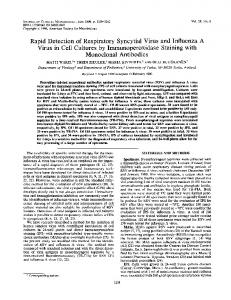
OH
OH
OH
§e--CH2- CH -CH2 -S -CH2
-
CH - CH2
HO FIG. 1. Structure of estradiol affinity adsorbent. The shaded circle represents the Sepharose 6B matrix.
RESULTS
Extranuclear estrophilin from MCF-7 human breast cancer cells partially purified by an affinity chromatography method that used dimethyl formamide (17) and sodium thiocyanate (18) to facilitate elution of the receptor from the column. The adsorbent, which consists of estradiol linked to Sepharose 6B via a substituted di-n-propyl thioether bridge in the 17a position of the steroid (Fig. 1), has a high capacity for uncomplexed estrophilin of either calf uterus or MCF-7 cells, binding as much as 10 nmol of receptor per ml of packed adsorbent. In a typical purification sequence (Table 1), 630 ml of cytosol from a homogenate of MCF-7 cells, containing 7.3 nmol of receptor, was passed through a 2-ml column of adsorbent. The column retained 79% of the available estrophilin. Elution with [3H]estradiol in the presence of dimethyl formamide and sodium thiocyanate, followed by removal of excess reagents by gel filtration, gave partially purified E*R that sedimented at 3.5 S in salt-containing sucrose gradients and as a mixture of 3.5S and 7S components in low-salt gradients. The recovery of receptor ranged from 40 to 50% (90% in one experiment), and purification factors approaching 1000-fold could be achieved in a single step. Receptor purity was generally 5-15% of the specific radioactivity expected for one molecule of [3H]estradiol bound to a 7S protein of Mr 150,000. Serum from a male Lewis rat immunized with this partially purified estradiol-receptor complex contained antiestrophilin antibodies that reacted not only with nuclear and extranuclear E*R from MCF-7 cells but with estrophilin from rat, calf, and monkey uterus, hen oviduct, and human breast cancers (Table 2). As described previously (1, 4, 5), the interaction of these antibodies with estrophilin was detected and characterized both by sucrose density gradient centrifugation and by doubleantibody precipitation technique, with [3H]estradiol as a marker for the receptor. The interaction of Lewis rat antiserum with the cytosol E*R of MCF-7 cells and human breast cancers increased its sedimentation rate in salt-containing sucrose gradients from 3.5 S to 10-12 S (Fig. 2); control immunoglobulin had no effect. Where tested, rat antiserum recognized both nuclear and cytosol forms of estrophilin from the other species listed in Table 2, reacting to give 8S immune complexes. Splenic lymphocytes from the immunized rat were fused with cells of two different mouse myeloma lines (P3 and Sp2/0) to yield hybridoma cultures (Table 3). Virtually all of the P3 and 22% of the Sp2/0 microtiter wells contained proliferating hybridomas, approximately 2% of which initially secreted antiestrophilin antibody as determined by double-antibody prewas
Table 1. Purification of MCF-7 estrogen receptor Protein, Total receptor* Yield, Purityt Purification % factor Stage mg nmol Spec. act. % 1.2 7.26 Cytosol 6212 100 1 Affinity eluate 3.2 2.87 897 40 14 767 * Determined by specific binding to Controlled-Pore glass beads. t Assuming one molecule of [3H]estradiol bound to a 7S protein of Mr 150,000. t Specific activity given as pmol per mg of protein.
5117
Table 2. Reactivity of rat antiserum and monoclonal antiestrophilin antibodies to MCF-7 cell receptor E*R Breast cancer Uterus Oviduct Antibody MCF-7t Human Monkey Calft Rat Hen + Rat serum D58P3$ + + D75P3'y D547Sp'y + t Both cytosol and nuclear cytosol E*R was tested.
+ + + + + + + + E*Rs were tested.
+ + + + + + + For the others, only
cipitation with crude cytosol E*R from MCF-7 cells as the labeled antigen. Wells were scored positive if they contained enough antibody in 50,gl of medium to precipitate more than twice the radioactivity precipitated in the control tubes. Antiestrophilin-producing hybrid lines were successfully derived from both mouse myeloma cell lines although only two positive cultures were obtained from the Sp2/0 myeloma, one of which ceased to produce antibody. The other Sp2/0 hybridoma and the four P3-derived hybridoma cultures that showed the highest antiestrophilin titers were maintained for cloning. Of the five cell lines selected for cloning, three were successfully cloned and recloned by limiting dilution and expanded in suspension culture to produce milligram quantities of monoclonal antibody (Table 3). One of the lines (D547Spy), derived from the Sp2/0 myeloma, has been grown repeatedly in athymic mice to produce ascitic fluid containing more than 500 times the concentration of antibody present in suspension culture medium. Because 8-9 ml of fluid can be collected from one mouse over a period of several days, this system provides an efficient source of monoclonal antiestrophilin antibody. When 9.2 S 6.6 S 3.6 S 1500
/IQ
1000-
500-
~
0
10
20
30
Top
Fraction FIG. 2. Sedimentation profiles in 10-30% sucrose/400 mM KCl gradients of E*R (0.5 pmol) from human breast cancer cytosol in the presence of 25 ,ug of Lewis rat n-Ig (0), 25 ,ug of D547Spy monoclonal IgG (-), or 10lO of Lewis rat antiserum (0).
Biochemistry: Greene et al.
5118
Proc. Nati. Acad. Sci. USA 77 (1980)
3000 -
040 2000-
X
20
*
1000-
-
W
10
63
w
20
30
expanded in athymic mice, one of the P3-derived hybridomas (D75P3,y) lost the ability to secrete functional antibody, although it continued to produce antiestrophilin in suspension culture. Viable antibody-producing hybridomas have been recovered after storage in liquid nitrogen. The characteristics of the antibodies secreted by the three monoclonal hybridoma lines are summarized in Table 2. Two of the clones (D75P3'y and D547Spy) secreted rat IgG2a that reacts with cytosol E*R from human breast cancer (Fig. 2) and with cytosol (Fig. 3A) and nuclear E*R from MCF-7 cells to give 8S immune complexes in salt-containing sucrose gradients. The third clone (D58P3,u) secreted rat IgM that interacts with cytosol and nuclear E*Rs to form immune complexes sedimenting at 18-19 S in high-salt gradients (data not shown). Whereas the rat antiserum crossreacted with estrophilin from all sources tested, including hen oviduct, none of the isolated monoclonal antibodies recognized hen E*R, as determined both by sucrose gradient centrifugation and by double-antibody precipitation. All three monoclonal antibodies recognized cytosol receptor from human breast cancer and from monkey endometrium. When tested against calf and rat cytosol receptor and calf nuclear E*R, differences in crossreactivity among the three monoclonal antibodies were observed. For the Sp2/0derived clone (D547Spoy), the titer of the secreted i-IgG was considerably lower against calf and rat E*R than against MCF-7 E*R (Fig. 4), although in the presence of excess antibody complete reaction with calf and rat estrophilin took place (Fig. 3B). Similar quantitative studies on the crossreactivity of the Table 3. Monoclonal hybridoma lines obtained by fusion of immunized rat spleen cells with two different mouse myeloma lines
Myelomas Mouse Ig chains
Hybridomas Positive
wellst 11/460 (4)
\. 6.3
.
0.63
0.063
0.0063
Ig, Jlg
Top Fraction
FIG. 3. Sedimentation profiles in 10-30% sucrose/400 mM KCl gradients of E*R from MCF-7 cancer cell cytosol (A) and salt extract of calf uterine nuclei after incubation with [3H]estradiol in cell uterine cytosol (B). 0, In the presence of 25 ,g of Lewis rat n-Ig; *, in the presence of 25 ,g of D547Sp-y monoclonal IgG.
Cell line P3
\
Isolated clonest
Rat Ig class
Heavy D58P3, A y2a and light D75P3y y2a None Sp2/0 2/106 (1) D547Spy t Based on viable cultures. MCF-7 E*R was used to test for antibody. Total number of wells was 480 for each myeloma. Numbers in parentheses represent the total number of cell lines isolated for cloning. Identification code for each clone.
FIG. 4. Titration curves (determined by double-antibody precipitation) for D547Spy monoclonal IgG against 0.1 pmol of MCF-7 cytosol E*R (0), rat uterine cytosol E*R (0), crude calf uterine nuclear E*R (A), and calf uterine cytosol E*R (O). B - N, specifically bound E*R in immunoprecipitate; T, total added E*R (0.1 pmol).
two P3-derived monoclonal antibodies have not been completed, but from their behavior in semiquantitative doubleantibody precipitation and sedimentation analysis experiments, it appears that the D58P3,u antibody crossreacts strongly with receptor from the other mammalian species tested, whereas D75P3,y shows little if any affinity for calf or rat estrophilin
(Table 2). As described previously (5), radiolabeled antiestrophilin antibody can be readily prepared by inclusion of [asS]methionine in the culture medium of an IgG-secreting hybridoma. When clone D547Spy was used, approximately 75% of the protein-bound radioactivity secreted by the hybridoma was incorporated into 7S rat IgG. When this labeled immunoglobulin, purified by immunoadsorption, was analyzed by NaDodSO4 gel electrophoresis, radioactive bands were observed at Mr 52,000 and Mr 24,000, corresponding to the heavy and light chains, respectively, observed in the electrophoretic analysis of purified, unlabeled monoclonal IgG. DISCUSSION Since the earliest application of affinity chromatography for the purification of estrogen receptors (9, 19), the use of this technique has been limited by the surprising resistance of the bound receptor to elution under conditions compatible with its stability. Attachment of the steroid to the supporting matrix by an extremely long molecular bridge has been reported to facilitate release of the bound receptor (20, 21) but, unless removed by chromatography over heparin-Sepharose (18), enzymes present in target tissue extracts tend to cleave the ester or amide groups in the macromolecular spacer linking the steroid to the supporting medium. The affinity chromatography system described here uses an adsorbent in which estradiol is connected to the supporting matrix by a short molecular bridge that is not susceptible to cleavage by hydrolytic enzymes (Fig. 1). When the eluting medium contains both sodium thiocyanate and dimethyl formamide, the adsorbed estrophilin of MCF-7 cancer cells exchanges readily with 50 IAM estradiol. Thus, a single purification step followed by gel filtration to remove excess reagents gives a high recovery of receptor in a form that was readily immunogenic in a Lewis rat, providing antibodies to a steroid hormone receptor of human origin. This affinity system proved even more effective when used for the purification of estrophilin of calf uterine cytosol (to be reported elsewhere).
Biochemistry:
Greene et al.
In contrast to the species-specific antiestrophilin obtained previously from a Lewis rat immunized with calf uterine estrophilin (5), the antibodies generated in the rat immunized with cytosol receptor of MCF-7 cells resemble those raised against calf receptor in rabbits and a goat (1, 3) in their crossreactivity with estrophilin from all animal species tested. The compound 10-12S sedimentation peak observed with E*R from human tumors (Fig. 2), in contrast to the 8S immune complexes obtained with estrophilin from other species, suggests that the immune serum of this rat, like that previously observed in a goat (3), contains antibodies that recognize both a common determinant and determinant(s) unique to the species from which the immunogen is derived. The antiestrophilin antibodies secreted by the three monoclonal hybridoma cell lines show interesting differences in their reactivity patterns. Unlike the antiserum of the parent rat, none of the monoclonal antibodies recognizes estrophilin from hen oviduct. All react strongly with receptor from primates but exhibit different degrees of affinity for estrophilin from calf and rat uterus (D58P3,g > D547Spy >> D75P3,y), with the D75P3y antibody showing essentially complete discrimination between primate and nonprimate E*R. These findings, taken with our previous observations, indicate that, in addition to the common antigenic determinant(s) recognized by rabbit and goat antiestrophilin (1, 3) and the calf-specific determinant recognized by previously described monoclonal antibodies (5), there is a determinant in mammalian estrophilin that is not present or available in hen receptor. They also suggest that there may be determinants characteristic of receptors from primate as compared to nonprimate sources. In any case, the assortment of antiestrophilin antibodies now available, including radiolabeled monoclonal reagents, should provide valuable tools for investigating similarities and differences in estrophilins of different animal species. An important potential application of monoclonal antibodies to human estrophilin is for the immunoradiometric analysis of estrogen receptors in breast cancers as a guide to therapy (6). For this purpose, clone D547Spy is particularly attractive. Not only is it derived from Sp2/0 myeloma cells that synthesize no contaminating mouse immunoglobulin (22), but this hybridoma line grows extremely well in athymic mice and, unlike D58P3su and D75P3'y, shows no tendency to drift on repeated cloning. By labeling with [35S]methionine, it has been prepared in radioactive form, and it is readily iodinated with 125I by standard techniques to provide an immunoradiometric reagent of high sensitivity.
Proc. Natl. Acad. Sci. USA 77(1980)
5119
We are grateful for the cooperation and advice of Prof. Frank Fitch and the help of Nancy Sobel. These investigations were supported by research grants from the American Cancer Society (BC-86) and Abbott Laboratories, by a research grant (CA-02897) and contract (CB-43969) from the National Cancer Institute, and by the Women's Board of the University of Chicago Cancer Research Foundation. 1. Greene, G. L., Closs, L. E., Fleming, H., DeSombre, E. R. & Jensen, E. V. (1977) Proc. Natl. Acad. Sci. USA 74, 36813685. 2. Radanyi, C., Redeuilh, G., Eigenmann, E., Lebeau, M. C., Massol, N., Secco, C., Baulieu, E. E. & Richard-Foy, H. (1979) C. R. Acad. Sci. Ser. D 288, 255-258. 3. Greene, G. L., Closs, L. E., DeSombre, E. R. & Jensen, E. V. (1979) J. Steroid Biochem. 11, 333-341. 4. Greene, G. L., Closs, L. E., DeSombre, E. R. & Jensen, E. V. (1980) J. Steroid Biochem. 12, 159-167. 5. Greene, G. L., Fitch, F. W. & Jensen, E. V. (1980) Proc. Natl. Acad. Sci. USA 77, 157-161. 6. DeSombre, E. R., Carbone, P. P., Jensen, E. V., McGuire, W. L., Wells, S. A., Jr., Wittliff, J. L. & Lipsett, M. B. (1979) N. Engl. J. Med. 301, 1011-1012. 7. Cerottini, J. C., Engers, H. D., McDonald, H. R. & Brunner, K. T. (1974) J. Exp. Med. 140,703-717. 8. Littlefield, J. W. (1964) Science 145, 709. 9. Vonderhaar, B. & Mueller, G. C. (1969) Biochim. Biophys. Acta ,
176,626-631. 10. Huffman, M. N., Reich, H. & Stacy, R. D. (1967) J. Chem. Eng. Data 12, 144-145. 11. Anderson, W. K. & Veysoglu, T. (1973) J. Org. Chem. 38, 2267-2268. 12. Bradford, M. M. (1976) Anal. Biochem. 72, 248-254. 13. McKearn, T. J., Fitch, F. W., Smilek, D. E., Sarmiento, M. & Stuart, F. P. (1979) Immunol. Rev. 47, 91-115. 14. Axen, R., Porath, J. & Ernback, S. (1967) Nature (London) 214, 1302-1304. 15. Bazin, H., Beckers, A. & Querinjean, P. (1974) Eur. J. Immunol. 4,44-48. 16. Laemmli, U. K. (1970) Nature (London) 227,680-685. 17. Musto, N. A., Gunsalus, G. L., Miljkovic, M. & Bardin, C. W. (1977) Endocr. Res. Commun. 4, 147-157. 18. Sica, V. & Bresciani, F. (1979) Biochemistry 18, 2369-2378. 19. Jungblut, P. W., Hatzel, I., DeSombre, E. R. & Jensen, E. V.
(1967) Colloq. Ges. Physiol. Chem. 18,58-86. 20. Sica, V., Parikh, I., Nola, E., Puca, G. A. & Cuatrecasas, P. (1973) J. Biol. Chem. 248,6543-6558. 21. Truong, H., Geynet, C., Millet, C., Soulignac, O., Bucourt, R., Vignau, M., Torelli, V. & Baulieu, E. E. (1973) FEBS Lett. 35, 289-294. 22. Shulman, M., Wilde, C. D. & K6hler, G. (1978) Nature (London)
276,269-270.