DOI 10.1007/s10517-017-3625-1
400
Bulletin of Experimental Biology and Medicine, Vol. 162, No. 3, January, 2017
MORPHOLOGY AND PATHOMORPHOLOGY Regenerative Potential of Stem and Progenitor Cells from Ischemic Testes of C57Bl/6 Mice in Culture and in the Model of Spermatogenesis Suppression Caused by Busulfan E. G. Skurikhin, A. V. Pakhomova, O. V. Pershina, L. A. Ermolaeva, N. N. Ermakova, V. A. Krupin, E. S. Pan, A. I. Kudryashova, O. Yu. Rybalkina, V. V. Zhdanov, V. E. Goldberg*, and A. M. Dygai
Translated from Byulleten’ Eksperimental’noi Biologii i Meditsiny, Vol. 162, No. 9, pp. 388-394, September, 2016 Original article submitted January 6, 2016 The regenerative potential of stem and progenitor cells from ischemic testes of C57Bl/6 mice was studied in vitro (cell culture) and in vivo (mouse model of busulfan-induced suppression of spermatogenesis). Spermatogonial stem cells with phenotypes CD117—CD90+ and CD51— CD24+CD52+ from ischemic testes demonstrated 33-fold and 7-fold increments of cell mass and generated colonies in vitro. Epithelial (CD45—CD31—Sca-1+CD49f+) and endothelial (CD45—CD31+) precursors exhibited lower self-renewal capacity. On day 30 after injection of stem and progenitor cells from ischemic testes to the rete testis zone of the testes of busulfantreated animals, an increase in the count of CD117—CD90+ spermatogonial stem cells, total count, and mobile sperm count in the testes of recipient mice was observed. In addition, we observed an increase in Sca-1+ cell count, recovery of the spermatogenic epithelium in the seminiferous tubules, and appearance of immature Leydig cells in “busulfan” testes; the level of tissue testosterone and fertility index also increased. Key Words: busulfan; spermatogonial stem cells; Sca-1 + cells; endothelial precursors; epithelial precursors Spermatogonial stem cells (SSC) maintain spermatogenesis in men throughout life by continuously producing spermatogonias, which, in turn, are differentiated into spermatozoa. Ideally, the population of stem cells can be distinguished by the expression of specific and unique surface markers. The following surface markers are described for SSC: CD9, CD52, CD90, ckit (CD117), CD51, CD24, and Sca-1 [5-7,15]. However, there is no consensus about the role of various populations of SSC in spermatogenesis recovery in E. D. Goldberg Research Institute of Pharmacology and Regenerative Medicine, *Tomsk Research Medical Center, Tomsk, Russia. Address for correspondence:
[email protected]. Pakhomova A. V.
various pathologies. This is largely explained by the lack of standard approaches to evaluation of functional activity of SSC. The potential of SSC to regenerate the testes is recommended to be evaluated by cell culturing methods, transplantation technologies, studies of surface marker expression in combination with morphological studies of tissues [6,7,9-12]. The diversity of undifferentiated cells is not confined to SSC. Mesenchymal, endothelial, and epithelial precursors are detected in the testes. It seems that these cells can be involved in the regeneration of the tubular spermatogenic epithelium and in neovasculogenesis after traumas to the testicular tissues [2,3]. Marker Sca-1 is of particular interest in this respect. Prostatic
0007-4888/16/16230400 © 2017 Springer Science+Business Media New York
401
E. G. Skurikhin, A. V. Pakhomova, et al.
Sca-1+ cells exhibit the properties of stem cells [14] and can regenerate the prostatic duct de novo [13]. The prostate is closely connected to the testes and hence, Sca-1+ cells can be involved in regeneration of the seminiferous tubules. We study the functional activities of various populations of stem and progenitor cells of ischemic testes in vitro and on the model of busulfan suppression of spermatogenesis.
MATERIALS AND METHODS Experiments were carried out on certified 8-10-weekold C57Bl/6 mice (n=40) from Breeding Center of Biosimulation Department, E. D. Goldberg Research Institute of Pharmacology and Regenerative Medicine. All procedures with animals were carried out in accordance with the European Convention for the Protection of Vertebrate Animals used for Experimental and Other Scientific Purposes. The study was approved by the Committee for Regulation of the Use and Handling of Laboratory Animals, E. D. Goldberg Research Institute of Pharmacology and Regenerative Medicine. The animals were distributed into 4 groups, 10 per group. Group 1 were intact mice, group 2 were animals with testicular ischemia, group 3 with busulfaninduced spermatogenesis disorders, and group 4 were animals with busulfan-induced spermatogenesis disorders receiving transplantation of stem and progenitor cells from ischemic testes. Testicular ischemia was induced under general anesthesia (pentobarbital) by temporary ligature of the distal part of the funiculus spermaticus, which led to partial occlusion of the arteria testicularis [4,8]. Devastation of the testes in mice was induced by a single intraperitoneal injection of busulfan in a dose of 40 mg/kg [1,10]. Busulfan injection was taken for day 0 of experiment. The morphology of the testes, level of testicular testosterone, counts of SSC (CD117 —CD90 +, CD117+CD90+, and CD51—CD24+CD52+), spermatogonias (CD9+), stromal cell precursors (Sca-1+CD45— CD31—), endothelial precursors (CD45—CD31+), and epithelial precursors (CD45 —CD31—Sca-1+CD49f+) in the testes were studied in mice on day 30 after busulfan injection. The expression of surface markers CD9, CD24, CD51, CD52, CD90, CD117 (c-kit), CD45, CD31, Sca-1, and CD49f was evaluated on a FACSCanto II device with FACS Diva software (BD Biosciences). The fraction of undifferentiated cells from ischemic testes of donor mice was injected (104 cells/20 µl PBS) was injected to the rete testis zone of recipient mice (group 4) according to a previous protocol [10] in our modification on day 30 after busulfan injection.
The self-renewal potential of donor mouse undifferentiated cells was evaluated in vitro by a previously described method [7] in our modification before the injection. The growth medium components were as follows: DMEM, L-glutamine, BSA, antibiotics, Dglucose, β-estradiol, progesterone, fetal calf serum, bovine holo-transferrin, insulin, ascorbic acid, sodium pyruvate, human bFGF, EGF (all components from Sigma), and 2-mercaptoethanol (Thermo Scientific). The final concentration of cells in the medium was 0.5×10⁶/ml. The cells were incubated at 37oC, 100% humidity, and 5% CO2 for 7 days. After culturing, the colonies were collected, mechanically separated into individual cells, and washed in DMEM. The expression of markers CD9, CD24, CD51, CD52, CD90, CD117 (c-kit), CD45, CD31, Sca-1, and CD49f was evaluated in cell fractions before and after culturing, and the self-renewal potential of donor mouse stem and progenitor cells was evaluated by the results of cytometry. Fertility of recipient mice (group 4) in comparison with intact control (group 1) and animals injected with busulfan without donor cell transplantation (group 3) was studied on days 50-60 after busulfan injection (days 20-30 after transplantation) [3]. The animals were sacrificed in a CO2 box on day 60 after busulfan injection. Testosterone was measured in testicular tissue homogenate by EIA. Spermatogenesis productivity of mice was evaluated by the total count of spermatozoa (TCS) per epididymidis and the percentage of their mobile forms, and the morphology of the testes was studied (spermatogenic epithelium, Leydig cells and inflammations, convoluted tubules, hemodynamic disorders, interstitial edema) [2]. The expression of surface markers CD9, CD24, CD51, CD52, CD90, CD117 (c-kit), CD45, CD31, Sca-1, and CD49f in the testicular cell fraction was studied in groups 1, 2, and 4. The data were statistically processed by standard methods of variation statistics.
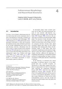
RESULTS The effect of busulfan on spermatogenesis (group 3) was evaluated at stage I of the study on day 30 of experiment. The cytostatic reduced significantly the concentration of tissue testosterone, caused hemodynamic disorders in the testes, increased the area of interstitial tissue at the expense of edema and infiltration of the interstitium mainly by lymphocytes and macrophages (Fig. 1, a, b). Total devastation of the seminiferous tubules was paralleled by necrobiosis, destruction of spermatogenic epithelium cells and their desquamation into the lumen of seminiferous tubules.
402
Bulletin of Experimental Biology and Medicine, Vol. 162, No. 3, January, 2017 MORPHOLOGY AND PATHOMORPHOLOGY
Fig. 1. Morphology of mouse testis: intact (a) and 30 days after intraperitoneal injection of busulfan (b); fertility index (c) and testosterone level in testicular tissue homogenate (d) in male C57Bl/6 mice of various groups. Hematoxylin and eosin staining, ×200 (a, b). p