A morphological mutant of Escherichia coli K-12 that grows as round cells at. 30, 37, or 42 C in a variety of ... EM medium (21) and EMB medium (21) were used for determination and ... (Toshiba GL-15) at a distance of 50 cm. After dilution.
JOURNAL OF BACTERIOLOGY, JUlY 1973, p. 436-442 Copyright 0 1973 American Society for Microbiology
Vol. 115, No. 1
Printed in U.SA.
Characterization and Genetic Analysis of a Mutant of Escherichia coli K-12 with Rounded Morphology HIROSHI MATSUZAWA,' KOU HAYAKAWA, TETSUJI SATO, AND KAZUTOMO IMAHORI Department of Agricultural Chemistry, University of Tokyo, Bunkyo-ku, Tokyo, Japan Received for publication 12 December 1972
A morphological mutant of Escherichia coli K-12 that grows as round cells at 30, 37, or 42 C in a variety of complex and synthetic media has been isolated and characterized. The gene concerned, designated rodA, has been shown to be on the chromosome between the purE and pyrC loci and to be located at about minute 15. The rodA gene has been found to be co-transducible with the lip gene at a frequency of 95%. The rodA mutant showed an increased resistance to ultraviolet irradiation and a changed sensitivity to drugs. The resistance to ultraviolet irradiation and mitomycin C appears to be co-transducible with the rodA gene.
It is of great interest to study the genetic control and biochemical determinant(s) of bacterial morphology. Conditional or nonconditional rod mutants have been described in Escherichia coli (2, 10, 14, 17), Bacillus subtilis (3, 19, 20), Bacillus licheniformis (19, 20), and Agrobacterium tumefaciens (8). Adler et al. have found a morphological mutant of E. coli K-12 which is radiation resistant, and it is not known whether the unusual cell morphology and radiation resistance are a result of the same genetic alteration (2). Normark has reported a morphological envB mutant that has a mutated gene near the strA gene and shows a changed resistance to several antibiotics as well as an increased tolerance to ultraviolet (UV) irradiation (17). We have found a morphological mutant of E. coli K-12 among a group of acridine orange-sensitive mutants. The cells grow as round cells at 30, 37, or 42 C in either liquid or solid media. In this report we describe a new gene, designated rod A, which controls the morphology of E. coli cells and is located at about minute 15 close to the lip gene (11). Resistance to UV irradiation and mitomycin C is co-transducible with the rodA gene. Gene designation and time scale in this report are described according to Taylor (22).
ducing phage Plkc was obtained from Y. Sugino. Media. Bacteria were usually cultured at 37 C with shaking in nutrient broth or L-broth. Nutrient broth contained (per liter of deionized water): polypeptone (Daigo Eiyo Kagaku Co., Osaka, Japan), 10 g; Ehrlich meat extract (Kyokuto Seiyaku Co., Tokyo, Japan), 10 g; and NaCl, 2 g. L-broth contained (per liter of deionized water): polypeptone, 10 g; yeast extract (Kyokuto Seiyaku Co., Tokyo, Japan), 5 g; NaCl, 5 g; glucose, 1 g; and thymine, 10 g supplemented. Media were adjusted to pH 7.2 by addition of approximately 0.5 ml of 10 N NaOH. Davis minimal medium (21) was used for determination of auxotrophic markers. EM medium (21) and EMB medium (21) were used for determination and selection of defective sugar fermentation. K10 medium (6) was used for transduction experiments. For solidifying media 1.5% agar (Wako Pure Chemical Co., Osaka, Japan) was used. Isolation of the morphological mutant. E. coli K-12 strain JE1011 was treated with N-methyl-N'nitro-N-nitrosoguanidine (NTG), essentially under the conditions described by Adelberg et al. (1). The cells were then allowed to grow for about three generations in nutrient broth, and 0.1-ml samples were spread on plates of nutrient agar at a concentration giving about 300 colonies per plate (master plate). The plates were incubated at 37 C, and colonies were replicated on nutrient agar plates containing 50 ,g of acridine orange (Chroma-Gesellschaft Schmid & Co., Germany) per ml. Colonies which did not grow on the replica plates were collected from the master plates, and the morphology of acridine orangesensitive mutants was observed with a phase-contrast microscope. MATERIALS AND METHODS UV sensitivity test. Bacteria in the logarithmic Bacterial strains. The properties of E. coli K-12 phase of growth (about 2 x 10⁸ cells per ml) were strains used are summarized in Table 1. The transdiluted to 2 x 10⁶f cells per ml in pH 6.8 buffer (0.13 M 1 Present address: Laboratory of Biochemical Genetics, NaCl, 0.02 M sodium phosphate) and irradiated in National Heart and Lung Institute, Bethesda, Md. 20014. petri dishes (84 mm in diameter) with a UV lamp 436
VoL. 115, 1973
E. COLI rod MUTANT
TABLE 1. Strains of Escherichia coli K-12 used Strain
Sexa
Genotype
[
Origin"
thr, leu, trp, his, thy, Obtained from Y. Sugino thi, ara, lac, gal, xyl, mtl, strA AOS15 thr, leu, trp, his, thy, Isolated from JE1011 after thi, ara, lac, gal, treatment xyl, mtl, strA, with NTG rodA, acridine orange-sensitive AOS151 thr, leu, trp, his, thy, Acridine orange-resistant thi, ara, lac, gal, recombinant xyl, mtl, strA, from AOS15 rodA x W2252 W2252 Hfr C met Obtained from Y. Sugino Hfr H met, thi JE1031 Obtained from Y. Sugino Lederberg LabW3807 Hfr 6 metBi, mal-20, oratory mtl-8, mut-2 KL99 Hfr K. B. Low thi-1, rel-1 strain AT1325 lip9 Fthi-1, his-4, purB15, J. R. Guest strain proA2, mtl-I, xyl-5, galK2, lacYl, lip-9, str-35 thi-1, metE70, R. Curtiss strain lysA23, trpE38, purE42, proC32, leu-6, mtl-l, xyl-5, ara-14, lacZ36, tonA23, azi-6, tsx-67, sup-45, str-109
JE1011
a Each Hfr strain has the point of origin and direction of chromosome transfer as shown in Fig. 4. °Strains W3807, KL99, AT1325 lip9, and X478 were obtained from the collection of Coli Genetic Stock Center, Department of Microbiology, Yale University School of Medicine, New Haven, Conn. CGSC numbers of the strains are 4236, 4242, 4286, and 4212, respectively. (Toshiba GL-15) at a distance of 50 cm. After dilution
of the irradiated suspension (0.7 mm in depth), 0.1-ml samples were spread on plates of L-broth agar. The plates were incubated at 37 C overnight in the dark, and the colonies formed were counted. Quantitative evaluation of sensitivity to drugs. After culturing overnight, the strains were diluted and 0.1-ml samples were spread on plates of L-broth agar containing different concentrations of each drug. The plates were incubated at 37 C for 1 or 2 days and the colonies were counted. The concentrations of drugs required to inhibit the colony formation to half that in the control plate were calculated. Mating procedure. The method used was essentially that of Taylor and Thoman (23), which is described in full detail (21). Transduction technique. The procedure used was based on that described by Lennox (15), and was as described previously (16) except that the temperature was kept at 37 C and the multiplicity of infection was 0.1. The cell shape of recombinants and transductants was examined with a phase-contrast microscope. Electron microscopy. The preparative method of Kellenberger et al. (13) was used. Sections were cut on a Porter-Blum MT-2 ultramicrotome with a glass
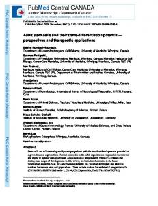
437
knife. The sections were collected on Formvar-coated copper grids and stained first with uranyl acetate and then with lead citrate (7). They were examined with a JEM-7A electron microscope.
RESULTS Genetic characteristics of acridine orangesensitive and morphological mutant strain AOS15. Forty-one acridine orange-sensitive mutants were isolated from strain JE1011 treated with NTG. Among them one mutant, strain AOS15, had rounded morphology and the same genetic markers as the parental strain. We determined whether the changed sensitivity to acridine orange and disturbed morphology were due to the same genetic alteration. Acridine orange-resistant revertants from AOS15 retained the disturbed morphology and could grow on plates containing 50 ,ug of acridine orange per ml. Acridine orange-resistant recombinants obtained by mating strain AOS15 with strain W2252 (Hfr C) also retained the rounded morphology. These results showed that the mutated gene(s) concerned with acridine orange sensitivity and cell morphology were not identical. Apparently this morphological mutant was obtained by chance out of a group of acridine orange-sensitive mutants. The acridine orange-sensitive locus of strain AOS15 is presumably identical or close to; the mtc gene (minute 12) (22), because in the interrupted mating between strain AOS15 and strain W2252 (Hfr C) the donor determinant of the acridine orange-resistant trait entered at about 5 min after the start of mating, and the lac+ allele was transfered at about 10 min (see Fig. 4). NTG is known to induce multiple mutations at the replicating region in the length between minute 1.5 and 2.0 (9). Therefore, the changed sensitivity to acridine orange and rounded morphology could be a result of comutation with NTG, taking into account that the mutated gene concerned with morphology of strain AOS15 is closely linked with the lip gene (minute 15) (11) as described below (see Fig. 4). In the following experiments we used one of the acridine orange-resistant recombinants with disturbed morphology, strain AOS151, derived from a 10-min mating between strain AOS15 and strain W2252 (Hfr C). Strain AOS151 has the same genetic markers as the parental strain JE1011 except rodA. General properties of strain AOS151. Strain AOS151 grows in the rounded morphology at 30, 37, or 42 C in a variety of complex and synthetic media (Fig. 1B), in contrast to the parental strain JE1011 which grows as a normal rod (Fig. 1A). At 25 C the cell shape of strain AOS151 becomes slightly lengthened and ab-
438
~r
MATSUZAWA ET AL.
normal (Fig. 1 C). These cells do not require isotonic osmotii c conditions for their growth and under these coinditions did not convert to rods. Strain AOS151 forms normal rough colonies on complex or syrithetic agar plates. In complex and synthetic liiquid cultures at 37 C, lysed cell debris can be observed, especially in the stationary phase )f growth. However, the growth curve of strain AOS151 is normal in nutrient or L-broth, and tthe visible cells are almost all viable in any phase of growth. In L-broth cultures the genieration time of strain AOS151 is 50 min, and thait of the parental strain JE1011 is
A
w
B*@
%
oo
w
J. BACTERIOL.
35 min. In the case of rodA+ and rodA- transductants selected with lip+ from strain AT1325 lip9, the generation time is 29 and 33 min, respectively. In view of the long generation time of strain AOS151 in contrast to the parent strain, it is likely that strain AOS151 still contains additional NTG-induced mutation(s). Electron microscopy examinations of ultrathin sections of strain AOS151 cells (Fig. 2) show no apparent defect in the cell membrane and wall. Normal symmetrical septum formation is observed in dividing cells of strain AOS151 (Fig. 2), which differs from that reported for the envB mutant with disturbed
~~~morphology (17).
Sensitivity of strain AOS151 to bacteriophages or colicins was examined by a cross* streak method. Strain AOS151 and the parental % * 0 ) @ strain JE1011 were both sensitive to phages T2, t t ks T3, T4, T6, T7, BF23, X, and Plkc and to -$ t e: colicins to E2 and K. 9 r ** Sensitivity of strain AOS151 to UV irrao . s :2 r ^diation and drugs. The morphological mutants nM,b