139
Abstract.–On the eastern seaboard of
the United States, populations of the blue crab, Callinectes sapidus, experience recurring outbreaks of a parasitic dinoflagellate, Hematodinium perezi. Epizootics fulminate in summer and autumn causing mortalities in highsalinity embayments and estuaries. In laboratory studies, we experimentally investigated host mortality due to the disease, assessed differential hematological changes in infected crabs, and examined proliferation of the parasite. Mature, overwintering, nonovigerous female crabs were injected with 103 or 105 cells of H. perezi. Mortalities began 14 d after infection, with a median time to death of 30.3 ±1.5 d (SE). Subsequent mortality rates were greater than 86% in infected crabs. A relative risk model indicated that infected crabs were seven to eight times more likely to die than controls and that decreases in total hemocyte densities covaried significantly with mortality. Hemocyte densities declined precipitously (mean=48%) within 3 d of infection and exhibited differential changes in subpopulations of granulocytes and hyalinocytes that lasted throughout the course of the infection. Crabs that did not present infections after injection (i.e. “immune” hosts) did not show hemocytopenia and exhibited significant long-term (21–27 d) granulocytemia. Detection of the parasite in the hemolymph of infected crabs increased from approximately 30% after 14 d to 60% after 21 d to 100% after 35 d. Plasmodial stages were, however, detectable in histological preparations of the heart within 3 days of infection and increased in number over 5 and 7 days. Sporulation of the parasite occurred over a short time (at least 4 d, after 43 d of infection) and did not culminate in the immediate death of the host. Hematodinium perezi represents a significant threat to the blue crab fisheries in high-salinity estuaries. Although the parasite infects male and female crabs, it may have a greater impact on mature females as they move to higher salinities to breed.
Manuscript accepted 23 August 1999. Fish. Bull. 98:139–152 (2000).
Mortality and hematology of blue crabs, Callinectes sapidus, experimentally infected with the parasitic dinoflagellate Hematodinium perezi* Jeffrey D. Shields Christopher M. Squyars Department of Environmental Sciences Virginia Institute of Marine Science The College of William and Mary P.O. Box 1346, Gloucester Point, VA 23602, USA E-mail address (for J. D. Shields):
[email protected]
Hematodinium perezi is a parasitic dinoflagellate that proliferates in the hemolymph of several crab species. In the blue crab, Callinectes sapidus, H. perezi is highly pathogenic and usually kills the host. The main symptom of the infection is lethargy. Heavy infections are characterized by discolored (brown, yellow, milky or chalky) hemolymph that does not clot. The disease occurs in blue crabs in high-salinity (>11‰) waters from Delaware to Florida, and in the Gulf of Mexico (Newman and Johnson, 1975; Messick and Sinderman, 1992). In 1975, Newman and Johnson (1975) reported a prevalence of 30% in blue crabs from Florida; the effect of this disease on the blue crab population was thought to be high. In 1991 and 1992, prevalences of infection up to 100% were found in blue crabs (mean prevalence=43%, several locations from 70% to 100%, n=971) from coastal bays in Maryland and Virginia (Messick, 1994). Commercial watermen reported reduced catches, lethargic and moribund crabs in pots and shedding facilities, and crabs that died soon after capture (Rux, Oesterling1). In 1996 and 1997, 10% to 40% of adult crabs from the eastern portions of Chesapeake Bay in Virginia were infected.2 The disease has a low prevalence or does not occur in the
larger, riverine (“bayside”) fishery; it appears most detrimental to the coastal (“seaside”) crab fisheries. Outbreaks of infestation by Hematodinium spp. have caused concerns to several major crustacean fisheries. Significant population declines and economic losses have been reported for the Tanner (Chionoecetes bairdi) and snow (C. opilio) crab fisheries of Alaska and Newfoundland (Meyers et al., 1987, 1990; Taylor and Khan, 1995),3 the Norway lobster (Nephrops norvegicus) fishery of western Scotland (Field et al., 1992), and the velvet crab (Necora puber) fishery of western France (Wilhelm and Miahle, 1996). The parasite causes a condi-
* Contribution 2241 from the Virginia Institute of Marine Science, The College of William and Mary, Gloucester Point, VA 23602. 1 Rux, S. 1993. Red Bank Seafood Co., Box 37 Marionville, VA 23408. Personal commun.; Oesterling, M. 1993. VASG, Virginia Inst. Marine Science, Gloucester Point, VA 23062. Personal commun. 2 Shields, J. D. 1997. An investigation into the epizootiology of Hematodinium perezi, a parasitic dinoflagellate in the blue crab, Callinectes sapidus. Saltonstall-Kennedy Program, National Marine Fisheries Service, NOAA. Final Report. 3 Prevalences in Newfoundland are now at 1–15% in the northern bays. Taylor, D. 1998. DFO, CP 5567, White Hills, St. Johns, Newfoundland, Canada, A1C 5X1. Personal commun.
140
tion known as bitter crab disease in snow and Tanner crabs (Meyers et al., 1987). Low prevalences (1–4%) of another species, H. australis, have been reported in sand (Portunus pelagicus) and mud (Scylla serrata) crabs from Australia (Shields, 1992; Hudson and Shields, 1994). Infections of Hematodinium spp. or Hematodinium-like species have been reported from a variety of different hosts (see Shields, 1994, for review). There are, however, only two described species of Hematodinium: H. perezi Chatton and Poisson, 1931, and H. australis Hudson and Shields, 1994. By convention (Newman and Johnson, 1975; MacLean and Ruddell, 1978) and from its distinct morphological features, we concur that Hematodinium perezi is the infectious species in the American blue crab. Blue crabs sustain one of the largest fisheries in Chesapeake Bay. Current management plans and state regulations are based on population assessments that include numbers of juvenile and adult crabs found during winter, spring, and summer surveys (Lipcius and Van Engel, 1990; Abbe and Stagg, 1996; Rugolo et al., 1998). Although these projections include estimates of natural mortalities, they do not account for the potential epizootics and mortalities caused by Hematodinium perezi. In this study, we examined host mortality in controlled laboratory experiments and documented changes in the hemograms (total cell counts, and differential counts) of inoculated crabs versus uninfected crabs. We also examined proliferative growth of H. perezi at approximately weekly intervals and made observations on the biology and life history of the parasite.
Materials and methods Blue crabs were collected from Chesapeake Bay and several of its subestuaries during the annual VIMS Winter Dredge Survey (part of the Chesapeake Bay Stock Assessment Program) with a 1.83-m-wide Virginia crab dredge fitted with 0.5-inch (1.25-cm) Vexar mesh dragged on the bottom for one minute at three knots. Crabs were also taken with commercial crab pots from two reference locations on the Delmarva Peninsula, Red Bank and Hungars Creeks, Virginia. Uninfected crabs were housed together for three to seven days prior to treatment to ensure acclimation and absence of overt bacterial or protozoal diseases (as assayed below). During the experiments, crabs were fed fish and squid semiweekly and held individually in aquaria (5 gal., 19 liter) at 20° to 21°C, and 24 ppt salinity. Although H. perezi infects both sexes, only mature, nonovigerous female crabs (healthy, orange maturing gonads, little to no shell damage, 120–160
Fishery Bulletin 98(1)
mm carapace width including epibranchial spines) were used in the experiments. Females were used to limit the number of treatment effects (e.g. potential differences between sexes) and to improve sample sizes given the laborious nature of the experiments. Hematodinium perezi was maintained in the laboratory by serial passage of infected hemolymph. Hemolymph from naturally infected crabs was injected directly into uninfected crabs. Naïve (unexposed) crabs and crabs used for inoculation experiments were obtained from low-salinity non-enzootic locations. Infected and inoculated crabs were housed separately and used as hemolymph donors to inject naïve hosts (105–106 parasites per host). Injections were given in the arthrodial membrane of the fifth leg at the juncture of the basis with the carapace. We have maintained H. perezi for over seven months using this method with no apparent loss from pathogenicity. Two mortality experiments and one early life history experiment were undertaken. The mortality-I experiment used raw, infected hemolymph as the inoculant. Although appropriate for maintaining infections in the laboratory, raw hemolymph cannot be adjusted to manipulate parasite densities without the use of physiological buffers, nor can it be guaranteed as sterile without appropriate assessment (see Welsh and Sizemore, 1985). Preliminary experiments with sterile sea water, physiological buffers, and infected hemolymph indicated that buffer-washed parasites remained infectious, and could, therefore, be adjusted to consistent densities appropriate to controlled experiments. The mortality-II experiment used buffer-washed parasites adjusted to a density similar to that used in the mortality-I experiment. Mortality-II experiment closely resembled mortality-I experiment except for 1) handling (buffer washes with centrifugation) and 2) the use of plasmodial versus uninucleate stages of the parasite. Uninfected crabs served as controls in both experiments. Controls were used to assess handling effects and to establish baseline densities of hemocytes. The early infection experiment was designed to examine the effects of early infections on the hematology of the host and the early life history of the parasite. Experimental densities in the early infection experiment were four times higher than those in the previous experiments (4.1 × 10⁵ vs. approx. 1.0 × 10⁵ parasites/crab, respectively) and were arbitrarily higher to insure observation of parasites prior to their proliferation. In the mortality-I and mortality-II experiments different proportions of trophonts and plasmodia were used (for definitions see below). The mortality-I experiment consisted of a control group of uninfected crabs (n=22) injected individually with 100 µL
Shields and Squyars: Mortality and hematology of Callinectes sapidus infected with Hematodinium perezi
of hemolymph from an uninfected donor crab and an experimental group (n=20) injected individually with 100 µL of infected hemolymph from a donor crab containing an estimated 1.3 × 10⁶ trophonts/mL (1.3 × 10⁵ trophonts per crab). The mortality-II experiment consisted of a control group (n=8) injected individually with 100 µL of physiological saline buffer (modified from Appleton and Vickerman, 1998; NaCl, 19.31 g/L; KCl 0.65 g/L; CaCl2 . 2H2O 1.38 g/L; MgSO4 . 7H2O 1.73 g/L; Na2SO4 0.38 g/L; HEPES 0.82 g/L;) adjusted to pH 7.8, with added glucose (1.0 mg/mL) and two experimental treatments (high dose=1.0 × 10⁵ parasites/ crab; low dose=1.0 × 10³ parasites per/crab, n=10, 10 respectively). To prepare the inoculum for the experimental treatments, 2.0 mL of infected hemolymph were drawn from a donor crab infected with 6.15 × 10⁷ parasites/mL (comprising 97% plasmodia; 3% trophonts). The infected hemolymph was diluted 1:1 with buffer, centrifuged at 4000 rpm for 10 minutes, the supernatant was decanted, and the cells were resuspended in buffer. The cells were then adjusted to 1.0 × 10⁷ parasites/mL, centrifuged through two more washes, and serially diluted to attain densities of 1.0 × 10⁶ parasites/mL and 1.0 × 10⁴ parasites/mL (for inoculum of 100 µL, 1.0 × 10⁵ parasites/crab and 1.0 × 10³ parasites/crab, respectively). In both experiments, crabs were monitored daily for mortalities. Deaths within the first nine days of each experiment were excluded because of handling stress arising from infrequent, bacterial infections (e.g. Johnson, 1976). None of the crabs in the experiments were infected with amoebae, microsporans, or overt bacterial infections (but see Welsh and Sizemore, 1985 for background levels of Vibrio spp. in hemolymph of C. sapidus). Ten crabs from each treatment in the mortality-I experiment, and all of the crabs in the mortality-II experiment were bled approximately weekly to assess infection status. In the mortality-I experiment, the same ten crabs were bled approximately weekly until they died; other crabs from within the experiment were added as replacements. Crab hemolymph was taken by using a tuberculin syringe (1 mL) with a 25.5-ga. needle from the arthrodial membrane at the juncture of the basis and the ischium of the 5th pereopod (swimming leg). Ethanol (70%) was used to sterilize the site of inoculation and blood letting. Total and differential counts of host hemocytes and estimates of parasite density were obtained from individual crabs with a hemocytometer (Neubauer improved, Bright Line, two counts per crab) with phase contrast microscopy at 400×. Host hemocytes were identified as granulocytes, semigranulocytes (intermediate cells with relatively few granules, Bodammer, 1978; Johnson,
141
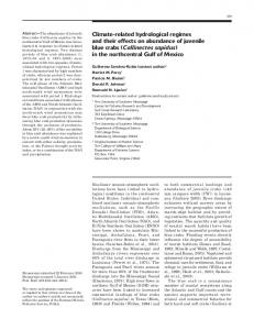
1980) and hyalinocytes (cell types defined in Söderhäll and Cerenius, 1992). Hemocyte and parasite densities higher than 1.0 × 10⁷ cells/mL were diluted 1:5 with buffer and recounted to provide better estimates of cell density. For comparative purposes, total hemocyte densities and differential counts from naturally infected male and female crabs were also obtained. Parasites were easily distinguished from host cells by using phase contrast microscopy (Fig. 1): uninucleate trophonts (9–15 µm) possessed few small, refractile vacuoles and were rounded or amoeboid, without filopodia; multinucleate plasmodia (20–100 µm) were slender, vermiform, and motile. The density of infection refers to the number of parasites per mL of hemolymph. Total hemocyte density refers to the number of hemocytes per mL of hemolymph. Mean intensity refers to the mean number of parasites per quantity of infected host tissue (Margolis et al., 1982). Permanent preparations of hemolymph were processed and stained as described in Messick (1994). Briefly, acid-cleaned, poly-l-lysine-coated microslides were smeared with fresh hemolymph, allowed to stand for 2–3 minutes, and fixed in Bouin’s fixative. The smears were processed through a routine Harris hematoxylin and eosin-Y procedure (Humason, 1979, p. 123 without acid destain). The early infection experiment consisted of a control group (n=5 crabs) injected individually with 100 µL of hemolymph from an uninfected donor crab and an experimental group (n=20) injected with 100 µL of hemolymph from a donor crab containing an estimated 4.1 × 10⁶ parasites/ml (4.1 × 10⁵ parasites per crab; comprising 79% plasmodia, 21% trophonts). Three days prior to infection, cell counts were conducted on all crabs to serve as a benchmark (presample) for before-after comparisons. On days 3, 5, and 7 after inoculation, five infected crabs were bled and dissected. Differential cell counts were conducted and tissue samples taken for histological analysis. Tissue samples were processed through a routine hematoxylin and eosin procedure and included muscle, hepatopancreas, heart, and, in some cases, foregut. The control crabs were bled and tissue samples taken 10 days after injection. For statistical analyses, the proportional hazards model with the Weibull distribution was used to examine survival data and associated variables (Cox and Oakes, 1984). The Tarone-Ware log-rank test was used to examine differences between survival curves (Wilkinson, 1997). ANOVA was used to analyze relationships in hemocyte densities and proportion of cell type (cell type density divided by total hemocyte density) between inoculated and uninfected crabs. Simi-
142
Fishery Bulletin 98(1)
Figure 1 Hematodinium perezi from the blue crab, Callinectes sapidus. (A and B) Vermiform plasmodia (p) in hemolymph (granulocytes, star). Bar = 10 µm. (C) Amoeboid trophonts (arrows) with few refractile granules. Bar = 10 µm. (D) Round trophonts (arrows) with many refractile granules. Bar = 10 µm.
lar densities and proportions of cell types were noted in hematology and survival between the mortality-I and mortality-II experiments; hence, data were combined a posteriori for the analyses. Where similar trends were noted between statistics for injection dosage (103 vs. 105), data were also combined for the analysis (i.e. survivorship, hematology). SYSTAT (Wilkinson, 1997) and SAS (SAS, 1988) were used for the analyses. A probability level of P < 0.05 was accepted as significant.
Results Inoculated crabs that became infected with Hematodinium perezi began dying two weeks after inoculation (Fig. 2). Mortalities peaked at three weeks after injection and continued to accumulate from weeks 3 through 5. The mortality rate of the infected crabs was 86%, whereas less than 20% of the controls died. Crab mortalities were similar over the time course of infection between mortality-I (infected
Shields and Squyars: Mortality and hematology of Callinectes sapidus infected with Hematodinium perezi
143
Figure 2 Survivorship curves for uninfected crabs and crabs infected with H. perezi (high dose=105 parasites/crab, low=103 parasites/crab). Sample sizes were 22 and 8 uninfected controls; 20 (mortality-I experiment), 10 and 10 (high and low dose, mortality-II experiment) respectively.
hemolymph, uninucleate trophonts) and mortality-II (buffer-washed parasites, vermiform plasmodia) experiments (Tarone-Ware, χ2=1.21 with 1 df, P=0.27), even between different initial doses of the parasite (Fig. 2; Tarone-Ware, χ2=0.74 with 1 df , P= 0.39). Uninfected crabs (controls) experienced significantly fewer mortalities than did infected hosts (Fig. 3; Tarone-Ware, χ2=, 19.27 with 1 df , P