Department of Genetics, University of Adelaide,. Adelaide S.A. 5000 Australia. K.H. Neldner. Department of Dermatology,. Texas Tech University Health ...
J Mol Med (2000) 78:282–286 Digital Object Identifier (DOI) 10.1007/s001090000114
R A P I D C O M M U N I C AT I O N
Berthold Struk · Li Cai · Stéphanie Zäch · Wan Ji Joon Chung · Amanda Lumsden · Markus Stumm Marcel Huber · Lori Schaen · Chung-Ah Kim Lowell A. Goldsmith · Denis Viljoen · Luis E. Figuera Wayne Fuchs · Francis Munier · Raj Ramesar Daniel Hohl · Robert Richards · Kenneth H. Neldner Klaus Lindpaintner
Mutations of the gene encoding the transmembrane transporter protein ABC-C6 cause pseudoxanthoma elasticum Received: 22 May 2000 / Accepted: 25 May 2000 / Published online: 26 May 2000 © Springer-Verlag 2000
B. Struk · L. Cai · W. Ji · J. Chung · C.-A. Kim K. Lindpaintner Department of Cardiology, Children’s Hospital, Cardiovascular Division, Brigham and Women’s Hospital, Department of Cardiology, and Department of Medicine, Harvard Medical School, Boston, MA 021152, USA B. Struk · K. Lindpaintner Max Delbrück Center for Molecular Medicine, 13122 Berlin, Germany L. Cai · W. Ji · K. Lindpaintner F. Hoffmann-La Roche Ltd, Roche Genetics, Pharmaceuticals Division, 4070 Basel, Switzerland S. Zäch · M. Huber · D. Hohl Department of Dermatology, University of Lausanne, 1011 Lausanne, Switzerland A. Lumsden · R. Richards Department of Cytogenetics and Molecular Genetics, Women’s and Children’s Hospital, 71 King William Road, North Adelaide S.A. 5006, Australia M. Stumm Human Genetics Institute, Otto-von-Guericke-University, Magdeburg, Germany L. Schaen · L.A. Goldsmith Department of Dermatology, University of Rochester, Rochester, New York 14642, USA D. Viljoen South African Institute for Medical Research, Johannesburg, South Africa L.E. Figuera Divisions of Genetics and Molecular Medicine, CIBO-IMSS, University of Guadalajara Medical School, Guadalajara, Jal Mexico W. Fuchs Department of Ophthalmology, Mount Sinai School of Medicine, New York, New York 10029, USA F. Munier Department of Ophthalmology, University of Lausanne, 1011 Lausanne, Switzerland
Abstract We recently published the precise chromosomal localization on chromosome 16p13.1 of the genetic defect underlying pseudoxanthoma elasticum (PXE), an inherited disorder characterized by progressive calcification of elastic fibers in skin, eye, and the cardiovascular system. Here we report the identification of mutations in the gene encoding the transmembrane transporter protein, ABC-C6 (also known as MRP-6), one of the BERTHOLD STRUK LI CAI received her Ph.D. degree in received his M.D. degree from genetics from Texas A&M the University of Münster, University in College Station, Germany. He was a resident in Texas, USA. She obtained her internal medicine and cardiolpostdoctoral and medical geogy at the German Heart Instinetics training at Children’s tute in Berlin. He is currently a Hospital, Harvard Medical research fellow at the Max School, Boston, MassachuDelbrück Center in Berlin and setts, USA. She is currently a at the Department of Medicine staff scientist at Roche Genet- at Brigham and Women’s Hosics, Hoffmann–La Roche in pital, Harvard Medical School, Basel, Switzerland. Her reBoston, Massachusetts, USA. search interests include genet- His research interests include ics of human diseases and ge- the genetics of cardiovascular nome resource development in diseases. human and mammalian systems. B. Struk and L. Cai contributed equally to this manuscript. R. Ramesar Department of Human Genetics, Medical School, University of Cape Town, Observatory 7925, South Africa R. Richards Department of Genetics, University of Adelaide, Adelaide S.A. 5000 Australia K.H. Neldner Department of Dermatology, Texas Tech University Health Sciences Center, Lubbock, Texas 79430
283
four genes located in the region of linkage, as cause of the disease. Sequence analysis in four independent consanguineous families from Switzerland, Mexico, and South Africa and in one non-consanguineous family from the United States demonstrated several different mis-sense mutations to cosegregate with the disease phenotype. These findings are consistent with the conclusion that PXE is a recessive disorder that displays allelic heterogeneity, which may explain the considerable phenotypic variance characteristic of the disorder.
processes that we know to be of relevance in a multitude of different conditions, and thus could all reasonably be viewed as “candidate” genes involved in a the pathogenesis of PXE. Genomic and c-DNA sequencing of these four genes in a number of affected and unaffected members of PXE families revealed ABC-C6 as the disease-causing gene.
Key words Pseudoxanthoma elasticum · Membrane transporter proteins · ATP binding cassette proteins · ABC-C6
Collection of PXE-families and selection of samples for mutation screen
Materials and methods
Abbreviations ABC: ATP binding cassette · ATP: Adenosine triphosphate · PCR: Polymerase chain reaction · PXE: Pseudoxanthoma elasticum
The establishment of a large repository of 192 affected and 380 nonaffected individuals from a total of 81 families, and the techniques to extract DNA from blood samples obtained after appropriate informed consent have been previously described [3, 4]. Haplotypic homozygous affected and haplotypic homozygous unaffected members of four consanguinous families were selected for systematic sequence analysis of pM5, MRP1, NPIP, and ABC-C6.
Introduction
Sequence and genotype analysis
Pseudoxanthoma elasticum (PXE), the prototypical Mendelian disorder of elastic tissue, is characterized by progressive calcification of elastic fibers in skin, retina, and the cardiovascular system. Whereas the cutaneous lesions are mainly of cosmetic concern, the ocular manifestations result in various degrees of visual impairment in about half of all cases, brought about by retinal hemorrhage due to vascular friability, and the cardiovascular lesions, albeit rare, can lead to serious morbidity [1, 2] Based on our previous genetic mapping studies in which we identified a 500-kb region on chromosome 16p13.1 as the localization of the gene associated with PXE [3, 4], we identified four putative candidate genes, among them NPIP, a nuclear core complex interacting protein, pM5, a gene with homologies to metalloproteinases, and MRP1 and ABC-C6 (MRP6), two members of the ATP-binding cassette (ABC) superfamily of polytopic, integral membrane transport proteins [5]. Whereas none of these genes was known to play any role in connective tissue morphogenesis or function, both transmembrane transport as well as protein degradation are
Fluorescent dideoxyterminator sequencing was carried out on ABI 377 and ABI 3700 automated sequencing devices, on appropriate polymerase chain reaction (PCR) amplified fragments of all four genes. Quality score based sequence comparisons used the Sequencher 4.0 (Gene Codes, Ann Arbor, Mich., USA) software tool. For two of the mutations identified in ABC-C6 we developed PCR restriction fragment length polymorphism screening assays: (a) The exon 24 C3421T single nucleotide polymorphism is associated with the loss of a BslI restriction endonuclease recognition site, resulting in major digestion products that differ by 13 bp in length. Briefly, 20 ng genomic DNA was PCR-amplified using primer pair E24 (Table 1) and incubated with 7 U BslI (New England Biolabs) in the appropriate buffer at 55°C for 6 h, with a subsequent inactivation at 80°C for 20 min, fractionation by submarine agarose (4% MetaPhor, FMC Bioproducts, Rockland, Me., USA) gel electrophoresis, and visualization by ethidium bromide staining. (b) The exon 28 C4015T single nucleotide polymorphism is associated with the gain of a novel BsiHKAI restriction endonuclease recognition site, resulting in major digestion products that differ by 72 bp in length. Briefly, 20 ng genomic DNA was PCR-amplified using primer pair E28 (Table 1) and incubated with 5 U BsiHKAI (New England Biolabs) in the appropriate buffer with addition of bovine serum albumin at 65°C for 6 h, with a subsequent inactivation at 80°C for 20 min, fractionation by submarine
Table 1 ABC-C6 mutations Exon
Mutation
Primers
Annealing temperature (°C)
Product size (bp)
15
nt 1896 C→A aa 632 His→Gln
E15F: TCC CTA AAA ACA TGA GGC TGG TTA CTA C E15R: CGG CCA GGT CAG GGG TCT C
60
166
24
nt 3421 C→T aa 1141 Arg→stop nt 3490 C→T
E24F: AGG TCT TCT CTG CCC TGG CTC TTC E24R: CTG GAA TCC TGT ACT TGG GGC TCT C
65
322
28
aa 1164 Arg→stop nt 4015 C→T aa 1339 Arg→Cys
E28F: CCC ACC ATG CCT CCC ATC TT E28R: GTA CAG CAG AAA GAT CTC CCC AAT AAA
60
282
284
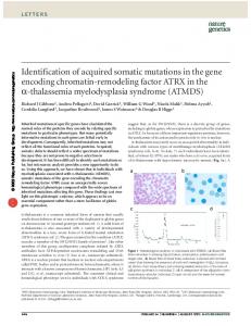
Fig. 1a,b Family A. a Haplotype analysis for single sequence length polymorphisms on chromosome 16p depicting a haplotypephenotype correlations. Squares Males; circles females; black symbols diseased individuals. (To protect study subjects’ and their family’s anonymity, the pedigree structure has been altered in a way not affecting the relevant genetic information concerning PXE; however, the original pedigrees were presented for review purposes.) b Results of mutation screening for variant C3421T. Lane numbers numbered individuals in the pedigree/haplotype rendition above
agarose (3.5% MetaPhor, FMC Bioproducts) gel electrophoresis and visualization by ethidium bromide staining.
Results No disease-cosegregating polymorphisms were encountered in either pM5, MRP1, or NPIP. Examination of the genomic sequence of ABC-C6 in an affected and unaffected member of several consanguineous families yielded four mutations that unambiguously cosegregated with both the disease and the presence of the disease-associated marker haplotype as determined earlier. The first of these mutations represents a C→T transition at nucleo-
tide position 3421 (C3421T) in exon 24, with a predicted precocious truncation of the translation product at amino acid position 1141 (Arg→stop). This mutation was encountered in two unrelated consanguineous Swiss families. The second mutation was found in a consanguineous family from the United States, represented a C→T transition, also in exon 24, at nucleotide position 3490 (C3490T), also resulting in a predicted premature termination of the translation product. The third mutation, observed in both a consanguineous Mexican and in three consanguineous South African families, represents a C→T transition at nucleotide position 4015 (C4015T), resulting in a predicted Arg→Cys substitution at amino acid position 1339. A fourth mutation, represented by a
285
Fig. 2a,b Family B. a Haplotype analysis for single sequence length polymorphisms on chromosome 16p depicting haplotypephenotype correlations. Squares Males; circles females; black symbols diseased individuals. (To protect study subjects’ and their family’s anonymity, the pedigree structure has been altered in a way not affecting the relevant genetic information concerning PXE; however, the original pedigrees were presented for review purposes.) b Results of mutation screening for variant C4015T. Lane numbers numbered individuals in the pedigree/haplotype rendition above
C→A transversion at nucleic acid position 1896 (C1896A) in exon 15, resulted in a predicted His→Gln exchange at amino acid position 632. Screening of additional family members in these pedigrees using either specific PCR restriction fragment length polymorphism assays or minisequencing demonstrated complete correlation of the C3421T, C3490T, and C4015T mutations, respectively, with the family-specific, disease-associated haplotypes, and – if homozygous – with clinical disease (Figs. 1, 2). Thus among different families the same mutation was found to cosegregate with the disease in the context of different marker haplotypes. Conversely, none of the heterozygous carriers in these families had been diagnosed with the disease, and in most of these probands PXE had been positively ruled out by skin biopsy. A survey among 100 random individ-
uals without history or family history of PXE revealed not a single C3421T allele. The C1896A variant was found in families with the C3421T and the C4015T variants but was not in complete linkage disequilibrium with the former, whereas for the latter the wild-type and variant were in complete linkage disequilibrium with the variant and the wildtype, respectively. The C1896A variant is therefore a polymorphism. In a family without known consanguinity we found affected offspring to be complex compound heterozygotes. They were hemizygous for the C3421T variant allele (that they inherited from the father), reflecting the fact that they carried in trans an ABC-C6 allele with a partial deletion that they had inherited form their mother (who was herself hemizygous for the wild-type allele).
286
Discussion Having previously mapped the gene associated with PXE into a 500-kb chromosomal region, and indicated four potential disease genes in this region, we have now found mutations that lead to obvious functional changes cosegregating with the disease in one of them, ABC-C6. In two cases, C3421T and C3490T, the mutations resulted in premature stop codons, with predicted truncated translation products that lack both the functionally crucial nucleotide binding Walker B motif and two putative transmembrane domains [6]. The same effect(s) may of course be predicted for partial deletions in this region. In the third case, C4015T, a basic amino acid, arginine, is supplanted by a cysteine, with potential structural consequences. Of note, this mutation occurs in a phylogenetically highly conserved region of the gene that shows significant homology to mouse, rat, Drosophila melanogaster, Caenorhabditis elegans, and Saccharomyces. cerevisiae, further emphasizing the functional importance of this region of the gene. The demonstration, on the other hand, that the C1896A mutation does not appear to be of pathogenic importance underscores the power of the collected pedigrees to discern disease-relevant mutations and highlights the relevance of those recognized as disease causing. Taken together, the evidence presented leaves no doubt that molecular variation in ABC-C6 causes PXE. In keeping with all our haplotyping data [4], our present observations strongly suggest that PXE is exclusively a recessive disorder, despite previous suggestions that an autosomal dominant form does exist as well [7]. Given the number of different mutations observed in the very limited sample that was the subject of this study, we predict that the majority of cases will be found to be compound heterozygotes. ABC-C6 is a member of the ubiquitous ABC superfamily characterized structurally by the presence of one or two nucleotide binding folds of approximately 200 well-conserved residues each, comprising the Walker A and B domains. Over 100 such proteins have been recognized to date, and while their primary role seems to be transmembrane transport, little is known about their substrates and energy coupling. ABC proteins are involved in such disorders as cystic fibrosis (CFTR) [8], Dubin-Johnson syndrome (MRP2) [9], and Tangier’s disease (ABC-1) [10] – all of which are transmitted, as is PXE, as autosomal recessive diseases – and in various drug-resistance scenarios in which they are thought to catalyze the ATPdependent removal of cytotoxic compounds from malignant target cells, and likewise in parasitic (such as Plasmodium falciparum and Entamoeba histolytica) and microbial (Staphylococcus aureus) pathogens [6]. ABC-C6 contains 31 exons, spans a 75-kb genomic region, and is predicted to encodes a protein of 1503 amino acids. No specific knowledge exists currently about the function of the ABC-C6 gene-product. ABC-C6 mRNA
has been found expressed in kidney and liver. Of potential relevance is the gene’s close physical association with MRP1, as well as evidence for a possible coregulation with MRP1. However, its structural similarity with MRP1 does suggest a role in transmembrane transport; there is also evidence that MRP1 and MRP6 are coregulated [6]. Significant additional basic investigation as well as human molecular genetic work is now required to allow us to use the key we discovered to understand the pathomechanism of the disease, and possibly develop a therapeutic or preventive approach. Acknowledgements We thank the members of PXE families from the United States, Canada, Switzerland, South Africa, United Kingdom, Mexico, and Germany for their participation in this study. Most of the North American families included into this study were obtained from the database of the National Association for Pseudoxanthoma elasticum (NAPE) (http://www.napxe.org). This work was supported by project grant 695-0209 from the March of Dimes Birth Defects Foundation, White Plains, N.Y. (http://www.modimes.org), and by a pilot and feasibility grant from the Harvard Skin Disease Research Center at Brigham and Women’s Hospital. K.L. is the recipient of a Research Career Development Award (K04-HL03138-01) from the National Heart, Lung, and Blood Institute. L.C. is the recipient of a National Research Service Award HL09783 from the National Institutes of Health. B.S. is the recipient of a National Research Fellowship Award from the Association of Clinical Pharmacology Berlin/Brandenburg and from the Ministry of Education, Science, Research and Technology of the Federal Republic of Germany. M.H.’s salary originates from a grant to D.H. by the SNFR (31–55849.98).
References 1. McKusick VA (1972) Pseudoxanthoma elasticum. In: McKusick VA (ed) Heritable disorders of connective tissue, 4th edn. Mosby, St. Louis, pp 475–520 2. Neldner KH (1988) Pseudoxanthoma elasticum. Clin Dermatol 6:1–159 3. Struk B, Neldner KH, Rao VS, St. Jean P, Lindpaintner K (1997) Mapping of both autosomal recessive and dominant variants of pseudoxanthoma elasticum to chromosome 16p13.1. Hum Mol Genet 6:1823–1828 4. Cai L, Struk B, Adams M, et al (2000) A 500-kb region on chromosome 16p13.1 contains the pseudoxanthoma elasticum locus: high resolution mapping and genomic structure. J Mol Med 78:36–46 5. Higgins CF (1992) ABC transporters: from microorganisms to man. Annu Rev Cell Biol 8:67–113 6. Kool M, van der Linden, de Haas M, Baas F, Borst P (1999) Expression of human MRP6, a homologue of the multidrug resistance protein gene MRP1, in tissues and cancer cells. Cancer Res 59:175–182 7. Pope FM (1974) Autosomal dominant pseudoxanthoma elasticum. J Med Genet 11:152–157 8. Zielenski J, Tsui, LC (1995) Cystic fibrosis: genotypic and phenotypic variations Annu Rev Genet 29:777–807 9. Paulusma CC, et al (1996) Congenital jaundice in rats with a mutation in a multidrug resistance-associated protein gene. Science 271:1126–1128 10. Orsoacute E, Broccardo C, Kaminski W (1999) Transport of lipids from Golgi to plasma membrane is defective in Tangier disease patients and Abc1-deficient mice. Nat Genet 24:192–196