□
CASE REPORT
□
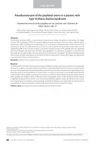
Mycotic Pseudoaneurysm of the Femoral Artery in a Patient with Psoriasis Basheer Tashtoush 1, Fernando Gonzalez-Ibarra 1 and Mary Abed 2
Abstract Mycotic aneurysms of the common femoral artery are rare and usually occur in intravenous drug abusers who use the femoral vessels for injection. We herein describe a case of mycotic aneurysm of the common femoral artery caused by methicillin-sensitive Staphylococcus aureus seeding of an atherosclerotic plaque in which the bacteria possibly originated from psoriatic skin lesions. A 67-year-old Caucasian man was admitted to the hospital after experiencing chest pain for two days. He was known to have psoriasis and coronary artery disease with a history of coronary artery bypass grafting surgery three years earlier. He was found to have methicillin-sensitive Staphylococcus aureus bacteremia and later developed a mycotic aneurysm of the femoral artery opposite to the site of catheterization access. Mycotic aneurysms are rare clinical conditions associated with significant morbidity and mortality. In patients with psoriasis, a high prevalence of Staphylococcus aureus colonization of the skin makes possible bacterial seeding of existing atherosclerotic plaques. Therefore, the risk of mycotic pseudoaneurysm formation in these patients should be considered. Key words: mycotic pseudoaneurysm, psoriasis, common femoral artery (Intern Med 51: 2831-2834, 2012) (DOI: 10.2169/internalmedicine.51.8195)
Introduction The term mycotic pseudoaneurysm refers to an arterial wall injury leading to an abnormal aneurysmal dilatation that is usually associated with local or systemic infection or direct damage of a vessel wall after an interventional procedure. The incidence of mycotic pseudoaneurysms is low and diagnosis is challenging, as patients with mycotic pseudoaneurysms may present with non-specific symptoms. Mortality is high if diagnosis and treatment are delayed (1). Mycotic pseudoaneurysms of the common femoral artery are rare and usually occur in intravenous drug abusers who use the femoral vessels for injection. In these patients, the development of mycotic pseudoaneurysms is most likely due to direct damage of the arterial wall and/or an increased incidence of inoculation with microorganisms, especially Staphylococcus aureus (S. aureus) (2). Patients with psoriasis are known to have aberrant immune responses and thus a higher incidence of various in-
fections, many of which are caused by Streptococcus or S. aureus (3). We herein present the case of a patient with psoriasis who developed a mycotic pseudoaneurysm of the common femoral artery due to methicillin-sensitive Staphylococcus aureus (MSSA) seeding of an atherosclerotic plaque. The source of the bacteremia was presumed to be colonization of the psoriatic skin lesions. To the best of our knowledge, this is the first case report in the literature to describe an association between a mycotic pseudoaneurysm and MSSA bacteremia in a patient with psoriasis.
Case Report The patient was a 67-year-old Caucasian man who presented to his primary medical doctor complaining of chest pain lasting for two days and one episode of fever with chills. His medical history included coronary artery disease with coronary artery bypass grafting performed three years earlier, plaque-type psoriasis, paroxysmal atrial fibrillation, hypertension, diabetes mellitus type 2 and gout. He had
1
Department of Internal Medicine, Mount Sinai School of Medicine, Jersey City Medical Center, USA and 2Department of Cardiology, Mount Sinai School of Medicine, Jersey City Medical Center, USA Received for publication May 21, 2012; Accepted for publication July 9, 2012 Correspondence to Dr. Fernando Gonzalez-Ibarra,
[email protected]
2831
Intern Med 51: 2831-2834, 2012
DOI: 10.2169/internalmedicine.51.8195
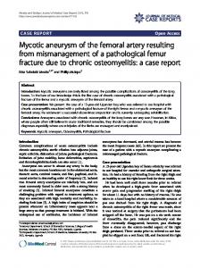
A B Figures 1. Computed tomography showing the presence of two aneurysms, one each on the medial and lateral aspects of the left common femoral artery, with irregular morphologies, suggesting arterial wall destruction (B, lateral view).
been an ex-smoker for more than 10 years with no history of intravenous drug use. His current medications included aspirin, clopidogrel, carvedilol, amlodipine, olmesartan, atorvastatin, metformin and allopurinol. His pain was moderate, intermittent and pressure-like with a gradual onset accompanied by one episode of fever (102°F) and chills the day prior to the admission. The patient was sent to the emergency department. A physical examination revealed an irregular heart rhythm (94110 bpm), a respiratory rate of 21 and a blood pressure of 99/60 mmHg. A pansystolic murmur with maximum intensity over the aortic area was noted. Additionally, the patient had plaque-type psoriasis with lesions over the extensor surface of both forearms and scalp. The lesions were raised and covered with silvery-white scales with minimal scratch marks; however, no signs of active inflammation were observed. The laboratory findings revealed a positive result for troponin I (1.03 ng/mL), an increased level of brain natriuretic peptide (5,380 pg/mL), a white blood cell count of 6,900 mm3 (differential: neutrophils 86%, bands 1%, lymphocytes 6%, monocytes 1%), a hemoglobin level of 12.5 g/dL, a hematocrit of 36.6%, a platelet count of 197,000 mm3, a glucose level of 352 mg/dL, a blood urea nitrogen level of 22 mg/dL, a creatinine level of 1.2 mg/dL, a sodium level of 131 meql/L, a potassium level of 4 meq/L, a chloride level of 94 meq/L, a bicarbonate level of 25 meq/L, a glomerular filtration rate of 64 mL/min, an aminotransferase aspartate level of 31 U/L, an aminotransferase alanine level of 22 U/ L, a total protein level of 6 mg/dL, an albumin level of 3.5 mg/dL and an alkaline phosphatase level of 4 U/L. Electrocardiography showed atrial flutter with a rapid ventricular rate of 110 bpm and no ST-T wave changes. The patient was admitted to the Cardiac Care Unit and administered treatment with diltiazem, carvedilol, clopidogrel, aspi-
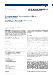
Figure 2. Image in vivo taken in the operating room before intervention of the left common femoral artery showing the morphology of the mycotic pseudoaneurism (arrow).
rin, therapeutic enoxaparin, a statin, cefepime and vancomycin. Transesophageal echocardiogram (TEE) showed severe aortic stenosis without vegetation and coronary angiography demonstrated the presence of patent grafts with no new lesions and an ejection fraction of 60% (coronary catheterization was performed with access through the right groin). Blood cultures drawn on admission grew MSSA in two different sets, and the patient began treatment with nafcillin. No foci of infection were identified; the only possible source of infection was the psoriatic skin lesions. On the fourth day of hospitalization, the patient suddenly experienced severe tearing pain and noticed a palpable, erythematous mass in the left groin. Doppler ultrasound showed a 2.5 cm outpouching from the left common femoral artery and CT-angiogram of the aorta, pelvis and both lower extremities showed the presence of two aneurysms, one each on the medial and lateral aspects of the left common femoral artery, with irregular morphologies suggestive of arterial wall destruction (Fig. 1). The findings noted on CT-angiogram along with persistent MSSA bacteremia detected in repeated blood cultures suggested a diagnosis of mycotic aneurysm of the common femoral artery. The patient underwent immediate vascular surgical intervention with aneurysmal resection, debridement of the periarterial necrotic tissue and placement of a great saphenous vein interposition graft (Fig. 2). The pathology reports confirmed the diagnosis of a common femoral artery pseudoaneurysm and moderate atherosclerosis at the site. Fluid collected intraoperatively from the tissue surrounding the aneurysm also grew MSSA. The clinical course of the patient was favorable.
Discussion Clinical suspicion in the diagnosis of mycotic aneurysms is fundamental, as it allows for early diagnosis and treat-
2832
Intern Med 51: 2831-2834, 2012
DOI: 10.2169/internalmedicine.51.8195
ment, which improves the likelihood of favorable outcomes. The possibility of aneurysmal rupture and death is high if treatment is delayed (4). Before the introduction of antibiotics, syphilis, tuberculosis and untreated endocarditis were among the most common causes of mycotic aneurysms (5, 6). Today, intravenous drug abuse, dental extraction and angiographic procedures are the most common predisposing conditions (2, 7-9). Mycotic aneurysms have also been reported in patients with immunodeficiency (10). The most common bacteria associated with mycotic aneurysms is S. aureus; however, the involvement of other organisms such as Candida albicans, Salmonella, Escherichia coli and Streptococcus pneumoniae have been reported (10-13). There are also reports of mycotic pseudoaneurysms associated with heart transplantation (14), alcoholic cirrhosis (15), Down syndrome (16) and Cushing’s disease (17); however, an association between mycotic aneurysms and psoriasis has not been previously described in the literature. In this case, the pathological process most likely responsible for the observed pseudoaneurysm formation was transient bacteremia originating from psoriatic lesions. As there was no evidence of infective endocarditis on TEE, no history of intravenous drug use and coronary catheterization was performed with access through the opposite groin, all other possible sources of infection could thus be ruled out. It has been well established that patients with psoriasis have a high prevalence of skin colonization with different microorganisms, especially Staphylococcus aureus (18). Balci and coworkers studied the prevalence of S. aureus and Staphylococcal enterotoxins in 50 patients with psoriasis. They found a high prevalence of colonization by S. aureus in this group of patients and reported that 96.8% of the strains isolated from lesional and non-lesional skin were toxigenic (p=0.01) (19). Even though the patient’s psoriatic lesions were not inflamed, there was evidence of a disruption of the integrity of the skin over the psoriatic lesions characterized by the presence of scratch marks. Additionally, the absence of signs of active inflammation does not rule out the skin as the source of the bacteremia. Patients with psoriasis have an impaired immune response to pathogens that can lead to an increased risk of infection (20). It has been reported that patients with psoriasis have an increased risk of persistent bacteremia caused by S. aureus despite an absence of actively inflamed skin lesions (21, 22). High rates of cutaneous colonization by S. aureus and subsequent S. aureus bacteremia have been reported to occur in several dermatological diseases. Published case reports have described atopic dermatitis lesions as important sources of bacteremia and have discussed the possibility of progression to serious complications, such as endocarditis due to invasive bacterial infection (23). Chronic inflammatory skin diseases, such as atopic dermatitis, psoriasis and rosacea are characterized by the dys-
regulation of cutaneous innate immunity and an increased incidence of bacteremia (24). Dysfunction of cathelicidin LL-37, a molecule proven to play a central role in innate cutaneous immunity, is expressed in all three of these diseases (25). This patient had type 2 diabetes mellitus. It is well known that many specific infections, including soft tissue infections, bacteremia and sepsis, tend to more commonly occur in diabetic patients. These infections occur with increased severity and are associated with a greater risk of complications in patients with diabetes (26). Several aspects of immunity are altered in patients with diabetes. For example, polymorphonuclear leukocyte function is depressed, particularly when acidosis is present. Leukocyte adherence, chemotaxis and phagocytosis may also be affected (27). Our patient presented to the hospital complaining of chest pain and was found to have elevated levels of the cardiac biomarkers cardiac muscle troponin I (cTnI) and brain natriuretic peptide (BNP). A non-ST elevation myocardial infarction (NSTEMI) was diagnosed; however, coronary angiography failed to demonstrate any significant stenosis of the coronary arteries or grafts. These findings can be attributed to sepsis induced myocardial injury, as there is a high prevalence of elevated serum levels of cTn and other cardiac biomarkers in septic patients (28). Approximately 50% of all patients presenting with severe sepsis and septic shock develop impairment of ventricular performance. Elevations in the cTn level correlate with the presence of left ventricular systolic dysfunction (29, 30). Elevated levels of cTn have been detected in 12-85% (median frequency: 43%) of patients treated in intensive care units for sepsis or systemic inflammatory response syndrome, according to a recent meta-analysis of 3,278 patients evaluated in 20 studies (31). Finally, to the best of our knowledge, this is the first case in which the formation of a mycotic pseudoaneurysm has been related to the presence of psoriasis as a risk factor. One report published by Gálvez et al. described five patients with mycotic pseudoaneurysms, one of whom had psoriatric arthritis. However, the formation of the pseudoaneurysm in that case was a direct complication of arteriography of the lower limbs, as the pseudoaneurysm developed 15 days after the procedure at the same site to establish arterial access (32). The clinical presentation in this case was not related to any interventional procedures, as the patient first presented with fever, thus suggesting that the bacteremia was present prior to angiography. Additionally, the aneurysm developed at the site opposite to the arterial access, suggesting no relationship with coronary catheterization as the cause of disruption of the arterial wall or inoculation with S. aureus. In conclusion, this case highlights various points that should be taken into account in patients with psoriasis, such as the high incidence of skin colonization with different bacteria, particularly S. aureus, and the possibility of bacteremia. Skin colonization may therefore represent a pathologi-
2833
Intern Med 51: 2831-2834, 2012
DOI: 10.2169/internalmedicine.51.8195
cal mechanism that increases the risk of many infectious complications, including the formation of mycotic pseudoaneurysms, in arteries with atherosclerotic lesions.
17.
The authors state that they have no Conflict of Interest (COI). 18.
References 1. Leo PL, Pearl J, Tsang W. Mycotic aneurysm: a diagnostic challenge. Am J Emerg Med 14: 70-73, 1996. 2. Cheng SW, Fok M, Wong J. Infected femoral pseudoaneurysms in intravenous drug abusers. Br J Surg 79: 510-512, 1992. 3. El Ferezli J, Jenbazian L, Rubeiz N, Kibbi AG, Zaynoun S, Abdelnoor AM. Streptococcus sp. and Staphylococcus aureus isolates from patients with psoriasis possess genes that code for toxins (superantigens): clinical and therapeutic implications. Immunopharmacol Immunotoxicol 30: 195-205, 2008. 4. Brown SL, Busuttil RW, Baker JD, Machleder HI, Moore WS, Barker WF. Bacteriologic and surgical determinants of survival in patients with mycotic aneurysms. J Vasc Surg 1: 541-547, 1984. 5. Felson B, Akers PV, Hall GS, Schreiber JT, Greene RE, Pedrosa CS. Mycotic tuberculous aneurysm of the thoracic aorta. JAMA 237: 1104-1108, 1977. 6. Christophe C, Burniat W, Spehl M, et al. Ruptured mycotic aneurysm of the superior mesenteric artery secondary to bacterial endocarditis in a 6-year-old-girl. Pediatr Radiol 15: 202-204, 1985. 7. Welch GH, Reid DB, Pollock JG. Infected false aneurysms in the groin of intravenous drug abusers. Br J Surg 77: 330-333, 1990. 8. Knouse MC, Madeira RG, Celani VJ. Pseudomonas aeruginosa causing a right carotid artery mycotic aneurysm after a dental extraction procedure. Mayo Clin Proc 77: 1125-1130, 2002. 9. McEntegart MB, Dalzell JR, Lindsay MM. An unusual complication of transradial coronary angiography. J Invasive Cardiol 21: E91-E92, 2009. 10. Zell SC. Mycotic false aneurysm of the superficial femoral artery. Delayed complication of Salmonella gastroenteritis in a patient with the acquired immunodeficiency syndrome. West J Med 163: 72-74, 1995. 11. Brunner S, Engelmann MG, Näbauer M. Thoracic mycotic pseudoaneurysm from Candida albicans infection. Eur Heart J 29: 1515, 2008. 12. McCann JF, Fareed A, Reddy S, Cheesbrough J, Woodford N, Lau S. Multiresistant Escherichia coli and mycotic aneurysm: two case reports. J Med Case Reports 3: 6453, 2009. 13. Kalavrouziotis D, Dagenais F. Giant mycotic pseudoaneurysm of the left main coronary artery after pneumococcal pneumonia. J Thorac Cardiovasc Surg 140: e50-e52, 2010. 14. Fraser CD 3rd, Arnaoutakis GJ, George TJ, Owens JB, Conte JV, Shah AS. Acute cholecystitis preceding mycotic aortic pseudoaneurysm in a heart transplant recipient. J Card Surg 25: 749751, 2010. 15. Yoneda K, Shiraki K, Tanaka J, et al. Cervical mycotic aneurysm in a patient with alcoholic cirrhosis. Intern Med 46: 1693-1695, 2007. 16. Naughton PA, Wang TT, Keeling AN, Moneley D, Kelly CJ.
19.
20.
21.
22.
23.
24.
25.
26.
27.
28.
29.
30.
31.
32.
Down syndrome: a risk factor for mycotic aneurysm? Vascular 18: 297-298, 2010. Bowden DJ, Hayes PD, Sadat U, Choon See T. Mycotic pseudoaneurysm of the superficial femoral artery in a patient with Cushing disease: case report and literature review. Vascular 17: 163-167, 2009. Flytström I, Bergbrant IM, Bråred J, Brandberg LL. Microorganisms in intertriginous psoriasis: no evidence of Candida. Acta Derm Venereol 83: 121-123, 2003. Balci DD, Duran N, Ozer B, Gunesacar R, Onlen Y, Yenin JZ. High prevalence of Staphylococcus aureus cultivation and superantigen production in patients with psoriasis. Eur J Dermatol 19: 238-242, 2009. Wakkee M, de Vries E, van den Haak P, Nijsten T. Increased risk of infectious disease requiring hospitalization among patients with psoriasis: a population based cohort. J Am Acad Dermatol 65: 1135-1144, 2011. Munz OH, Sela S, Baker BS, Griffiths CE, Powles AV, Fry L. Evidence for the presence of bacteria in the blood of psoriasis patients. Arch Dermatol Res 302: 495-498, 2010. Bakri FG, Al-Hommos NA, Shehabi A, Naffa RG, Cui L, Hiramatsu K. Persistent bacteraemia due to methicillin-resistant Staphylococcus aureus with reduced susceptibility to vancomycin in a patient with erythrodermic psoriasis. Scand J Infect Dis 39: 457-456, 2007. Mohiyiddeen G, Brett I, Jude E. Infective endocarditis caused by Staphylococcus aureus in a patient with atopic dermatitis: a case report. J Med Case Rep 2: 143, 2008. Villaseñor-Park J, Wheeler D, Grandinetti L. Psoriasis: Evolving treatment for a complex disease. Cleve Clin J Med 79: 413-423, 2012. Reinholz M, Ruzicka T, Schauber J. Cathelicidin LL-37: An antimicrobial peptide with a role in inflammatory skin disease. Ann Dermatol 24: 126-135, 2012. Joshi N, Caputo GM, Weitekamp MR, Karchmer AW. Infections in patients with diabetes mellitus. N Engl J Med 341: 1906-1912, 1999. Valerius NH, Eff C, Hansen NE, et al. Neutrophil and lymphocyte function in patients with diabetes mellitus. Acta Med Scand 211: 463-467, 1982. Agewall S, Giannitsis E, Jernberg T, Katus H. Troponin elevation in coronary vs. non-coronary disease. Eur Heart J 32: 404-411, 2011. Mehta NJ, Khan IA, Gupta V, Jani K, Gowda RM, Smith PR. Cardiac troponin I predicts myocardial dysfunction and adverse outcome in septic shock. Int J Cardiol 95: 13-17, 2004. ver Elst KM, Spapen HD, Nguyen DN, Garbar C, Huyghens LP, Gorus FK. Cardiac troponins I and T are biological markers of left ventricular dysfunction in septic shock. Clin Chem 46: 650657, 2000. Lim W, Qushmaq I, Devereaux PJ, et al. Elevated cardiac troponin measurements in critically ill patients. Arch Intern Med. 166: 2446-2454, 2006. Gálvez J, Almendro M, Valenzuela LF, Méndez I, Gallego P. Catheterization and vascular infection. Rev Esp Cardiol 59: 391395, 2006.
Ⓒ 2012 The Japanese Society of Internal Medicine http://www.naika.or.jp/imonline/index.html
2834