transduction pathways of this allosteric motor. Structure 2007, 15, (5), 553-564. 23. Rayment, I.; Rypniewski, W. R.; Schmidt-Base, K.; Smith, R.; Tomchick, D. R.; ...
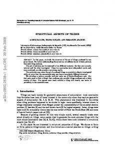
Selected myosin X-ray structures in the PDB database (www.rcsb.org). Myosin class
Organism
Motor domain 1 Dd
Ligand 1 (active site)
Ligand 2
State (ATPase cycle)
cleft
active site
lever arm position
Resolution [Å]
# amino acids
Mg · ADP·VO4
-
Pre-power stroke
-
post-rigor post-rigor post-rigor post-rigor post-rigor post-rigor
partially closed open open open open open open
closed
up
3.0
open open open open open open
down down down down down down
2.3 2.3 2.8 2.1 2.15 2.0
-
post-rigor post-rigor post-rigor
open open open
open open open
down down down
-
post-rigor
open
open
-
post-rigor
open
-
post-rigor post-rigor post-rigor post-rigor post-rigor post-rigor post-rigor post-rigor Pre-power stroke
open open open open open open open open partially closed partially closed partially closed partially closed partially closed partially closed partially closed partially closed partially closed partially closed partially closed partially closed partially closed closed closed closed open closed closed closed open open partially closed
2 2 2 2 2 2
Dd Dd Dd Dd Dd Dd
2 2 2
Dd Dd Dd
2
Dd
2
Dd
2 2 2 2 2 2 2 2 2
Dd Dd Dd Dd Dd Dd Dd Dd Gg
Mg · ADP Mg · ADP Mg · ADP / R238E Mg · ATP Mg · mNPhAE·BeF3 Mg · o-NPhAE·BeF3 Mg · p-NPhAE·BeF3 Mg·o,pNPhAE·BeF3 Mg·o,pNPhAP·BeF3 Mg·N-methylNPhAE·BeF3 Mg · MNT Mg · ADP Mg · ATPγS Mg · AMP·PNP Mg · ADP·BeF3 Mg · PPi Mg · ADP·BeF3 Mg · ADP·BeF3 Mg · ADP·AlF4
2
Gg
Mg · ADP·AlF4
-
Pre-power stroke
2
Gg
Mg · ADP·VO4
-
Pre-power stroke
2
Dd
Mg · ADP·AlF4
-
Pre-power stroke
2
Dd
Mg · ADP·VO4
Blebbistatin
Pre-power stroke
2
Dd
Mg · ADP·VO4
BL4
Pre-power stroke
2
Dd
Mg · ADP·VO4
BL6
Pre-power stroke
2
Dd
Mg · ADP·VO4
BL7
Pre-power stroke
2
Dd
Mg · ADP·AlF4
-
Pre-power stroke
2
Dd
Mg · ADP·AlF4
-
Pre-power stroke
2
Dd
Mg · ADP·BeF3
-
2
Dd
Mg · ADP·VO3
-
2
Dd
Mg · ADP·VO3
PBP
2 5 5 5 5 6 6 6 6 6
Dd Gg Gg Gg Gg Ss Ss Ss Ss Ss
2SO4 2SO4 Mg · ADP·BeF3 Mg · ADP SO42Mg · ADP·BeF3 Mg · ADP·BeF3 Mg · ADP·VO4
-
Post-recovery stroke Post-recovery stroke Post-recovery stroke rigor-like rigor-like rigor-like post-rigor rigor-like rigor-like rigor-like post-rigor post-rigor Pre-power stroke
S1 2 2 2 2 2 2 2 2 2 2 2 2 2 2 2 2 2
Lp Lp Lp Pm Lp Lp Lp Pm Gg Lp Pm Lp Ai Ai Ai Ai Ai
SO42SO42SO42Mg · ADP Mg · ADP Mg · ADP Mg · ADP / SO422SO4 Mg2+ · SO42Mg · ADP·VO4
-
rigor-like rigor-like rigor-like rigor-like rigor-like rigor-like rigor-like rigor-like post-rigor post-rigor post-rigor post-rigor post-rigor post-rigor post-rigor post-rigor Pre-power stroke
2
Ai
Mg · ADP·VO4
-
Pre-power stroke
2
Ai
Mg · ADP
-
2
Ai
Mg · AMP·PNP
-
2
Ai
Mg · ATPγS
p-PDM
2
Ai
Mg · ADP
p-PDM
2
Ai
Mg · ADP·BeF3
-
internally uncoupled internally uncoupled internally uncoupled internally uncoupled internally uncoupled
PDB id
Ref.
697
1LKX
1
776 776 1010 761 761 761
1JWY 1JX2 1G8X 1FMV 1FMW 1D0X
2
2.0 2.0 2.0
761 761 761
1D0Y 1D0Z 1D1A
5
down
2.0
761
1D1B
5
open
down
2.3
761
1D1C
5
open open open open open open open open closed
down down down down down down down down up
1.9 2.1 2.1 2.1 2.0 2.7 1.75 2.0 3.5
762 762 762 762 762 762 770 770 820
1LVK 1MMA 1MMG 1MMN 1MMD 1MNE 1W9I 1W9K 1BR1
6
closed
up
2.9
791
1BR2
11
closed
up
1.9
762
1VOM
12
closed
up
2.6
762
1MND
8
2 3 4 4 5
5 5
7 7 7 8 9 10 10 11
closed
up
2.0
762
1YV3
13
closed
up
2.0
762
3BZ7
14
closed
up
2.2
762
3BZ8
14
closed
up
2.1
762
3BZ9
14
closed
up
2.0
770
1W9J
10
closed
up
1.95
770
1W9L
10
closed
up
3.6
820
1BR4
11
closed
up
2.3
788
2JJ9
15
closed
up
2.8
788
2JHR
15
closed closed closed open closed open open open open closed
down down down down down down down down down up
1.9 2.7 2.05 2.0 3.0 2.4 2.9 2.4 2.3 1,75
776 766 795 795 795 814 858 788 788 784
2AKA/1Q5G 1W8J 1OE9 1W7J 1W7I 2BKH 2BKI 2VAS 2VB6 2V26
16
closed closed closed closed closed closed closed closed open open open open open open open open partially closed partially closed open
closed closed partially closed closed closed partially closed closed open ? open open open open open open open ? closed
down down down down down down down down down ? down down down down down down down up
2.6 3.4 3.3 3.25 3.4 3.3 2.6 3.3 2.8 3.1 3.1 3.0 3.1 2.75 3.2 4.2 2.5
839 839 839 838 839 839 839 840 843 839 840 839 840 840 837 830 840
3I5G 3I5H 3I5I 2EC6 2EKV 2EKW 2OVK 2OS8 2MYS 3I5F 2OTG 2OY6 1S5G 1SR6 1KK7 1DFK 1QVI
22
?
up
4.2
831
1DFL
26
?
uncoupled
2.5
835
1B7T
28
open
?
uncoupled
3.0
835
1KQM
25
open
?
uncoupled
3.8
835
1KWO
25
open
?
uncoupled
2.8
835
1L2O
25
open
?
uncoupled
2.3
837
1KK8
25
17 18 17 17 19 19 20 20 21
22 22 22 22 22 22 22 23 22 22 22 24 24 25 26 27
1. 2. 3. 4. 5. 6.
7. 8. 9. 10. 11. 12. 13. 14. 15.
16. 17. 18. 19. 20. 21. 22.
23. 24.
25.
26. 27. 28.
Kollmar, M.; Dürrwang, U.; Kliche, W.; Manstein, D. J.; Kull, F. J., Crystal structure of the motor domain of a class-I myosin. EMBO J. 2002, 21, (11), 2517-2525. Niemann, H. H.; Knetsch, M. L.; Scherer, A.; Manstein, D. J.; Kull, F. J., Crystal structure of a dynamin GTPase domain in both nucleotide-free and GDP-bound forms. EMBO J. 2001, 20, (21), 5813-5821. Kliche, W.; Fujita-Becker, S.; Kollmar, M.; Manstein, D. J.; Kull, F. J., Structure of a genetically engineered molecular motor. EMBO J. 2001, 20, (1-2), 40-46. Bauer, C. B.; Holden, H. M.; Thoden, J. B.; Smith, R.; Rayment, I., X-ray structures of the apo and MgATP-bound states of Dictyostelium discoideum myosin motor domain. J. Biol. Chem. 2000, 275, (49), 38494-38499. Gulick, A. M.; Bauer, C. B.; Thoden, J. B.; Pate, E.; Yount, R. G.; Rayment, I., X-ray structures of the Dictyostelium discoideum myosin motor domain with six non-nucleotide analogs. J. Biol. Chem. 2000, 275, (1), 398-408. Bauer, C. B.; Kuhlman, P. A.; Bagshaw, C. R.; Rayment, I., X-ray crystal structure and solution fluorescence characterization of Mg.2'(3')-O-(N-methylanthraniloyl) nucleotides bound to the Dictyostelium discoideum myosin motor domain. J. Mol. Biol. 1997, 274, (3), 394-407. Gulick, A. M.; Bauer, C. B.; Thoden, J. B.; Rayment, I., X-ray structures of the MgADP, MgATPγS, and MgAMPPNP complexes of the Dictyostelium discoideum myosin motor domain. Biochemistry 1997, 36, (39), 11619-11628. Fisher, A. J.; Smith, C. A.; Thoden, J. B.; Smith, R.; Sutoh, K.; Holden, H. M.; Rayment, I., X-ray structures of the myosin motor domain of Dictyostelium discoideum complexed with MgADP.BeFx and MgADP.AlF4. Biochemistry 1995, 34, (28), 8960-8972. Smith, C. A.; Rayment, I., X-ray structure of the magnesium(II)-pyrophosphate complex of the truncated head of Dictyostelium discoideum myosin to 2.7 Å resolution. Biochemistry 1995, 34, (28), 8973-8981. Morris, C. A.; Coureux, P. D.; Wells, A. L.; Houdusse, A.; Sweeney, H. L., To be Published. Dominguez, R.; Freyzon, Y.; Trybus, K. M.; Cohen, C., Crystal structure of a vertebrate smooth muscle myosin motor domain and its complex with the essential light chain: visualization of the pre-power stroke state. Cell 1998, 94, (5), 559-571. Smith, C. A.; Rayment, I., X-ray structure of the magnesium(II).ADP.vanadate complex of the Dictyostelium discoideum myosin motor domain to 1.9 A resolution. Biochemistry 1996, 35, (17), 5404-5417. Allingham, J. S.; Smith, R.; Rayment, I., The structural basis of blebbistatin inhibition and specificity for myosin II. Nat. Struct. Mol. Biol. 2005, 12, (4), 378-379. Lucas-Lopez, C.; Allingham, J. S.; Lebl, T.; Lawson, C. P.; Brenk, R.; Sellers, J. R.; Rayment, I.; Westwood, N. J., The small molecule tool (S)-(-)-blebbistatin: novel insights of relevance to myosin inhibitor design. Org. Biomol. Chem. 2008, 6, (12), 2076-2084. Fedorov, R.; Böhl, M.; Tsiavaliaris, G.; Hartmann, F. K.; Taft, M. H.; Baruch, P.; Brenner, B.; Martin, R.; Knölker, H. J.; Gutzeit, H. O.; Manstein, D. J., The mechanism of pentabromopseudilin inhibition of myosin motor activity. Nat. Struct. Mol. Biol. 2009, 16, (1), 80-88. Reubold, T. F.; Eschenburg, S.; Becker, A.; Leonard, M.; Schmid, S. L.; Vallee, R. B.; Kull, F. J.; Manstein, D. J., Crystal structure of the GTPase domain of rat dynamin 1. Proc. Natl. Acad. Sci. U S A 2005, 102, (37), 13093-13098. Coureux, P. D.; Sweeney, H. L.; Houdusse, A., Three myosin V structures delineate essential features of chemo-mechanical transduction. EMBO J. 2004, 23, (23), 4527-4537. Coureux, P. D.; Wells, A. L.; Ménétrey, J.; Yengo, C. M.; Morris, C. A.; Sweeney, H. L.; Houdusse, A., A structural state of the myosin V motor without bound nucleotide. Nature 2003, 425, (6956), 419-423. Ménétrey, J.; Bahloul, A.; Wells, A. L.; Yengo, C. M.; Morris, C. A.; Sweeney, H. L.; Houdusse, A., The structure of the myosin VI motor reveals the mechanism of directionality reversal. Nature 2005, 435, (7043), 779-785. Ménétrey, J.; Llinas, P.; Cicolari, J.; Squires, G.; Liu, X.; Li, A.; Sweeney, H. L.; Houdusse, A., The post-rigor structure of myosin VI and implications for the recovery stroke. EMBO J. 2008, 27, (1), 244-252. Ménétrey, J.; Llinas, P.; Mukherjea, M.; Sweeney, H. L.; Houdusse, A., The structural basis for the large powerstroke of myosin VI. Cell 2007, 131, (2), 300-308. Yang, Y.; Gourinath, S.; Kovacs, M.; Nyitray, L.; Reutzel, R.; Himmel, D. M.; O'Neall-Hennessey, E.; Reshetnikova, L.; SzentGyorgyi, A. G.; Brown, J. H.; Cohen, C., Rigor-like structures from muscle myosins reveal key mechanical elements in the transduction pathways of this allosteric motor. Structure 2007, 15, (5), 553-564. Rayment, I.; Rypniewski, W. R.; Schmidt-Base, K.; Smith, R.; Tomchick, D. R.; Benning, M. M.; Winkelmann, D. A.; Wesenberg, G.; Holden, H. M., Three-dimensional structure of myosin subfragment-1: a molecular motor. Science 1993, 261, (5117), 50-58. Risal, D.; Gourinath, S.; Himmel, D. M.; Szent-Gyorgyi, A. G.; Cohen, C., Myosin subfragment 1 structures reveal a partially bound nucleotide and a complex salt bridge that helps couple nucleotide and actin binding. Proc. Natl. Acad. Sci. U S A 2004, 101, (24), 8930-8935. Himmel, D. M.; Gourinath, S.; Reshetnikova, L.; Shen, Y.; Szent-Györgyi, A. G.; Cohen, C., Crystallographic findings on the internally uncoupled and near-rigor states of myosin: further insights into the mechanics of the motor. Proc. Natl. Acad. Sci. U S A 2002, 99, (20), 12645-12650. Houdusse, A.; Szent-Györgyi, A. G.; Cohen, C., Three conformational states of scallop myosin S1. Proc. Natl. Acad. Sci. U S A 2000, 97, (21), 11238-11243. Gourinath, S.; Himmel, D. M.; Brown, J. H.; Reshetnikova, L.; Szent-Györgyi, A. G.; Cohen, C., Crystal structure of scallop Myosin S1 in the pre-power stroke state to 2.6 A resolution: flexibility and function in the head. Structure 2003, 11, (12), 1621-1627. Houdusse, A.; Kalabokis, V. N.; Himmel, D.; Szent-Györgyi, A. G.; Cohen, C., Atomic structure of scallop myosin subfragment S1 complexed with MgADP: a novel conformation of the myosin head. Cell 1999, 97, (4), 459-470.