View Article Online
Nanoscale
View Journal
Accepted Manuscript This article can be cited before page numbers have been issued, to do this please use: A. H. Mo, P. Landon, B. Meckes, M. Yang, G. V. Glinsky and R. Lal, Nanoscale, 2013, DOI: 10.1039/C3NR05365B.
Volume 2 | Number 1 | 2010
This is an Accepted Manuscript, which has been through the RSC Publishing peer review process and has been accepted for publication.
www.rsc.org/nanoscale
Volume 2 | Number 1 | January 2010 | Pages 1–156
Nanoscale
Accepted Manuscripts are published online shortly after acceptance, which is prior to technical editing, formatting and proof reading. This free service from RSC Publishing allows authors to make their results available to the community, in citable form, before publication of the edited article. This Accepted Manuscript will be replaced by the edited and formatted Advance Article as soon as this is available. To cite this manuscript please use its permanent Digital Object Identifier (DOI®), which is identical for all formats of publication. More information about Accepted Manuscripts can be found in the Information for Authors.
ISSN 2040-3364
Pages 1–156
COVER ARTICLE Graham et al. Mixed metal nanoparticle assembly and the effect on surface-enhanced Raman scattering
REVIEW Lin et al. Progress of nanocrystalline growth kinetics based on oriented attachment
2040-3364(2010)2:1;1-T
Please note that technical editing may introduce minor changes to the text and/or graphics contained in the manuscript submitted by the author(s) which may alter content, and that the standard Terms & Conditions and the ethical guidelines that apply to the journal are still applicable. In no event shall the RSC be held responsible for any errors or omissions in these Accepted Manuscript manuscripts or any consequences arising from the use of any information contained in them.
www.rsc.org/nanoscale Registered Charity Number 207890
Page 1 of 6
Nanoscale View Article Online
Using an inosine substitution, a junction structure can independently control the activity of a split dnazyme through repeated cycles.
Nanoscale Accepted Manuscript
Published on 06 November 2013. Downloaded by University of California - San Diego on 06/11/2013 17:24:13.
DOI: 10.1039/C3NR05365B
Journal Name
Nanoscale
Page 2 of 6 Dynamic Article Links ► View Article Online
DOI: 10.1039/C3NR05365B
Cite this: DOI: 10.1039/c0xx00000x
ARTICLE TYPE
On-demand Four-way Junction DNAzyme Nanoswitch Driven by Inosine-Based Partial Strand Displacement Alexander H. Mo*, 1 , Preston B. Landon*, #, 2,3, Brian Meckes3, Max M. Yang3, Gennadi V. Glinsky4 and Ratnesh Lal#,1,2,3 5
10
Received (in XXX, XXX) Xth XXXXXXXXX 20XX, Accepted Xth XXXXXXXXX 20XX DOI: 10.1039/b000000x A DNA four-way junction device capable of junction expansion and contraction cycles using an inosine-based partial strand displacement scheme is reported. These nanoscale positioning capabilities are used to provide on-demand activation and deactivation of a pair of split E6 DNAzymes on the device. The device also demonstrates a combined catalytic rate significantly higher than the original E6 DNAzyme under similar operational conditions. This approach can provide structural organization and spatially control other multicomponent molecular complexes. platform for DNA-based biosensing because its conformational switching abilities give it the requisite functionality to operate like those reported in literature.20-24
Introduction 15
20
25
30
35
40
45
DNA four-way junctions (4WJ) are found naturally as mobile junctions during DNA replication1, and are also synthesized as immobile structural components of 2D DNA lattices. The 4WJ motif presents a way to precisely place different molecules in close proximity with a dynamic control over their spacing. Mobile 4WJ can spontaneously switch between different conformations.2-5 Immobile 4WJ, however are usually static and have limited switchability, if any.6-9 With controllable shifts in conformation, a DNA 4WJ can be useful for the precise positioning of chemical moieties or a nanoscale switch to regulate other processes such as activity of E6 DNAzymes10. One way to design such a 4WJ is to make parts of the DNA junction stimuli responsive. DNA-based nanostructures with active elements responsive to light11, metal ions12, pH13, 14, temperature15, 16, and toehold strand displacement 17, 18 have been designed. The toehold strand displacement that has been used in actuation of various DNA nanomachines rely on the invasion of a singlestranded DNA into a double-stranded oligonucleotide until the toehold-less strand is completely displaced.19 However if this is applied to a 4WJ, full displacement of any portion of a 4WJ will disassociate the structure. Here we report a 4WJ capable of repeatable junction point expansion and contraction via an inosine-based partial strand displacement scheme. The cycles of the 4WJ are monitored using a fluorophore and quencher pair. Furthermore, the nanoscale positioning capability of this structure was used for ondemand switching of a pair of opposing split E6 DNAzymes. A combined catalytic rate obtained with this device is significantly higher than a previously reported E6 DNAzyme10 under similar conditions. The switching system of this device turns a chemical cue into a physical displacement, which in turn facilitates defined structural organization and enables controlling the behaviour of enzyme complexes. In addition it may also be applied as reusable This journal is © The Royal Society of Chemistry [year]
50
55
60
65
70
75
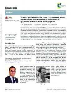
Results and Discussion In order to develop the controllable 4WJ nanodevices, we have designed an inosine-based strand displacement system (SDS). This SDS is capable of partial strand displacement by introducing energetic differences between the original and the displaced duplex.25 In this system, one strand is limited to inosines and thymines, while its complimentary strand is limited to adenines and cytosines. The duplex is held together by two hydrogen bonds across its entire length. A third strand called a displacement strand (the opening strand) consists of thymines, and guanines in place of inosines (similar to the above mentioned complementary strand but with guanine instead of inosine).25 Because the guanine-cytosine bond is more energetically favorable than the inosine-cytosine bond, the displacement strand is able to dislodge the inosine containing strand without use of a toehold. By using this inosine-rich active region, a contractile system capable of maintaining a stable, partially-displaced state with multiple opening and closing cycles is accomplished. (Figure 1).
Figure 1. 4WJ Layout The closed DNA 4WJ (A) and the open DNA 4WJ (B). Two inosine containing strands (red/blue duplex) were on opposite sides of the 4WJ. The device can be cycled between closed state and open state by the introduction of the appropriate strands. The conformation change is monitored by fluorophore (red dot) and quencher (black dot) pair placed near the junction point of the 4WJ. The insets show how a difference in bond strength between I-C and G-C allows the
[journal], [year], [vol], 00–00 | 1
Nanoscale Accepted Manuscript
Published on 06 November 2013. Downloaded by University of California - San Diego on 06/11/2013 17:24:13.
www.rsc.org/xxxxxx
Page 3 of 6
Nanoscale View Article Online
DOI: 10.1039/C3NR05365B
55
purely by the collision of the opening strand with the inosine section as no toehold was present to further speed up the reaction. Closing of the device was mediated by a 6 nucleotide (nt) toehold. . It took about 8-10 minutes to reach half fluorescence
5
10
15
20
25
30
35
40
45
50
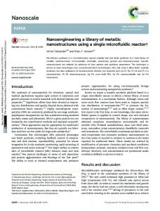
Figure 2. 4WJ Cycling A) Real time monitoring of the four-way junction is performed by measuring the fluorescence intensity upon introduction of an opening or closing strand. The device can be cycled between closed and open states by introduction of successively higher amounts of opening and closing strands at 37°C. In this figure the opening strands were introduced at 10, 30, and 50 times the amount of dual-spring actuators. And closing strands were introduced at 20 and 40 times the amount of dual-spring actuators. Additionally approximately 10% of opened strands never fully close again. This behavior was attributed to competition between the closing strand and device with the opening strand. B) Independent functionality of the two inosine-rich sequences was confirmed by sequentially adding opening strands for each side and then sequentially closing each side at 37°C. Upon addition of side 1’s opening strands the fluorescence increased approximately 50% and leveled off. Upon addition of side 2’s opening strand, the fluorescence rises sharply again and levels off. This behavior is confirmed again when sequential addition of closing strands yields the same behavior. The appearance of these fluorescence levels indicates that inosine-rich sequences were sufficiently different in sequence to be independently operated. It also demonstrates that both inosine-rich sequences contribute to the opening of the 4WJ. Error bars represent one standard deviation.
To demonstrate the thermodynamic stability of the 4WJ in their different conformations, a full opening and closing cycle were annealed in separate batches and compared using gel electrophoresis. (Figure S1) A comparison of the 4WJ annealed by itself to 4WJs annealed with excess opening strands showed an increase in the molecular weight and increased FAM fluorescence of the latter due to incorporation of opening strands into the device. Comparison of the first two samples with 4WJs annealed with excess of opening and closing strand revealed 4WJ switched back to its closed state. This was indicated by a drop in the molecular weight and the disappearance of the FAM fluorescence signal. The structure of the 4WJ shape was imaged by transmission electron microscope. Figure 1 showed a four arm X-shape about 50 nm long and 30 nm wide. This confirms that the structure has assembled as designed. Additional TEM images can be found in Figure S2. The kinetic behavior of the 4WJ was observed using time lapsed fluorescence measurements at 37°C. In order to determine the range of mole excess appropriate for kinetic cycling experiments, various concentrations of opening strands were attempted. (Figure S3). At mole excesses tested (10, 50, 75 times) the fluorescence intensity levels out to the same place indicating that a population of 4WJs is fully opened. The device was then cycled multiple times by alternating addition of opening and closing strands at successively higher concentrations (Figure 2A). Opening of the device was driven
60
65
70
75
80
85
90
95
2 | Journal Name, [year], [vol], 00–00
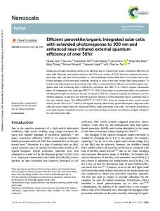
Figure 3. 4WJ DNAzyme A) The DNAzymes attached to the 4WJ can be inactivated by introduction of opening strands that widen the structure and separate the two halves of the DNAzyme. Side 1’s DNAzyme strands (W1 E6, N2 E6) and Side 2’s DNAzyme strands (W2 E6, N2 E6) were now so far apart, any target strand that diffuses into the area may bind with one side of the DNAzyme but will never be cut. The target strand consists of a fluorophore and quencher at opposite ends of the nucleic acid strand. The strand is mostly DNA with the exception of one RNA adenine (rA) in the middle. B) The DNAzyme is active when the both sides of the 4WJ were in the closed position, bringing the two split parts of the DNAzyme close together. This close proximity at all times allows a target strand that diffuses into to be cut by the waiting DNAzymes. It is at the rA where the DNAzyme breaks the target strand into two pieces, resulting in an increase of the fluorescent signal.
when the device was opening but only less than 1 minute to drop to half when closing. Comparing rate constants between opening and closing legs of the cycle, the closing rate is 2 orders of magnitude faster than the opening strand. The 4WJ structure was faster when closing than when opening, however, not every device closed after each cycle. The fluorescence levels upon closing remain at 10% of the normalized fluorescence value (Figure 2A, B). This would indicate approximately 10% of 4WJ did not fully close indicating that the devices cannot be cycled with perfect efficiency. This behavior was consistent with other strand displacement devices where device cycling efficiency decreases with cycle number.18 We also examined the ability of each side of the 4WJ for independent actuation. This was tested by sequential addition of opening strands for side 1 followed by side 2 at 10 times excess followed by sequential addition of closing strands of side 1 and side 2 at 20 times excess. In Figure 2B, two sharp increases in fluorescence intensity followed by two sharp drops in fluorescence intensity demonstrated that each side contributed to roughly half the function of the device, as well as that each side’s opening and closing strands retained their strand specificity. Rate constants extracted by curve fitting of Figure 2B, using assumptions described in the supplemental section, were listed in This journal is © The Royal Society of Chemistry [year]
Nanoscale Accepted Manuscript
Published on 06 November 2013. Downloaded by University of California - San Diego on 06/11/2013 17:24:13.
4WJ to open. The opening strand is removed by a toehold (arrows coming off opening strands) that binds with the closing strand, thus resetting the device. C) A representative TEM images shows reveals the structure has assembled as designed. Scale bar is 20 nm.
Nanoscale
Page 4 of 6 View Article Online
Published on 06 November 2013. Downloaded by University of California - San Diego on 06/11/2013 17:24:13.
5
Table 1. The closing reaction was significantly faster (~100 times faster) than the opening reaction for both sides. This can be explained by the different mechanisms by which the opening and closing are achieved. The opening operates based on a currently unknown mechanism by which the opening strand presumably infiltrates the device’s helix and displaces the weak side. The
55
60
65
10
15
20
25
30
35
40
45
50
Figure 4. 4WJ DNAzyme Operation A) Demonstration of 4WJ E6-type DNAzyme function at 25°C. I) Addition of 1 µL active DNAzyme (0.5 µM) into 50 µL of target strand (0.1 µM) with Mg2+ (10 mM). II) Shut off DNAzyme activity with addition 1 µL of solution of Open N1 and Open N2 (both 5 µM). III) Restoration of DNAzyme activity with 1 µL of solution of Close N1 and Close N2 (both 10 µM). Error bars represent one standard deviation. B) Michaelis-Menten fitting of an active 4WJ DNAzyme with both sides closed at 25°C. Error bars represent two standard deviations.
closing strand operates on the better understood toehold strand displacement. This infiltration method takes a greater time to complete than the toehold mechanism. The 4WJ‘s utility as a switch was then demonstrated by annealing two identical split E6-type DNAzymes into the 4WJ in place of fluorophore/quencher attached strands of DNA. When the 4WJ was open the DNAzymes were inactive because the two halves of the split DNAzyme on both sides of the device were separated (Figure 3A). When the 4WJ was closed, the DNAzyme became active because the two halves of the DNAzyme were close enough together to cut its target: the reporter strand (Figure 3B). This reporter strand consisted of complementary DNA bases at the ends that bind to the DNAzyme, a single RNA adenine (rA) at the center of the strand, and a fluorophore and quencher on the 5’ and 3’ ends respectively. Thus, when DNAzymes cut the reporter after rA, the fluorophore separated from the quencher and allowed the reaction to be monitored. Initially active in the closed state, the 4WJ DNAzyme was allowed to cut reporter strands at 25°C (Figure 4A, I). The device was inactivated by injection of 10 times excess opening strands at 120 minutes (Figure 4A, II). The device gradually became inactive and remained so until 20 times excess closing strands were added at 390 minutes (Figure 4A, III) to reactivate the 4WJ DNAzyme. This behavior was consistent with opening and closing cycles found in Figure 2A. The DNAzyme was also able to cut with specificity when tested against a reporter with a scrambled sequence (Figure S4). Analysis of 4WJ annealed DNAzymes on one or both sides revealed that Side 1’s DNAzyme cutting velocity was about three times as fast as side 2’s (Figure S5). By design, both sides should have the same cutting speed because they were binding the same reporter strand and had the same catalytic core sequences. 4WJ DNAzymes with 0, 4, and 8 bp complementary stabilizer sections between the two halves of the DNAzyme showed direct gains in cutting velocity with This journal is © The Royal Society of Chemistry [year]
70
75
80
85
stabilizer sections length (Figure S6). Based on this data, the longer the stabilizer sections, the higher the melting point, the more stable the stabilizer section and the accompanying DNAzymes were. This strongly suggested that the conformational stability of the DNAzyme catalytic core was related to its cutting velocity. Upon inspection of the sequences in side 1 and side 2, it can be seen that 3 bases of the catalytic core for side 2 adjacent to the inosine-rich section on W2 was complementary to 3 bases in the inosine-rich section. The DNAzyme strand on W2 was partially competing with part of the normal side. This competition would lead to lower catalytic core stability and reduce the overall cutting velocity of side 2’s DNAzyme. No such complementary bases were present on side 1, and thus side 1’s catalytic core was more stable and able to cut reporter strands more often. This competition for bases on side 2 was the most likely reason for the cutting speed discrepancy between the two sides. Thus taking advantage of inosine’s wobble base nature and with careful placement in the inosine SDS sequence, the sequences can be used to modify the cutting velocity of other split DNAzymes. In addition to monitoring 4WJ DNAzyme switching ability, the kinetics of the device’s cutting ability was characterized with Michaelis-Menten kinetics (Figure 4B). Using substrate concentrations ranging from 0.1-1 µM and an enzyme concentration of 0.01 µM, initial velocities were found by a linear fit to the first 10% of data points in the reaction. MichaelisMenten constants were determined by plotting substrate concentration and initial velocities using the Hanes-Woolf plot. When both sides were activated, their combined catalytic rate (kcat) was 0.052 min-1, the effective binding constant (Km) is 110 nM, and their ratio (kcat/Km) is 480,000 M-1min-1. Under similar buffer (magnesium ion levels (10 mM vs. 12 mM), sodium ion levels (160 mM vs. 160 mM)), and temperature (23 vs. 25°C), the collective catalytic activity of the 4WJ DNAzymes was an order of magnitude higher than the originally developed E6 DNAzyme with catalytic rate of ~0.005 min-1.10 Table 1. Rate constants calculated from curve fitting Figure 2b Opening Cycle
ko (M-1s-1)
Side 1
190 ± 10
Side 2
380 ± 40
Closing Cycle
kc (M-1s-1)
Side 1
14000. ± 5000
Side 2
20000 ± 7000
Conclusions 90
We have successfully constructed a stable 4WJ with conformational switching abilities. The non-complementary sequences that make up the 4WJ prevent the device from spontaneously switching between conformations. Addition of the inosine rich sequences in two of the arms introduces switchable elements in the junction without disassociating the whole Journal Name, [year], [vol], 00–00 | 3
Nanoscale Accepted Manuscript
DOI: 10.1039/C3NR05365B
Page 5 of 6
Nanoscale View Article Online
DOI: 10.1039/C3NR05365B
10
15
20
60
65
70
75
drop cast on Formavar coated TEM grids (Tedpella, Redding, CA). The grids were incubated for 15 minutes before a laboratory tissue absorbed the solution. The samples were then stained by floating the TEM grids on a 100 µL droplet of 1% uranyl acetate for 5 minutes. Afterwards the excess stain was removed via filter paper and allowed to air dry for 5 minutes. TEM images were acquired using JEOL 1200 EX II at an accelerating voltage of 80 kV. Gel Electrophoresis 10 µL of 0.1 µM DNA solution was mixed with 2 µL of 10% v/v glycerin solution and placed in a 4% agarose gel (Lonza, Rockland, ME) preloaded with ethidium bromide (EtBr). DNA gel electrophoresis was performed with the gel placed in a 1X TBE Buffer (Fischer Scientific, Pittsburgh, PA) at temperatures between 20-25°C at with an electric field of 7.3 V/cm for 45 minutes. Gels are imaged with a Bio-Rad FX-Imager Pro Plus (Bio-Rad, Hercules, CA) and analyzed with the Quantity One software package (Bio-Rad). EtBr imaging is performed with the internal 532 nm laser and 555 nm band pass filter, while Cy5 650/670 nm (ex/em) imaging used an external 635 nm laser and a 690 nm long pass filter. FAM imaging relied on an external 488 nm laser with a 530 nm band pass filter.
Methods 25
30
35
40
4WJ design The device is driven by two independent inosine rich sequences indicated as “1” and “2”. Strands containing only inosines and thymines in the sequence are indicated as such with a “W”. Strands containing cytosines and adenines are denoted as such with an “N”. Four structural strands (N1, N2, W1, and W2) plus two cross over strands (N2/W1, N1/W2) assemble together to form the 4WJ (Figure 1). The crossover strands and all other regions do not contain any inosine. To monitor device functionality, two carboxyfluorescein (FAM) fluorophores and two Iowa Black FQ quenchers were attached to N2/W1 and N1/W2 respectively on both the 5’ and 3’ ends. (Figure 1) Device opening was triggered by introduction of a single stranded DNA complementary to the “N” strands. Opening strands (Open N1, Open N2) also contain a 6 nucleotide (nt) sticky end enabling removal by a closing strand (Close N1, Close N2).
80
4WJ Characterization
85
Measurements were performed at 480/530 (Ex/Em) in 96 well black plates (Nalgen Nunc, Rochester, NY) in a using Infinite M200 Pro (Tecan, San Jose, CA) spectrophotometer at 37°C. 50 µL of DNA 4WJs (0.1 µM) are placed in a well and 1 µL of increasingly concentrated solutions alternating between opening and closing strands (50, 100, 150, 200, 250 µM) were added to the solution as soon as the fluorescence signal starts to level out. The molar excess in the cycling progression are equivalent to opening/closing strands 10, 20, 30, 40, and 50 times excess.
90
DNAzyme Control
95
DNA strands
45
50
DNA strands were designed and ordered from Integrated DNA Technologies (Coralville, IA). The strands were resuspended in a buffer consisting of 30 mM of Tris (Fischer Scientific, Pittsburgh, PA) and 160 mM of NaCl (Sigma, St. Louis, MO) in MilliQ deionized water (Millipore, Billerica, MA) . DNA strands were assembled via thermal annealing using a PCR thermocycler (Mastercycler Personal, Eppendorf, Westbury, NY) programmed to raise the solution temperature to 94°C and then cool at rate of 1°C every 2 minutes until the final temperature is 4°C. A complete list of sequences is given in Table S1. Imaging
55
Annealed 4WJs were diluted to 1 nM in MilliQ water and 10 µL 4 | Journal Name, [year], [vol], 00–00
100
105
110
Using the sequences for a split E6-type DNAzyme adapted from Elbaz et al., two identical DNAzymes were annealed into the device, one on each side of the device. (Figure 3A, W1 E6, N1 E6, W2 E6, N2 E6) E6-type containing 4WJ were annealed at a concentration 0.5 µM. A reporter nucleic acid strand consisting mostly of DNA with a single RNA adenine base (rA) in the middle is used to monitor DNAzyme activity. A fluorescentquencher pair on the 5’ (Texas Red) and 3’ (Iowa Black RQ) of the reporter strand normally keeps the fluorescence of the molecule low. However in the presence of the 4WJ DNAzyme, the reporter strand is hydrolyzed just after the rA. This separates the fluorophore from the quencher, allowing the fluorescent signal to increase. Measurements were performed at 590/620 nm (Ex/Em) in black 96 well plates using the Infinite M200 Pro spectrophotometer at 25°C. 50-µL of substrate strand of known concentrations was added in at least triplicate with 1 µL of a buffered magnesium chloride solution (600 mM MgCl2, 30 mM Tris, 16 mM NaCl). This raised the Mg2+ concentration of the well to 12 mM. Incubation of the well plate took place at 25°C for at least 15 This journal is © The Royal Society of Chemistry [year]
Nanoscale Accepted Manuscript
Published on 06 November 2013. Downloaded by University of California - San Diego on 06/11/2013 17:24:13.
5
junction. Using gel electrophoresis and time lapsed fluorescence, the 4WJ is able to open and close multiple times and each inosine-rich section is able to function independently of the other. The 4WJ is demonstrated to be a competent nanoswitch, able to turn on and off the activity of split DNAzymes attached to it. The 4WJ DNAzyme structure also had an order of magnitude increase in its catalytic rate over its single stranded form with all else being the same. The inclusion of the inosine inside a four-way junction opens up the center to allow for a variety of processes to happen. These features can be readily expanded into other applications in quantitative biology, nanomedicine, and nanomanipulation. DNA strands dissociation and re-association processes are essential components of DNA double helix dynamics in vivo during replication, transcription, and reciprocal dynamic transitions between right-handed B-DNA and high-energy lefthanded Z-DNA conformations. Reported here successful development of nano-devices demonstrating on-demand dissociation and re-association of DNA strands in response to induced 4WJ expansion and contraction cycles should facilitate a single molecule level mechanistic analyses of DNA strand displacement reactions during these fundamental biological processes.
Nanoscale
Page 6 of 6 View Article Online
minutes followed by at least 30 minutes of background readings. 1 µL of 4WJ DNAzymes were added to each measurement well after background readings were completed. This raised the concentration of 4WJ DNAzyme to 0.01 µM. 60
Published on 06 November 2013. Downloaded by University of California - San Diego on 06/11/2013 17:24:13.
5
10
Acknowledgements We extend our thanks to Prof. Xiaohua Huang and Nirav Patel for helpful discussions and Timo Merloo for TEM sample preparation and imaging assistance. The work is supported by grants from National Institute on Drug Abuse [5R01DA02529604]; and departmental development funds from the Dept. of Mechanical and Aerospace Engineering, UCSD.
Notes and references
65
70
1
15
20
Materials Science and Engineering Program Department of Mechanical and Aerospace Engineering 3 Department of Bioengineering, University of California, San Diego, 9500 Gilman Drive, La Jolla, CA 92093, 4 Stanford University Medical School & Sanford-Burnham Medical Research Institute, 10901 North Torrey Pines Road, La Jolla, CA 92037. *Authors contributed equally to the paper. # For more information please contact PB,
[email protected], or RL,
[email protected] 2
† Electronic Supplementary Information (ESI) available: Supplemental Fig and Discussion. See DOI: 10.1039/b000000x
75
80
14. F. Xia, W. Guo, Y. Mao, X. Hou, J. Xue, H. Xia, L. Wang, Y. Song, H. Ji, Q. Ouyang, Y. Wang and L. Jiang, Journal of the American Chemical Society, 2008, 130, 8345-8350. 15. L. M. Dillenback, G. P. Goodrich and C. D. Keating, Nano Letters, 2005, 6, 16-23. 16. T. Goda and Y. Miyahara, Biosensors and Bioelectronics, 2011, 26, 3949-3952. 17. P. Yin, H. Yan, X. G. Daniell, A. J. Turberfield and J. H. Reif, Angewandte Chemie International Edition, 2004, 43, 49064911. 18. B. Yurke, A. J. Turberfield, A. P. Mills, F. C. Simmel and J. L. Neumann, Nature, 2000, 406, 605-608. 19. D. Y. Zhang and G. Seelig, Nature chemistry, 2011, 3, 103-113. 20. D.-L. Ma, H.-Z. He, D. S.-H. Chan and C.-H. Leung, Chemical Science, 2013, 4, 3366-3380. 21. D. Li, S. Song and C. Fan, Accounts of Chemical Research, 2010, 43, 631-641. 22. D.-L. Ma, H.-Z. He, K.-H. Leung, H.-J. Zhong, D. S.-H. Chan and C.-H. Leung, Chemical Society Reviews, 2013, 42, 3427-3440. 23. O. I. Wilner and I. Willner, Chemical Reviews, 2012, 112, 25282556. 24. C.-H. Leung, H.-J. Zhong, H.-Z. He, L. Lu, D. S.-H. Chan and D.-L. Ma, Chemical Science, 2013, 4, 3781-3795. 25. P. B. Landon, S. Ramachandran, A. Gillman, T. Gidron, D. Yoon and R. Lal, Langmuir, 2011, 28, 534-540.
25
1. 2. 30
3. 4.
35
5.
40
6. 7. 8.
45
9. 10. 11.
50
12. 13. 55
F. A. Hays, J. Watson and P. S. Ho, Journal of Biological Chemistry, 2003, 278, 49663-49666. S. Hohng, R. Zhou, M. K. Nahas, J. Yu, K. Schulten, D. M. J. Lilley and T. Ha, Science, 2007, 318, 279-283. S. A. McKinney, A. C. Declais, D. M. Lilley and T. Ha, Nature structural biology, 2003, 10, 93-97. A. R. Mount, C. P. Mountford, S. A. G. Evans, T. J. Su, A. H. Buck, P. Dickinson, C. J. Campbell, L. M. Keane, J. G. Terry, J. S. Beattie, A. J. Walton, P. Ghazal and J. Crain, Biophysical Chemistry, 2006, 124, 214-221. C. P. Mountford, A. H. Buck, C. J. Campbell, P. Dickinson, E. E. Ferapontova, J. G. Terry, J. S. Beattie, A. J. Walton, P. Ghazal, A. R. Mount and J. Crain, The journal of physical chemistry. B, 2008, 112, 2439-2444. N. C. Seeman, Journal of Theoretical Biology, 1982, 99, 237-247. Y. He, Y. Tian, Y. Chen, Z. Deng, A. E. Ribbe and C. Mao, Angewandte Chemie, 2005, 117, 6852-6854. L. Feng, S. H. Park, J. H. Reif and H. Yan, Angewandte Chemie International Edition, 2003, 42, 4342-4346. P. Sung Ha, Y. Hao, H. R. John, H. L. Thomas and F. Gleb, Nanotechnology, 2004, 15, S525. R. R. Breaker and G. F. Joyce, Chemistry & biology, 1995, 2, 655660. H. Liu, Y. Xu, F. Li, Y. Yang, W. Wang, Y. Song and D. Liu, Angewandte Chemie International Edition, 2007, 46, 25152517. Z. Wang, J. Heon Lee and Y. Lu, Chemical Communications, 2008, 6005-6007. L. Wang, X. Liu, Q. Yang, Q. Fan, S. Song, C. Fan and W. Huang, Biosensors and Bioelectronics, 2010, 25, 1838-1842.
This journal is © The Royal Society of Chemistry [year]
Journal Name, [year], [vol], 00–00 | 5
Nanoscale Accepted Manuscript
DOI: 10.1039/C3NR05365B