340
LETTER TO JMG
Naturally occurring mutations and functional polymorphisms in multidrug resistance 1 gene: correlation with microsatellite instability and lymphoid infiltration in colorectal cancers U Potocˇnik, M Ravnik-Glavacˇ, R Golouh, D Glavacˇ .............................................................................................................................
J Med Genet 2002;39:340–346
glycoprotein (Pgp), encoded by the MDR1 gene, is a transmembrane transporter that acts as an efflux pump in an ATP dependent fashion.1 Multidrug resistance, the main problem in efficient cancer chemotherapy, is mainly caused by increased expression and acquired mutations in the MDR1 gene.2 Pgp is expressed physiologically in epithelial cells of the kidney, liver, pancreas, and colon, suggesting its role in secretion of toxic compounds.3 Pgp is also expressed in the blood-brain barrier, adrenal glands, and lymphocytes where its role is still uncertain. Recently, additional functions for Pgp, including immune response4 and regulation of apoptosis,5 have been suggested in normal tissues and in cancers. High expression of Pgp at the apical surface of differentiated tubular structures was identified in previously untreated colorectal cancers (CRC)6 and its high expression at the leading edge of a colorectal carcinoma was associated with tumour progression.7 In contrast to the majority of CRC, which develop as a result of chromosomal instability (CIN), a proportion of sporadic CRC and 90% of cancers in patients with hereditary non-polyposis colorectal cancer (HNPCC) exhibit microsatellite instability (MSI).8 In MSI CRC, chromosomal aberrations such as large deletions, translocations, and gene amplifications are rare; the great majority of MSI tumours are usually diploid or near diploid. In MSI CRC, inactivation of the mismatch repair (MMR) system owing to mutations or methylation of MMR genes results in a 1000-fold accumulation of point mutations in oncogenes and tumour suppressor genes which trigger tumour progression. Therefore, in addition to genes mutated in microsatellite stable (MSS) CRC, such as APC, p53, and K-ras, there are other genes important in the development of MSI cancers. Interestingly, MSI CRCs are also more often resistant to several chemotherapy drugs; the selection of cells for resistance to cisplatin can result in the loss of DNA mismatch repair, and loss of DNA mismatch repair in turn contributes to resistance to cisplatin.9 Recently, a transcription factor complex TCF4/β catenin responsive element was identified in the MDR1 promoter region pointing to a direct link between the MDR1 gene and the WNT signalling pathway, the most important pathway altered in colorectal cancers.10 To determine the role of the MDR1 gene in the initiation and progression of CRC, we systematically screened the complete coding and promoter region of the MDR1 gene for alterations in a large cohort of patients with previously untreated colorectal cancer and in a normal control population. In this study, we report naturally occurring functional germline and somatic mutations in the MDR1 gene in patients with microsatellite unstable CRC and correlation of MDR1 functional polymorphisms with increased lymphoid infiltration in tumours with and without MSI.
P
www.jmedgenet.com
MATERIALS AND METHODS Patients Between 1996 and 2000, 400 newly diagnosed colorectal cancer (CRC) patients from clinics all over Slovenia participated in this study. None of these patients had received chemotherapy treatment before operation. Primary colorectal adenocarcinomas as well as corresponding normal colorectal mucosa taken from a site several centimetres distant from the tumour were used in the study. Tumours were histopathologically evaluated according to the classification of Jass et al.11 The lymphoid infiltration of the tumours was assessed semiquantitatively by two independent pathologists. Accordingly, tumours with well represented lymphocytes along the advancing margin of the tumour were scored as positive for lymphoid infiltration. Control samples To determine the potential pathogenicity of MDR1 alterations detected in our study, we also analysed DNA from 100 unrelated unaffected blood donors. DNA isolation Colorectal tumours and corresponding normal tissue samples were snap frozen in liquid nitrogen and stored at −70°C. DNA was isolated after tissue digestion using standard phenol/ chloroform extraction and ethanol precipitation. Analysis of microsatellite instability Microsatellite instability (MSI) analysis using a “reference panel” of microsatellite markers was performed in 400 unselected primary colorectal cancers (CRC) as described in our previous study.12 Thirty-eight tumours were defined as high microsatellite instability tumours (MSI-H) and analysed for MDR1 mutations. MDR1 mutational analysis We designed primers based on known genomic DNA sequences (Genbank accession numbers AC002457 and AC005068) specifically to amplify all 28 exons and exon/intron boundaries as well as the promoter region of the MDR1 gene in separate PCR reactions. Primer sequences and optimised PCR conditions are available at
[email protected]. For mutational analysis of the MDR1 gene, we used non-isotopic conformation analysis and silver staining. The basic principle of this method is a combination of three analyses which are all based on changes in ............................................................. Abbreviations: Pgp, P glycoprotein; CRC, colorectal cancer; CIN, chromosome instability; HNPCC, hereditary non-polyposis colorectal cancer; MSI, microsatellite instability; MMR, mismatch repair; MSS, microsatellite stable; SSCA, single strand conformation analysis; HA heteroduplex analysis; DSCA, double strand conformation analysis
Letters
341
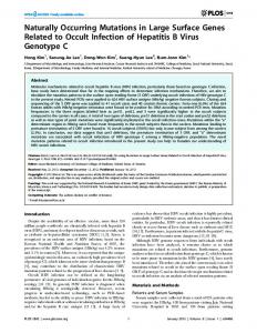
Figure 1 Immunohistochemical staining for P glycoprotein using JSB-1 monoclonal antibodies. We observed moderate to high (A) and in some samples low (B) Pgp staining in epithelial cells of normal colonic mucosa. High Pgp staining was present in lymphocytes of normal mucosa, in particular in those forming lymphoid follicles (C, D, E). Even higher intensity of Pgp staining was observed in tumours compared to normal mucosa (F). In some tumour samples, high Pgp staining was present in lymphocytes of the infiltrating tumour border (G). The intensity of Pgp staining in tumours correlated with grading of tumours, being most intense in well differentiated (H), moderate in moderately differentiated (I), and low in poorly differentiated tumours (J).
three dimensional DNA structures, that is, single strand conformation analysis (SSCA), heteroduplex analysis (HA), and double strand conformation analysis (DSCA). We conducted them simultaneously on the same thin polyacrylamide gel. With this
method, more than 95% of point mutations could be identified.13 Sequencing was performed with the BigDye Terminator Cycle Sequencing Ready Reaction Kit and ABI 310 sequencer (Perkin Elmer Cetus, Norwalk, CT, USA).
www.jmedgenet.com
342
Letters
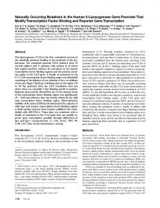
Figure 2 Sequence analysis of the MDR1 gene. (A) Germline mutation in exon 28 of the MDR1 gene identified in tumour sample MSI 11. Arrow indicates G>A substitution at position 3793 resulting in an amino acid change at position 1265 from non-polar glycine to polar serine (G1265S). (B) Somatic mutation in the MDR1 gene promoter region (−14 G>T) identified in tumour sample MSI 11. Arrow indicates C>A substitution (sequenced with reversed primer) 14 bp before the major transcription initiation site (numbering according to Kohno et al19 (MSI 11 (tumour)). Sequencing of the MDR1 promoter region in normal tissue from the same patient showed the somatic origin of this mutation (MSI 11 (normal)) .
www.jmedgenet.com
Letters
343
Table 1 Mutations and germline promoter functional polymorphism in the MDR1 gene identified in colorectal cancers with microsatellite instability Sample
Origin
Exon
Nucleotide change
Consequence
Amino acid change
Domain
Consensus
MSI 3
Germline Somatic
Promoter 25
+8 T>C* 3149 T>C
I1050T
Non-polar>polar
IC6
Yes
Germline Somatic
Promoter 4
+8 T>C* 209 T>C
L70P
Non-polar>non-polar
TM1
Yes
Somatic Germline
Promoter 28
−14 G>T 3793 G>A
G1265S
Non-polar>polar
IC6
Yes
Somatic Somatic
Promoter 20
−29 G>A 2422-2426 del A
Frameshift
MSI 22
Germline
Promoter
+8 T>C*
MSI 34
Germline
8
729 A>G
MSI 5
MSI 11
MSI 19
E243 no change
IC4
/
/
/
IC=intracelular domain, TM=transmembrane domain. *This polymorphism was also identified in microsatellite stable tumours and in controls. Nucleotide numbering according to Kohno et al.[19]
Methylation analysis Tumour DNA was treated with sodium bisulphite to selectively convert only unmethylated cytosines in CpG sites of the MDR1 promoter region to thymines as described previously.14 To determine the methylation status of the MDR1 promoter, sequencing of bisulphite modified DNA using previously reported primers was performed.15 For sequencing we used the BigDye Terminator Cycle Sequencing Ready Reaction Kit and ABI 310 sequencer (Perkin Elmer Cetus, Norwalk, CT, USA). We scored a sample as hypermethylated if signals for unconverted methylated cytosines were higher than those for thymines converted from unmethylated cytosines in the majority of CpG sites after sequencing. Immunohistochemical analysis of P glycoprotein expression Sections of formalin fixed and paraffin embedded tissue blocks of tumour and normal intestinal wall were used. In short, a mouse monoclonal antibody (JSB-1, Biogenex) at dilution 1:10 was applied after microwave antigen retrieval (citrate pH 6.0, 15 minutes, at 850 W). For detection of the antigen, the LSAB method, using DAKO Tech-mate stainer, and DAB were used. According to the intensity of the imunnohistochemical staining estimated by two independent pathologists, samples were divided into three groups of low, moderate, and high Pgp expression. Samples with an estimated intensity of more than 50% (fig 1A, I) and less than 50% (fig 1J) of the most intensively stained sample were scored as high and moderate Pgp expression, respectively. Samples with no distinctive membrane staining (fig 1B, J) were scored as low Pgp expression. For statistical comparisons, moderate and high Pgp expression was evaluated against low Ppg expression. Statistical analysis We used the χ2 test with software package SPSS to compare clinicopathological characteristics between colorectal tumours with and without MDR1 polymorphisms. We used the two sided Fisher exact test to compare MDR1 mutations and methylation status with MSI status of colorectal tumours. We also used the two sided Fisher exact test to compare Pgp expression in unselected tumours (controls), tumours with functional polymorphisms, and MSI-H tumours. In all tests, p values of less than 0.05 were considered to indicate statistical significance.
RESULTS Mutations and polymorphisms in the MDRI gene We identified 12 different germline and five different somatic alterations in the MDR1 gene in initial screening of 60 patients
with primary untreated colorectal cancer (CRC). Thirty patients with tumours exhibiting high microsatellite instability (MSI-H) and 30 patients with microsatellite stable (MSS) tumours were included in this initial screening. We confirmed the origin of the alterations by comparing DNA from tumour and from corresponding normal tissue. For exons with detected alterations, the study was extended to up to 350 patients with colorectal cancer and 100 unaffected blood donors. All alterations were identified as aberrant gel migration patterns with conformational analysis and further confirmed and characterised by direct sequencing of both DNA strands (forward, reverse) (fig 2). To exclude possible random errors, PCR and sequencing reactions were repeated at least three times for each alteration identified. Seven different MDR1 alterations, two germline and all five somatic, were only detected in 5/38 (13%) MSI-H tumours, but not in 400 patients with MSS tumours or in 100 controls (pC) but without MSI were included in mutation screening of the complete MDR1 coding region, but no additional mutations were found. Previously, this promoter polymorphism has been associated with haematological malignancies.17 Our results might support the previously suggested role for Pgp in immune response.4 18 Consistent with previous reports, we observed higher Pgp staining in tumour cells as compared to normal mucosa cells even in untreated tumours. Pgp expression was correlated with tumour differentiation. The lower Pgp expression we observed in MSI-H tumours might be associated with poor differentiation of MSI-H tumours and hypermethylation of the MDR1 promoter. We found hypermethylation of the MDR1 promoter in 10/12 (80%) MSI-H tumours, including 5/6 MSI-H tumours with MDR1 mutations, but in none of the 10 MSS tumours and corresponding normal mucosa (p