Journal of Translational Medicine
Li et al. J Transl Med (2017) 15:18 DOI 10.1186/s12967-016-1115-2
Open Access
RESEARCH
Near infrared fluorescent imaging of brain tumor with IR780 dye incorporated phospholipid nanoparticles Shihong Li1*, Jennifer Johnson2, Anderson Peck1 and Qian Xie2,3,4*
Abstract Background: Near-IR fluorescence (NIRF) imaging is becoming a promising approach in preclinical tumor detection and clinical image-guided oncological surgery. While heptamethine cyanine dye IR780 has excellent tumor targeting and imaging potential, its hydrophobic property limits its clinical use. In this study, we developed nanoparticle formulations to facilitate the use of IR780 for fluorescent imaging of malignant brain tumor. Methods: Self-assembled IR780-liposomes and IR780-phospholipid micelles were prepared and their NIRF properties were characterized. The intracellular accumulation of IR780-nanoparticles in glioma cells were determined using confocal microscopy. The in vivo brain tumor targeting and NIRF imaging capacity of IR780-nanoparticles were evaluated using U87MG glioma ectopic and orthotopic xenograft models and a spontaneous glioma mouse model driven by RAS/RTK activation. Results: The loading of IR780 into liposomes or phospholipid micelles was efficient. The particle diameter of IR780liposomes and IR780-phospholipid micelles were 95 and 26 nm, respectively. While stock solutions of each preparation were maintained at ready-to-use condition, the IR780-phospholipid micelles were more stable. In tissue culture cells, IR780-nanoparticles prepared by either method accumulated in mitochondria, however, in animals the IR780phospholipid micelles showed enhanced intra-tumoral accumulation in U87MG ectopic tumors. Moreover, IR780phospholipid micelles also showed preferred intracranial tumor accumulation and potent NIRF signal intensity in glioma orthotopic models at a real-time, non-invasive manner. Conclusion: The IR780-phospholipid micelles demonstrated tumor-specific NIRF imaging capacity in glioma preclinical mouse models, providing great potential for clinical imaging and image-guided surgery of brain tumors. Keywords: Near infrared fluorescence imaging, Liposomes, Phospholipid micelles, Brain tumor, Blood–brain barrier Background Non-invasive imaging modalities, such as computed tomography (CT), magnetic resonance imaging (MRI), single-photon emission computed tomography (SPECT), and positron emission tomography (PET) play key roles in clinical diagnosis, evaluation of disease status and treatment of tumor. The in vivo optical imaging *Correspondence:
[email protected];
[email protected] 1 Small Animal Imaging Facility, Van Andel Research Institute, Grand Rapids, MI 49503, USA 3 Department of Biomedical Science, Quillen College of Medicine, East Tennessee State University, Johnson City, TN 37614, USA Full list of author information is available at the end of the article
technology using near-infrared fluorescent (NIRF) probes, due to the low NIR absorption and scattering by the tissue, and minimal tissue auto-fluorescence in the NIR window (700–900 nm), is becoming a convenient alternative to the comprehensive imaging modalities in preclinical studies for tumor detection [1–4] and is showing promising results in clinical image-guided oncological surgery [5–10]. The NIRF dye indocyanine green (ICG) has been exploited for imaging of angiogenesis and hepatic segments after hepatectomy [5, 6], as well as for NIR image-guided surgery in a few cancer types [7]. Methylene blue (MB) showed good potential to aid pancreatic tumor resection [8]. Both dyes are approved
© The Author(s) 2017. This article is distributed under the terms of the Creative Commons Attribution 4.0 International License (http://creativecommons.org/licenses/by/4.0/), which permits unrestricted use, distribution, and reproduction in any medium, provided you give appropriate credit to the original author(s) and the source, provide a link to the Creative Commons license, and indicate if changes were made. The Creative Commons Public Domain Dedication waiver (http://creativecommons.org/ publicdomain/zero/1.0/) applies to the data made available in this article, unless otherwise stated.
Li et al. J Transl Med (2017) 15:18
for clinical use [9, 10]. Among other dyes, NIRF heptamethine cyanine dye IR780 was found to have excellent intrinsic tumor targeting and imaging properties without further modification [11–14], providing great potential for tumor NIRF imaging. IR780′s low cytotoxicity makes it of potential clinical use; however, it is also hydrophobic and insoluble in pharmaceutically acceptable solvents, thus an appropriate formulation is required for clinical use [15, 16]. Several formulations of IR780-encapsulated nanoparticles have been investigated, such as the heparin-folic acid conjugate [17], biodegradable human serum albumin nanoparticles [15], transferrin nanoparticles [16], poly(n-butyl cyanoacrylate) nanocapsules [18], poly(styrene-alt-maleic anhydride)-based diblock copolymer micelles [19], rhenium-188 labeled methoxy poly(ethylene glycol)-block-poly(ε-caprolactone) copolymeric micelles [20], pH-responsive polymeric prodrug micelles [21], phospholipid mimicking homopolymeric micelles [22], bubble-generating folate-targeted liposomes [23], and amsacrine analog-loaded solid lipid nanoparticle [24]. However, most of these carriers were designed for both diagnostic and therapeutic purpose, rarely for fulfilling the unique requirement of NIRF imaging or tumor detection. Glioblastoma multiforme (GBM) is the most common and lethal primary brain tumor lacking effective therapeutics due to the invasive growth. The migratory tumor cells penetrate into normal parenchyma preventing its complete surgical removal, and its high resistance to chemotherapy and radiotherapy contribute to GBM’s recurrence as a more invasive phenotype [25, 26]. Although it is well established that the degree of surgical resection directly correlated to patient survival [27, 28], most surgery is performed based on the surgeon’s direct visualization of the tumor without any image guidance. Blood–brain barrier (BBB) and blood–tumor barrier (BTB) further challenge the effective treatment of this brain tumor, as most chemotherapy reagents fail to benefit the patients due to the lack of penetration into tumor tissue [29, 30]. NIRF imaging is expected to benefit the preclinical study of GBM and the optical image-guided surgery. Phospholipid nanoparticles, including liposomes and phospholipid micelles are promising drug carriers, which are biocompatible and able to improve the pharmacokinetics of the encapsulated drug and accumulation in a solid tumor via the enhanced permeability and retention (EPR) effect [31–33] In this study, we generated two formulations, liposomes and phospholipid micelles (Schematic diagram in Fig. 1) to incorporate IR780 for in vivo brain tumor imaging using both the human GBM xenograft model and the spontaneous mouse GBM model. The goal was to develop an appropriate
Page 2 of 12
formulation of IR780 for pharmaceutically acceptable use for clinical imaging and brain tumor detection.
Methods Chemical and material
1,2-distearoyl-sn-glycero-3-phosphocholine (DSPC) and N-(carbonyl-methoxypolyethyleneglycol 2000)-1,2-distearoyl-sn-glycero-3-phosphoethanolamine sodium salt (DSPE-PEG2000) were purchased from NOF America Corporation. Cholesterol, IR780 iodide and other chemicals were purchased from Sigma-Aldrich. Hoechst 33,342, MitoTracker were purchased from Molecular Probes. Cell lines and culture
U87MG human glioma cells were from American Type Culture Collection (ATCC, Manassas, VA). U87M2/luc cells were derived from U87MG that stably overexpress firefly luciferase [34]. T98G human glioblastoma cells were from ATCC. All cell lines were grown in DMEM (invitrogen, CA) supplemented with 10% fetal bovine serum (FBS) (Hyclone, UT), 1% penicillin, and 1% streptomycin (invitrogen). Preparation of IR780‑liposomes and IR780‑phospholipid micelles
For IR780-liposome preparation, DSPC, cholesterol and DSPE-PEG2000 at a molar ratio of 54.8:40:5 were dissolved in chloroform (about 15 mg total lipids/ml); 1 mg/ ml IR780 iodide in ethanol then was added to final 0.2% molar ratio of dye to total lipids. The solution was rotary evaporated in the dark to dryness in vacuo. The dried lipid film was hydrated in nitrogen gas-flushed 0.9% saline to a final total lipid concentration of 30 mM, vortexed and ultra-sonicated under a nitrogen atmosphere for final suspension of the lipid particles. The suspension was repeatedly extruded 16 times through a 100 nm Whatman Nucleopore track-etched polycarbonate membrane at 56 °C. The acquired clear bluish suspension was flushed with nitrogen gas, sealed in a glass vial and stored at either 4 or −20 °C in the dark. To prepare IR780-phospholipid micelles, thin lipid film composed of DSPC and DSPE-PEG2000 containing IR780 (molar ratio, 59.5:40:0.5) was formed in the same way as for the liposomes preparation, then hydrated with nitrogen gas-flushed 0.9% saline to a final total lipid concentration of 15 mM, vortexed and ultra-sonicated at 56 °C for 15 min under a nitrogen gas atmosphere, and extruded through a 100 nm Whatman Nucleopore tracketched polycarbonate membrane. The acquired clear cyan micelle suspension was stored under the same conditions as the liposomes.
Li et al. J Transl Med (2017) 15:18
Page 3 of 12
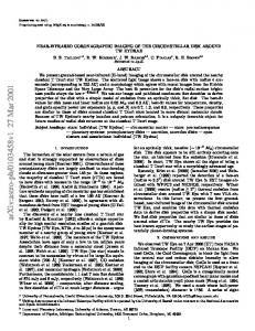
IR780-liposome H3C
CH3
H3C
N
Cl CH3
IR780 iodide
I
-
CH3
+
N
H3C
IR780-phospholipid micelle Phospholipid Phospholipid-PEG IR780
Fig. 1 Chemical structure of IR780 and schematic diagram of IR780-liposomes and phospholipid micelles. Phospholipid nanoparticles, including liposomes and micelles are promising drug carriers, which can improve the pharmacokinetic property of the encapsulated drug and its accumulation and retention in solid tumor via the enhanced intratumoral permeability. The liposomes vesicle is composed of a phospholipid bilayer membrane enclosing an aqueous compartment. The phospholipid micelle vesicle has a single layer of phospholipid core with hydrophilic PEG chain coating on the surface. The sizes of phospholipid micelles are generally smaller than liposomes. Hydrophobic IR780 can self-assemble into the phospholipid bilayer membrane of liposomes and the phospholipid core of micelles during the formation of IR780-nanoparticles
Characterization of IR780‑liposomes and IR780‑phospholipid micelles Particle sizing of IR780‑liposomes and IR780‑phospholipid micelles
The particle size distributions of IR780-liposomes and IR780-phospholipid micelles were measured with a DynaPro dynamic light-scattering system (Wyatt Technology, CA). Before measurement, the samples were diluted with 200 nm membrane-filtered saline to reach appropriate signal concentrations. Near infrared absorption and fluorescence spectra of IR780‑liposomes and IR780‑phospholipid micelles
Visible absorption spectra of IR780, IR780-liposomes and IR780-phospholipid micelles diluted in PBS, ethanol/PBS and PBS/FBS mixtures were measured by a preconfigured UV–visible spectrometer (StellarNet, Inc., FL). The near infrared fluorescent spectra were measured using a Synergy™ Neo HTS Multi-mode microplate reader (BioTek, VT).
Stability of IR780‑liposomes and IR780‑phospholipid micelles
IR780-liposomes and IR780-phospholipid micelles were aliquoted as stock solution (20×) and kept in the dark at 4 or −20 °C. At different time points the aliquots were equilibrated at room temperature and further diluted with PBS into a working solution (1×). The NIR fluorescent signal intensity was measured at Ex/ Em = 745/815 nm. The stability was determined using relative fluorescent intensity (FI/FI0) which was measured by fluorescent signal intensity of samples stored at 4 °C (FI) as compared with those stored at −20 °C (FI0). In vitro cellular uptake of IR780‑liposomes and IR780‑phospholipid micelles
T98G and U87MG cells were pre-cultured in a flask with DMEM medium supplied with 10% FBS at 37 °C with 5% CO2. For cellular uptake experiments, cells were trypsinized and re-seeded on glass coverslips at a density of 6 × 10⁴/cm2 and cultured for another 24 h to reach 60–80% confluency; the medium was replaced with fresh
Li et al. J Transl Med (2017) 15:18
medium supplied with free IR780 (IR780 stock solution in ethanol freshly diluted in PBS), IR780-liposomes or IR780-phospholipid micelles with the final IR780 concentration at 1 μM IR780. After 30 min of incubation, the cells were washed twice with PBS and supplied with fresh DMEM medium without phenol red followed by further staining with Hoechst 33,342 (10 μg/ml) for 1 h and MitoTracker (1 nM) for 10 min. Microscopic images of cells washed with cold PBS and then supplemented with medium were acquired using a confocal laser scanning microscope (Nikon A1 Plus-RSi, Japan). The excitation/ emission wavelengths for fluorescent imaging of Hoechst 33,342, IR780 and Mitotracker were 350/461, 650/780 and 554/576 nm, respectively. GBM tumor models
All studies involve animals were approved by the VARI Institutional Animal Care and Use Committee (IACUC). To establish U87M2/luc ectopic xenograft tumor model, 5 × 10⁵ cells in 100 μl of PBS were subcutaneously inoculated into the flank region of 6-week old nude mice to initiate tumor growth. Three weeks after inoculation, tumor initiation rate reached 90%. Tumor size was measured twice a week using a caliper with tumor volume (mm3) = width2 × length/2. The ability of IR780-nanoparticles to cross the BBB was tested using the U87M2/ luc orthotopic model as previously described [34]. Briefly, mice were inoculated using a stereotaxic frame. A burr hole was created through the skull 2 mm posterior to the bregma, 2 mm anterior to the central suture, and 3 mm below the meninges; U87M2/luc cells (5 × 10⁵ cells in 5 μl of PBS) were injected into the brain parenchyma. The orthotopic tumor growth was measured by bioluminescence signal intensity (BLI). Each mouse received an intraperitoneal injection of 100 μl of 30 mg/ ml D-luciferin sodium solution, and images were taken after 10 min using an AMI1000 optical imager (Spectral Instruments Imaging, Inc., Tucson, AZ). To induce mouse glioma, plasmids pT2/C-Luc/PGK-SB100, pT/ CAGGS-NRASV12, pKT2/CLP-AKT, and pT2/shP53/ GFP4 (provided by Dr. David Largaespada, University of Minnesota) were mixed and the intracerebroventricular injection was performed as previously described [35]. In brief, neonatal mice were placed on ice for 4 min to induce anesthesia before being secured in a cooled, “neonatal rat” stereotaxic frame (Stoelting, IL) maintained at 4–8 °C by a dry ice/ethanol reservoir. A 10 μl syringe fitted with a 30 gauge hypodermic needle (12.5° bevel; Hamilton, NV) attached to an automatic injector (Stoelting, IL) was used to inject plasmids at a flow rate of 0.7 μl/min into the right lateral cerebral ventricle. A total of 2 μg of plasmid DNA (mixed at 1:1:1:1) in 2 μl was injected into each mouse to induce spontaneous glioma.
Page 4 of 12
No incision was made for the injection. The skull of a neonate was penetrated with the needle for all injections. Growth of the orthotopic tumor was monitored by BLI as described above. NIRF imaging with IR780 incorporated nanoparticles in GBM mouse models
Fourteen nude mice bearing U87M2/luc tumors with volumes of 122–580 mm3 were divided into three groups based on balanced tumor volumes: (1) free IR780 (IR780 freshly prepared in ethanol/saline (V/V, 1:9) (n = 4), (2) IR780-liposomes, and (3) IR780-micelles (IR780liposomes or IR780-phospholipid micelles freshly diluted in saline containing 2 nmol of IR780, n = 5). Each mouse was intravenously injected via tail vein with 100 μl imaging agent. The sequential whole body NIRF images at different time points (5 min, 1, 4, 24, 48, 72, and 96 h post injection) were acquired using an AMI1000 optical imager (Ex/Em = 745/810 nm, acquisition time: 1 s). At two time points, 24 h and 96 h post injection of IR780 agents, bioluminescent images also were acquired (imaging acquisition time: 10 s) to determine tumor growth. Five nude mice with four bearing U87M2/luc orthotopic tumors and one tumor-free healthy control, and three FVB mice bearing spontaneous GBM induced by activation of RAS and AKT and Trp53 loss (FVB/NRAS/AKT/shP53) [35] were used to evaluate the imaging capacity of IR780phospholipid micelles in orthotopic brain tumors. With orthotopic tumors, each mouse was intravenously injected with 100 μl IR780-phospholipid micelles freshly diluted in saline containing 4 nmol of IR780 (U87M2/luc orthotopic model) or 6 nmol of IR780 (FVB/NRAS/AKT/shP53 spontaneous tumor model). The sequential whole body images at different time points were captured following the same procedure performed for U87M2/luc ectopic models. Ex vivo imaging and histological staining
At the end of the in vivo imaging, mice bearing tumors were sacrificed, and brain, heart, lung, liver, spleen, stomach, intestine, normal muscle and skin from lumbar were dissected for immediate fluorescent photography using the AMI1000 optical imager. For histology analysis, mice brains were harvested and fixed in 10% neutral-buffered formalin and embedded into paraffin blocks and slides (20 μm) were cut for H&E staining. For microscopic NIRF images, additional brain sections were cut from the paraffin blocks. Unstained slides were scanned using the Odyssey imager (LI-COR Biosciences) and software suite version 3. Settings were optimized for the highest resolution and power to allow for visualization of both the fluorescent target and non-fluorescent anatomy. Resolution was set to 21 microns with no focal offset and excitation intensity was set to 6.0 for the 800 nm channel only.
Li et al. J Transl Med (2017) 15:18
Page 5 of 12
IR780-liposomes and 793 nm for IR780-phospholipid micelles (Additional file 1: Figure S2). Adding ethanol to IR780-liposomes or IR780-phospholipid micelles resulted in maximal NIR absorption shifted to 783 nm, indicating the release of IR780 from dissolved nanoparticles. Both IR780-liposomes and IR780-phospholipid micelles showed broad NIR fluorescent spectrum with maximum excitation/emission wavelength at 745/815 nm. (Additional file 1: Figure S3).
1.2 1.0
FI/FI 0
0.8 0.6 0.4
IR780-liposomes IR780-phospholipid micelles
0.2 0.0
0
5
10
15
20
25
Stability of IR780‑liposomes and IR780‑phospholipid micelles 30
35
40
Time (d) Fig. 2 Relative fluorescent intensity of IR780-liposomes and IR780phospholipid micelles. FI/FI0 was measured by fluorescence signal intensity of samples stored in the dark at 4 °C as compared with those stored in the dark at −20 °C (n = 3, Ex/Em = 745/815 nm). Short bar refers to standard deviation
Statistical analysis
All experimental data were shown as mean ± SD unless stated otherwise. Comparisons of data between 2 groups or among 3 groups were analyzed using independentsamples t test or one-way analysis of variance (ANOVA) at P