leate (TIM; Merck, Sharp & Dohme, West Point,. Pa.) ..... Effect of TIM on the SCC stimulated with serotonin. ..... Candia OA, Lanzetta PA, Alvarez LJ, and Gaines.
Neural serotonin stimulates chloride transport in the rabbit corneal epithelium S. D. Klyce, K. A. Palkama, M. Harkonen,* W. S. Marshall, S.
Huhtaniitty,
K. P. Mann, and A. H. Neufeld** Evidence is presented that serotonin acts as a neurotransmitter in the cornea of the adult rabbit. Serotonin was localized to granules in a sparse population of subepithelial corneal nerves by an electron microscopic histochemical procedure. Significant endogenous levels of serotonin and its principal metabolite, 5-hydroxyindoleacetic acid, were detected in the central cornea by a fluorometric assay. Exogenous serotonin stimulated ion transport by the corneal epithelium. This effect was potentiated by monoamine oxidase inhibition and was unaffected by an a-adrenergic receptor antagonist. Serotonin-stimulated ion transport was inhibited by the specific antagonist, methysergide, and by the replacement of Cl~ with an impermeable anion. In tracer experiments, the serotonin-stimulated ion transport was shoion to be caused by increased epithelial Cl~ secretion. The serotonin response was partially inhibited by the ^•adrenergic antagonist, timolol. In a companion article,16 assay of corneal cyclic AMP showed stimulation of cyclic AMP synthesis by serotonin, inhibition by the specific antagonist, lysergic acid diethylamide, and potentiation by monoamine oxidase inhibition. We postulate that specific serotonergic receptors are present in the corneal epithelium and that activation of these receptors by serotonin released from serotonergic neurons increases the level of cyclic AMP, tohich stimulates active Cl~ secretion by the corneal epithelium. (INVEST OPHTHALMOL VlS SCI 23:181-192, 1982.) Keywords: serotonin, cornea, chloride, transport, corneal nerves, methysergide, epithelium, epinephrine, timolol, rabbit, nialamide
From the Lions Eye Research Laboratories, Louisiana State University Eye Center, New Orleans, La., *the Department of Clinical Chemistry, University of Helsinki, Helsinki, Finland, and **the Eye Research Institute of Retina Foundation, Boston, Mass. Supported by U.S.P.H.S. research grants EY03311, EY02360, and EY02377, the Louisiana Lions Eye Research Foundation, S. Jurelius Foundation, and the Medical Research Council of the Academy of Finland. Submitted for publication Aug. 20, 1981. Reprint requests: Stephen D. Klyce, Ph.D., LSU Eye Center, 136 South Roman St., New O.'-ans, La. 70112. Portions of this work were reported at the Annual Meeting of the Association for Research in Vision and Ophthalmology, May 2, 1981, Sarasota, Fla.1
he modulation of corneal epithelial function by an adrenergic pathway has been studied extensively. Stimulation of the superior cervical ganglion is known to suppress corneal mitotic activity.2' 3 The presence of adrenergic corneal nerves has been detected histochemically in a variety of species. 4 ' 5 Although adrenergic neurons have not been found in adult primate corneas with this histochemical procedure, this negative result could be due to inadequate sensitivity of the method. Recent neuroanatomic evidence has shown that intrastromally injected horserad-
0146-0404/82/080181+12$01.20/0 © 1982 Assoc. for Res. in Vis. and Ophthal., Inc.
181
Invest. Ophthalmol. Vis. Sci. August 1982
182 Klyce et al.
•
•
&
1 Fig. 1. Dense core vesicles associated with adrenergic nerves demonstrated with the KMnO4 reaction in the rabbit cornea. Nerves in the central stroma are unmyelinated and are often only partially ensheathed by Schwann cells. Several dense core vesicles appear in the fiber at the top of the figure. (Bar = 100 nm.) ish peroxidase appears a day later in the ipsilateral superior cervical ganglion cell bodies, lending additional support to the view that sympathetic fibers innervate the adult rabbit cornea.6 In addition to the antimitogenic effect of catecholamines, /3-adrenergic agonists have been shown to stimulate Cl~ transport in the frog, rabbit, and human corneal epithelia.7^9 The Cl~ transport response involves activation of adenylate cyclase and produces a specific Cl~ permeability increase at the outer epithelial surface.10 However, addi-
tional physiologic roles of the /3-adrenergic pathway in the cornea have not been forthcoming. Although cholera enterotoxin has been shown recently to stimulate epithelial wound healing through an action involving cyclic AMP, there is no evidence that /3-adrenergic stimulation is needed. 11 The sensory fibers may participate in the process of healing, since trigeminal ganglion ablation in the rabbit significantly delays corneal epithelial wound healing and increases epithelial fluorescein permeability.12 The superior cervical ganglion innervates a
* Volume 23 Number 2
Serotonin-stimulated chloride transport
183
Fig. 2, FGD reaction in the rabbit cornea. In this method for localizing 5-hydroxytryptamine, the electron density of the specimen is extremely low except at the dense core vesicles. The periphery of the vesicles (arrows) shows only a weak electron density. Collagen fibers (F) and a stromal keratocyte (K) are detectable. (Bar = 100 nm.) variety of tissues, including the pineal gland. Sympathetic fibers in the pineal gland have been shown to contain both norepinephrine and serotonin13"15; thus serotonin was examined as a potential neurotransmitter in the cornea. In this article we present the evidence that serotonin functions as a neurotransmitter in the cornea and thereby influences corneal epithelial function. The accompanying article describes the evidence that serotonin stimulates the synthesis of cyclic AMP, which mediates the physiologic response. 16 The com-
bined evidence supports the novel finding of specific serotonergic receptor-activated, cyclic AMP-mediated stimulation of chloride transport by the corneal epithelium. Methods Adult New Zealand White rabbits (3 to 5 kg) were killed with an overdose of phenobarbital immediately prior to enucleation of the globes. The following pharmacologic agents were used: serotonin oxalate or HC1 (salt) (Sigma Chemical Co., St. Louis, Mo.), nialamide (NM; Sigma), methysergide bimaleate (MSD, Sansert; Sandoz Pharmaceuticals, E. Hanover, N. J.), timolol ma-
Invest. Ophthalmol. Vis. Sci. August 1982
184 Klyce et al.
Table I. Concentrations of serotonin (5-HT) and 5-HIAA in the cornea compared with levels in other tissues* Rat18
Rabbit (n = 8) 30
Iris-ciliary body
Compound
Cornea
5-HT 5-HIAA
0.41 ± 0.01 0.11 ± 0.01
1.17 ± 0.06 0.16 ± 0.02
Rabbit uvearetina 4.0
Cerebral cortex
Hypothalamus
3.0 1.0
15.0 5.6
* Values are expressed as means ± S.E.M. in /uM/kg wet weight.
Table II. Effect of serotonin on corneal electrica parameters* Control period Serotonin (10"4M) Change p value
SCC (fiA/cm2 hr)
PD (mV)
Resistance (kit • cm2)
2.7 ± 0.2 3.4 ± 0 . 2 0.7 ± 0.1 < 0.001
26.3 ± 2.3 27.6 ± 2.4 1.3 ± 0.9 >0.1
10.2 ± 1.1 8.9 ± 1.2 - 1 . 3 ± 0.4 0.6 mM K 2 HPO 4) 25 mM Na/HEPES (pH 7.4), 1.4 mM Ca gluconate, 0.61 mM MgSO4, and 26 mM glucose (all from Sigma). Cl~-free medium was produced by SO42~ substitution, with added sucrose to maintain solution osmolarity (305 mOsm). The media were bubbled with 95% O 2 /5% CO 2 and had a pH of 7.4. Corneal short-circuit current (SCC) and resting potential (PD) were measured with dual automatic current/voltage clamps (D. Lee, Inc., Sunnyvale, Calif). The SCC and PD measured across the whole cornea accurately reflect transport processes arising from the epithelial cell layer, since these properties are not altered when the endothelial layer is removed.17 Unidirectional fluxes of 36C1 were measured as described previously,8 with 200 /A samples taken from the less radioactive side every 15 min. During the flux experiments, the specific activity of the more radioactive side (nominally 30 /xCi/mEq Cl~) varied by less than 1%.
Biochemistry Serotonin and 5-hydroxyindoleacetic acid (5HIAA) assay in cornea and iris-ciliary body. Tissue concentrations of serotonin and its principal metabolite, 5-HIAA, were determined by the method introduced by Curzon and Green, 18 with modifications. Central mil-thickness corneal tissue samples and pieces of the iris-ciliary body complex (50 to 70 mg) were rinsed, rapidly frozen, weighed at — 25° C, and then homogenized in 1 ml of cold acidified n-butanol with a small glass homogenizer. After centrifugation for 5 min at 1100 X g, 0.9 ml of the supernatant was transferred to glass-stoppered tubes containing 1.8 ml ofn-heptane and 145 fi\ of 0.1M HC1 with 0.1% L-cysteine. After shaking for 5 min, the two phases were separated by centrifugation as before. For the determination of serotonin levels, 25 /AI of the lower aqueous phase were pipetted into 0.004% o-phthalaldehyde (OPT) in 10M HC1. This solution was mixed and the tubes were heated in a boiling water bath for 15 min. The tubes were then cooled in water and fluorescence was measured in microcuvettes (150 fj\) with a spectrofluorophotometer (Jasco FP-4). Appropriate standards, reagent blanks, and tissue blanks were used. For the determination of 5-HIAA, 2.3 ml of the organic phase (n-heptane-n-butanol) were acid washed by shaking with 115 /xl of 0.01M HC1 and were centrifuged as before. The organic phase (2 ml) was pipetted into a tube containing 240 fx\
Volume 23 Number 2
Serotonin-stimulated chloride transport
185
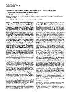
EPINEPHRINE 10" 7 M
12 10
/ S .C.C.
5-HT
i 8 I : 6 40 OOOOOOOOOOOOoOOOOOo
noo°°°o°oooooooooooooe
°ooooooooooooo! |
Ooo
PDJ
^
E 20 ~ 0
30
60 TIME (min)
Fig. 3. Effect of serotonin (5-HT) and epinephrine on epithelial SCC and PD in the isolated rabbit cornea.
EPINEPHRINE 10 8
10" 7 M
"NIALAMIDE
5-HT 10~5M
4
10" M
40
4 »OOOOOO 000 O 0 O < K,OOOOOOOO«OOOO««>C O oo000«ooooooooo«oooo 0oooooo