Pharmacological Reports
Copyright © 2013
2013, 65, 14791488
by Institute of Pharmacology
ISSN 1734-1140
Polish Academy of Sciences
Review
Neurochemical modulation of stress-induced cognitive inflexibility in a rat model of an attentional set-shifting task Agnieszka Nikiforuk, Piotr Popik Department of Behavioral Neuroscience and Drug Development, Institute of Pharmacology, Polish Academy of Sciences, Smêtna 12, PL 31-343 Kraków, Poland Correspondence: Agnieszka Nikiforuk, e-mail:
[email protected]
Abstract: It is widely accepted that chronic stress, which is considered a risk factor for several neuropsychiatric disorders, may have detrimental effects on prefrontal functions. In animal models, chronic stress produces morphological, physiological and functional alternations in the rat medial prefrontal cortex (mPFC). Specifically, repeated restraint stress results in mPFC dendritic atrophy that is associated with deficits in the prefrontal cortex-dependent attentional set-shifting task (ASST). Thus, restraint-induced cognitive inflexibility may serve as a model for the study of the mechanisms, prevention and treatment of stress-related disorders. The current article provides a summary of the literature on stress-related effects on cortical functions, as assessed in the rodent ASST. The neurochemical substrates underling stress-evoked frontal-like disturbances, as well as pharmacological targets for potential treatment, are briefly discussed. Key words: stress, prefrontal cortex, cognition, set-shifting, corticosteroids, depression, monoamines, ketamine
Abbreviations: 5-HT – 5-hydroxytryptamine (serotonin), ACTH – adrenocorticotropic hormone, ASST – attentional set-shifting task, CD – compound discrimination, ED – extradimensional shift, ID – intradimensional shift, ID/ED – intradimensional/extradimensional shift, MDD – major depressive disorder, mPFC – medial prefrontal cortex, mTOR – the mammalian target of rapamycin, NMDA – N-methyl-D-aspartic acid, OFC – orbitofrontal cortex, PKC – protein kinase C, Rev – reversal, SD – simple discrimination, SSRI(s) – selective serotonin reuptake inhibitor(s)
Introduction Chronic stress may precipitate or exacerbate many psychiatric disorders, including depression, due to its detrimental effects on many brain functions. The impact of stress on the brain is complex, though some regions, such as the hippocampus, amygdala and prePharmacological Reports, 2013, 65, 14791488
1479
frontal cortex, seem to be key targets for stressinduced structural, physiological and functional alterations [25]. Experimental evidence suggests that the prefrontal cortex may be particularly susceptible to stress exposure [21]. Of particular note is the fact that many frontal-governed cognitive processes show declines as a result of stress-related disorders. Animal models of the effects of stress on frontal functions have been developed to elucidate the mechanisms underlying these disorders as well as for the development of novel strategies for their treatment.
Stress-induced dendritic remodeling in the rat prefrontal cortex Chronic stress produces profound changes in the morphology of neurons in the rodent medial prefrontal cortex (mPFC). Indeed, the initial findings of Cook and Wellman [13], demonstrating that 3 weeks of restraint evoked apical dendritic retraction and debranching in mPFC neurons in rats, have been confirmed in a number of subsequent studies [9, 28, 44]. Moreover, it has been demonstrated that the morphology of the mPFC is exquisitely sensitive to stress, as dendritic remodeling occurred in response to just 1 week of brief daily restraint [9]. The fact that stress-induced dendritic atrophy and spine retraction appear to be restricted to the most apical dendrites of the pyramidal cells may provide a link between altered mPFC morphology and physiological changes. Accordingly, Liu and Aghajanian [30] demonstrated that stress-induced apical dendritic atrophy resulted in diminished responses to apically targeted excitatory inputs. However, it is an important question as to how these structural and electrophysiological changes may affect cortical functions.
Stress and prefrontal functions It is likely that dendritic remodeling in the prefrontal cortex may underlie functional deficits. Specifically, an impairment in set-shifting ability, which is a sensitive indicator of cortical processing, has been connected to stress-evoked disruption [28]. The next
1480
Pharmacological Reports, 2013, 65, 14791488
paragraphs briefly describe the attentional set-shifting task and the impact of stress procedures on rats’ performance on the task. The assessment of frontal-dependent functions: the attentional set-shifting task
The prefrontal cortex subserves higher order executive functions, including cognitive flexibility, i.e., the ability to modify behavior in response to the altering of environmental demands. This aspect of executive functioning is commonly assessed in humans using the Wisconsin Card Sorting Test and its modified version the intradimensional/extradimensional shift (ID/ED) task [45]. Impaired performance on these tasks is demonstrated in patients with frontal cortex damages [15] and with psychiatric disorders known to involve the PFC, such as schizophrenia [16]. Cognitive flexibility may also be assessed in a rodent version of the ID/ED task, that is, in the attentional set-shifting task (ASST) [6]. In this paradigm, rats must select a bowl containing a food reward based upon their ability to discriminate among odors and the media covering the bait. The ASST session consists of series of discriminations that require rats to initially learn a rule and form an attentional “set” within the same stimulus dimensions (Tab. 1). In the simple discrimination (SD) involving only one stimulus dimension, the pots differ along one of two dimensions (i.e., a digging medium). For the compound discrimination (CD), a second (irrelevant) dimension (i.e., an odor) is introduced, but the correct and incorrect exemplars of the relevant dimension remain constant. For the reversal of this discrimination (Rev 1), the exemplars and relevant dimension are unchanged, but the previously correct exemplar is now incorrect and vice versa. The intradimensional shift (ID) is then presented, comprising new exemplars of both the relevant and irrelevant dimensions, with the relevant dimension remaining the same as before. The ID discrimination is then reversed (Rev 2) so that the formerly positive exemplar now becomes the negative exemplar. Finally, for the extradimensional shift (ED) a new pair of exemplars is again introduced, but this time a relevant dimension is also changed. Thus, at the ED stage, animals must switch their attention to a new, previously irrelevant stimulus dimension and, for ex-
Stress-induced cognitive inflexibility Agnieszka Nikiforuk and Piotr Popik
Tab. 1. Order of discriminations performed
Phase
Relevant dimension
SD
medium
crumpled tissue
cotton wool
cotton wool
crumpled tissue
CD
medium
crumpled tissue
cotton wool
cotton wool
crumpled tissue
lemon
almond
lemon
almond
crumpled tissue
cotton wool
cotton wool
crumpled tissue
lemon
almond
lemon
almond
Rev 1 ID Rev 2 ED Rev 3
medium medium medium
Discrimination 1
Discrimination 2
clay pellets
silk
silk
clay pellets
rum
cream
rum
cream
clay pellets
silk
silk
clay pellets
rum
cream
rum
cream
vanilla
spicy
spicy
vanilla
shredded paper
metallic filler
shredded paper
metallic filler
odor odor
vanilla
spicy
spicy
vanilla
shredded paper
metallic filler
shredded paper
metallic filler
An example of the cue combinations used in the attentional set shifting task (ASST) in rats that were shifted from digging medium to odor as the relevant dimension. Rats performed a series of 7 discriminations: simple discrimination (SD), compound discrimination (CD), reversal 1 (Rev1), intradimensional shift (ID), reversal 2 (Rev 2), extradimensional shift (ED), reversal 3 (Rev 3). The correct exemplar (shown in bold) was paired with either of two exemplars from the irrelevant dimension (i.e., at the CD phase, the crumpled tissue was paired with either lemon or almond odor, etc.). In the ID and ED, there were novel pairs of exemplars of each dimension. The left-right positioning of the pots in the test apparatus on each trial were randomized
ample, discriminate between the odors and no longer between the media covering the bait. The ED phase, which is regarded as an index of cognitive flexibility, is impaired by lesions of the mPFC [6]. Hence, the ASST measures specific frontal-dependent cognitive functions in a way that is homologous to human tests, and it therefore represents a useful translational approach along the continuum from purely animal models to the clinic [23]. Stress-induced cognitive inflexibility
Liston et al. [28] demonstrated that rats subjected for 21 days to 6 h daily restraint stress exhibited cognitive inflexibility, as demonstrated by a selective impairment in ED set-shifting and a corresponding retraction (20%) of the apical dendritic arbors in layer II/III of the anterior cingulate region of the mPFC. This decrease in mPFC dendritic arborization predicted impaired ED set-shifting performance. In parallel to this rodent model, chronic psychosocial stress-induced disruption of prefrontal functional connectivity in human subjects predicted their decline in attentional
set-shifting ability [29]. As this finding suggests, the utility of the rodent restraint-stress procedure in modeling clinical aspects of stress, this paradigm was employed in our studies [43]. However, taking into account the particular vulnerability of the PFC to stress, our experimental procedure included only 1 h daily exposure to restraint stress for 7 days. Nevertheless, this relatively mild stress protocol significantly and specifically impaired rats’ ED set-shifting capacity. The novel finding of our study was that stress apparently exerted long-lasting negative consequences on prefrontal cognitive processes because restraintinduced ED set-shifting deficits were still evident 3 weeks following stress cessation. Stress induced cognitive-inflexibility is not restricted to the restraint paradigm. As demonstrated in the study of Bondi et al. [7], a rat model of chronic unpredictable stress also induced a cognitive impairment in ED set-shifting capability in the ASST, suggesting an alteration in function of the mPFC. On the other hand, rats subjected to chronic intermittent cold stress, a potent metabolic stressor, exhibited a selective impairment on the first reversal stage of the Pharmacological Reports, 2013, 65, 14791488
1481
ASST [24]. The demonstrated deficit of reversal learning was suggested to involve dysregulation of serotonin modulatory functions in the orbitofrontal cortex (OFC), a brain region known to mediate reversal learning. Cold stress-induced cognitive disturbances persisted only 3 days after the last stress exposure, as no impairment was evident in rats tested 7, 14 or 21 days after termination of the stress protocol [14]. Interestingly, repeated restraint stress did not adversely affect either reversal learning or dendritic morphology in the lateral OFC [28]. Recent experimental work also aimed to elucidate the effects of acute stress on cognitive flexibility. Using an operant-based set shifting task in rats, Butts et al. [10] demonstrated that exposure to acute stress (15 min of mild tail-pinch stress) immediately before testing significantly disrupted set-shifting, but it had no effect on reversal learning. On the contrary, Thai et al. [50] found that acute restraint stress did not affect rats’ setshifting performance, but it did significantly facilitate reversal learning. Together, these findings suggest that various types of acute or chronic stress do not produce exactly the same effects on cognitive flexibility.
Neurochemical basis of stress-induced prefrontal deficits The mechanisms that mediate morphological and functional changes in the mPFC are interrelated and
complex. Exposure to stressors results in a variety of neurochemical changes in prefrontal cortex, including glucocorticoids and the monoaminergic and glutaminianergic systems. The following paragraph will briefly summarize experimental data on the potential involvement of these neurochemical substrates in stress-induced cognitive inflexibility.
Corticosteroids
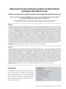
A wide body of evidence suggests that the PFC might be influenced by corticosteroids released during stress. Accordingly, corticosterone administration mimics the impact of stress on the mPFC morphology [53]. This cortical reorganization may contribute to stress-induced changes in cognition, as chronic exposure to corticosterone evokes both impairments in reversal learning and a reduction in the volume of layer II of prefrontal areas [11]. Moreover, systemic administration of the glucocorticoid receptor blocker, RU38486, prevented this restraint-induced dendritic retraction in the rat mPFC [30]. It has also been demonstrated that pharmacological blockade of corticosterone synthesis may prevent stress-induced dendritic retraction, at least in the hippocampus [31]. Accordingly, administration of metyrapone, an inhibitor of corticosteroid synthesis, prior to restraint sessions prevented ASST deficits (Fig. 1) [43]. This finding supports the involvement of endogenous corticosteroids in stress-induced cognitive inflexibility.
Fig. 1. The effect of metyrapone ad-
ministration prior to restraint on stressinduced deficits in the attentional setshifting task in rats. Animals were exposed to restraint stress for 1 h daily for 7 days. Metyrapone (0 or 50 mg/kg) was administered 60 min before each stress exposure. The attentional setshifting task was performed 14 days after stress cessation. Unstressed groups were treated according to the same experimental schedule. The results represent the mean ± SEM of the number of trials required to reach the criterion of 6 consecutive correct trials for each of the discrimination phases. Symbols: * p < 0.001 vs. ED performance in the unstressed vehicle-treated group, #p < 0.001 vs. ED performance in the stressed vehicle-treated group, NewmanKeuls post-hoc test. Adapted from [43]
1482
Pharmacological Reports, 2013, 65, 14791488
Stress-induced cognitive inflexibility Agnieszka Nikiforuk and Piotr Popik
Dopamine
Experimental data suggest that prefrontal cortical dopaminergic dysfunction may underlie chronic stressinduced cognitive deficits. Mizoguchi et al. [35] demonstrated that chronic stress induced working memory impairments via a dopamine D1 receptor-mediated hypodopaminergic mechanism in the rat mPFC. In the latter study, stress-induced impairments in spatial working memory were accompanied by a marked reduction in dopaminergic transmission, which is concomitant with an increase in D1 receptor density in the PFC. This cognitive deficit was reversed by the intraPFC infusion of a D1 receptor agonist. Likewise, our study demonstrated that acute administration of the dopamine reuptake inhibitor nomifensine ameliorated stress-induced impairments in ED set-shifting [43]. Interestingly, nomifensine, administered at a dose that was inactive in unstressed controls, still enhanced ED performance in stressed animals. Therefore, the follow-up study sought to explore the hypothesis that activation of D1 receptors may be responsible for preferential dopamine-induced facilitation of cognitive flexibility in stressed rats. To this end, the effect of the selective D1 receptor agonist, SKF 81297, on ASST performance was investigated in control and stressed rats [39]. SKF 81297 at doses of 0.1 and 0.3 mg/kg, but not lower (0.01 mg/kg) or higher (1 mg/kg) doses, improved ED performance in the unstressed control group. Interestingly, the drug administered at the lowest dose (0.01 mg/kg) was still capable of promoting set-shifting performance in stressed rats. Therefore, it may be concluded that stressinduced changes at the D1 receptor level may underlie the hyperresponsiveness to dopaminergic enhancement in the rat’s ASST paradigm. This issue might be of special interest because dopaminergic enhancement represents one of the therapeutic strategies for ameliorating frontal-like cognitive disturbances [33]. Noradrenaline
PFC-dependent cognitive functions are also modulated by stress-evoked alterations in noradrenergic transmission. Accordingly, a high level of noradrenaline during stress exposure [17] stimulates a1-adrenoceptors and activates the phosphatidylinositol signaling pathway and protein kinase C (PKC). Consistent with the role of a1-adrenoceptor-PKC signaling in the detrimental effects of stress, PFC-related cognitive
deficits were observed after exposure to a pharmacological stressor, stimulation of a1-adrenoceptors or direct activation of PKC [2, 4, 5]. Subsequently, the inhibition of either a1-adrenoceptor or PKC restored cognitive functions [4, 5]. Moreover, Hains et al. [20] suggested that PKC overactivity might be responsible for alterations in prefrontal dendritic morphology and in cognitive functions in rats subjected to the chronic restraint-stress paradigm. Therefore, it may be assumed that stress-evoked excessive PKC signaling, arising from a1-adrenoceptor stimulation, accounts for the ED set-shifting deficits observed in restrained rats. In line with this hypothesis, our previous study demonstrated that prazosin (1 mg/kg) given to rats prior to restraint sessions completely prevented stress-induced cognitive inflexibility (Fig. 2) [40]. This finding corroborates the results of Jett and Morilak [22], demonstrating that noradrenaline signaling during chronic unpredictable stress compromised mPFC functions. In this study, the infusion of a cocktail of a1-, b1-, and b2-adrenergic receptor antagonists into the mPFC prior to each stress session protected rats from stress-induced ED deficits. However, although stress-evoked excessive noradrenaline release may have detrimental effects on frontal functions, it is also widely accepted that noradrenaline transmission is involved in the regulation of prefrontal cortical functions, including those related to attentional set-shifting. An elevation of noradrenaline neurotransmission in the mPFC by acute administration of the a2-adrenergic autoreceptor antagonist, atipamezole, or chronic treatment with the noradrenaline reuptake inhibitor, desipramine, abolished setshifting deficits in rats subjected to chronic unpredictable stress [8]. It has also been suggested that a1adrenoceptor stimulation may play a role in noradrenaline-induced facilitation in the ASST responding in stressed rats. Indeed, acute blockade of postsynaptic a1-adrenergic receptors in mPFC prior to testing blocked the beneficial effect of desipramine on setshifting [8]. Correspondingly, our previous study demonstrated that acute administration of desipramine reversed the restraint-induced set-shifting deficit and promoted cognitive flexibility in control rats [43]. Collectively, these results suggest that noradrenergic modulation of mPFC may retain enough of its functional activity to reverse the effects of chronic stress, and they support the notion that elevated noradrenergic tone in mPFC may represent a therapeutic target for treating stress-related frontal dysfunction. Pharmacological Reports, 2013, 65, 14791488
1483
Fig. 2. The effect of prazosin administration prior to restraint on stressinduced deficits in the attentional setshifting task in rats. Animals were exposed to restraint stress for 1 h daily for 7 days. Prazosin (0 or 1 mg/kg) was administered 60 min before each stress exposure. The ASST was performed 14 days after stress cessation. Unstressed groups were treated according to the same experimental schedule. The results represent the mean ± SEM of the number of trials required to reach the criterion of 6 consecutive correct trials for each of the discrimination phases. Symbols: * p < 0.001 vs. ED performance in the unstressed vehicle-treated group, # p < 0.001 vs. ED performance in the stressed vehicle-treated group, NewmanKeuls post-hoc test. Adapted from [40]
Serotonin
It is well established that serotonin (5-HT) modulates the hypothalamic-pituitary-adrenal axis response to stress. Recent data suggest that chronic restraintinduced endocrine disruption, involving decreased ACTH and sensitized corticosterone responses to acute restraint, may be associated with the increased function and expression of 5-HT7 receptors [18]. Consequently, pharmacological blockade of adrenocortical 5-HT7 receptors could be of therapeutic benefit to overcome endocrine disruption in stress-related diseases. Moreover, 5-HT7 receptors may also be involved in the response of neural circuits to repeated stress. Repeated corticosterone administration, a model known to mimic some aspects of stress exposure, increased the reactivity of rat CA3 hippocampal circuitry to the activation of 5-HT7 receptors [52]. Moreover, the selective 5-HT7 receptor antagonist SB-269970 counteracted restraint stress-induced attenuation of long-term potentiation in the rat frontal cortex [51]. It has also been demonstrated that exposure to chronic mild stress evokes up-regulation of 5-HT7 receptor mRNA in the rat hippocampus and hypothalamus [27]. Based upon this evidence, we evaluated the effectiveness of SB-269970 for ameliorating stress-induced cognitive impairments in the ASST. Acute administration of SB-269970 restored cognitive flexibility in stressed rats [41]. In line with data demonstrating that selective blockade of 5-HT7 receptors augmented the behavioral effects of antidepres1484
Pharmacological Reports, 2013, 65, 14791488
sants [54], SB-269970, given at an inactive dose, enhanced the pro-cognitive efficacy of the selective serotonin reuptake inhibitor (SSRI) escitalopram. Thus, stress-evoked enhancement of 5-HT7 receptor-mediated responses or up-regulation of their expression may also account for the efficacy of the 5-HT7 receptor antagonist in ameliorating stress-induced frontal-like cognitive impairments. Interestingly, amisulpride, an antipsychotic drug with a high affinity for D2/D3 and 5-HT7 receptors, administered to rats subjected to repeated restraint mimicked the action of the selective 5-HT7 receptor antagonist and reversed stress-evoked set-shifting impairments [42]. As this effect was fully blocked by the 5-HT7 receptor agonist AS19, the amisulpride-induced cognitive enhancement was likely mediated through the drug’s antagonistic action at 5-HT7 receptors. Glutamate
Stress preferentially increases levels of glutamate in the prefrontal cortex [36]. According to Martin and Wellman [32], glutamatergic transmission at N-methyl-D-aspartic acid (NMDA) receptors may play a role in stress-induced dendritic reorganization in the mPFC. The latter authors demonstrated that the administration of the competitive NMDA receptor antagonist, CPP (3-(2-carboxypiperazin-4-yl) propyl-1phosphonic acid), prevented stress-induced mPFC dendritic atrophy. This finding raises the possibility that stress, acting through NMDA receptors, may pro-
Stress-induced cognitive inflexibility Agnieszka Nikiforuk and Piotr Popik
duce changes in PFC-mediated processes such as setshifting. To explore this hypothesis further, a noncompetitive antagonist of NMDA receptors, ketamine, was administered before each of the restraint stress sessions. The results of this experiment demonstrate that NMDA receptor blockade during stress exposure prevented stress-induced cognitive inflexibility (our unpublished data). The precise mechanisms mediating the involvement of NMDA receptors in stress-evoked dysfunction remain unknown. One possibility is that stressinduced increases in corticosterones are responsible for the role of NMDA receptors in the stress response. For example, glucocorticoids have been demonstrated to mediate the stress-induced extracellular accumulation of glutamate in the rat brain [37]. Likewise, corticosterones prolong NMDA receptor-mediated Ca2+ elevation in cultured rat hippocampal neurons [49]. In line with this hypothesis, pharmacological blockade of corticosteroid synthesis mimicked the effects of NMDA receptor blockade and also prevented the stress-induced cognitive inflexibility [43]. Nevertheless, other mechanisms of ketamine action in this stress paradigm cannot be excluded. For example, Li et al. [26] demonstrated that acute administration of ketamine rapidly ameliorated deficits in the depressive-like behavior, spine density and synaptic function of PFC neurons in rats subjected to the chronic unpredictable stress. These effects were mediated by activation of the mammalian target of rapamycin (mTOR) pathway.
Antidepressant drugs and stress-induced cognitive inflexibility Because prolonged stress is a major risk factor for depression, stress-based animal models represent a useful instrument for mimicking depressive-like symptomatology [1] and have been used to find novel antidepressants [55]. Most of these studies have been focused on the anti-anhedonic action of compounds with potential antidepressant-like activity. It should, however, be noted that, in addition to mood disturbances, cognitive deficits represent an integral feature of depressive disorder. Depressed patients demonstrate impairments in psychological tasks thought to reflect executive prefrontal functions [12, 34]. Particularly, deficits reflecting reduced cognitive flexibility are demonstrated independently of the disease subtype and subjects’ motivation, and they may persist after the remission of other clinical symptoms [3, 46]. Therefore, it is important to investigate the procognitive efficacy of compounds with purported antidepressant activity. Deficits in attentional set-shifting performance induced by chronic unpredictable stress were prevented by chronic treatment with antidepressant drugs of two different classes, a noradrenaline reuptake inhibitor, desipramine, and an SSRI, escitalopram [7]. Similar effectiveness was demonstrated for milnacipran, a serotonin and noradrenaline reuptake inhibitor [38]. Moreover, both chronic and acute treatment with the
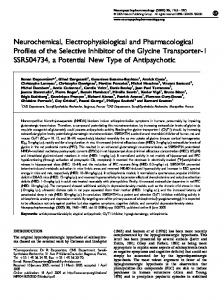
Fig. 3. The effect of acute administra-
tion of desipramine, nomifensine and fluoxetine on stress-induced deficits in the attentional set-shifting task in rats. Animals were exposed to restraint stress for 1 h daily for 7 days. The attentional set-shifting task was performed 14 days after stress cessation. The drugs were administered 30 min before testing. Results represent the mean ± SEM number of trials required to reach the criterion of 6 consecutive correct trials for each of the discrimination phases. Symbols: * p at least < 0.05 vs. ED performance in the unstressed vehicle-treated group, # p at least < 0.05 vs. ED performance in the stressed vehicle-treated group, NewmanKeuls post-hoc test. Adapted from [43]
Pharmacological Reports, 2013, 65, 14791488
1485
SSRI, citalopram, but not with desipramine, attenuated the detrimental effects of chronic intermittent cold stress on the first reversal learning stage in the ASST [14, 24]. Our results have also indicated that restraint-induced cognitive inflexibility was fully reversed by acute administration of antidepressants acting at various monoamine systems, i.e., desipramine, nomifensine, fluoxetine and escitalopram (Fig. 3) [43]. It remains an unresolved issue, however, whether the effects of acute treatment with antidepressants in the rat may accurately predict a compound’s clinical efficacy because the clinical effects are typically achieved only after chronic drug administration. Nonetheless, acute administration of a variety of antidepressants produces antidepressant-like effects in screening procedures such as the forced swim test. If an effect of an acute treatment with antidepressant in the rat predicts its therapeutic action in humans, our study provides evidence that this may also partially restore cognitive flexibility.
Concluding remarks
The available research tools have only begun to reveal all of the possible stress-brain interactions and their consequences. Recent studies have also found gender differences in the response of cortical morphology to stress [47]. It remains to be determined whether this diversity in structural changes may also be translated to altered functions. Another unresolved question is the potential functional implication of the recently demonstrated circuit specificity of stress-evoked morphological changes within mPFC [48]. An intriguing possibility is the reversibility of either morphological or functional alterations [19], as well as possibly determining the factors that may facilitate or hamper these recovery processes. Further research is needed to better understand the complex impact of stress on the brain structure and functions.
Acknowledgments:
This work was supported by the grant POIG.01.01.02-12-004/09 Depression mechanisms therapy, which was co-financed by the European Union from the European Fund of Regional Development (EFRD).
1486
Pharmacological Reports, 2013, 65, 14791488
References: 1. Anisman H, Matheson K: Stress, depression, and anhedonia: Caveats concerning animal models. Neurosci Biobehav Rev, 2005, 29, 525–546. 2. Arnsten AF, Mathew R, Ubriani R, Taylor JR, Li BM: Alpha-1 noradrenergic receptor stimulation impairs prefrontal cortical cognitive function. Biol Psychiatry, 1999, 45, 26–31. 3. Austin M-P, Ross M, Murray C, O’Carroll RE, Ebmeier KP, Goodwin GM: Cognitive function in major depression. J Affect Disord, 1992, 25, 21–30. 4. Birnbaum S, Gobeske KT, Auerbach J, Taylor JR, Arnsten AF: A role for norepinephrine in stress-induced cognitive deficits: a-1-adrenoceptor mediation in the prefrontal cortex. Biol Psychiatry, 1999, 46, 1266–1274. 5. Birnbaum SG, Yuan PX, Wang M, Vijayraghavan S, Bloom AK, Davis DJ, Gobeske KT et al.: Protein kinase C overactivity impairs prefrontal cortical regulation of working memory. Science, 2004, 306, 882–884. 6. Birrell JM, Brown VJ: Medial frontal cortex mediates perceptual attentional set shifting in the rat. J Neurosci, 2000, 20, 4320–4324. 7. Bondi CO, Rodriguez G, Gould GG, Frazer A, Morilak DA: Chronic unpredictable stress induces a cognitive deficit and anxiety-like behavior in rats that is prevented by chronic antidepressant drug treatment. Neuropsychopharmacology, 2008, 33, 320–331. 8. Bondi CO, Jett JD, Morilak DA: Beneficial effects of desipramine on cognitive function of chronically stressed rats are mediated by a1-adrenergic receptors in medial prefrontal cortex. Prog Neuropsychopharmacol Biol Psychiatry, 2010, 34, 913–923. 9. Brown SM, Henning S, Wellman CL: Mild, short-term stress alters dendritic morphology in rat medial prefrontal cortex. Cereb Cortex, 2005, 15, 1714–1722. 10. Butts KA, Floresco SB, Phillips AG: Acute stress impairs set-shifting but not reversal learning. Behav Brain Res, 2013, 252C, 222–229. 11. Cerqueira JJ, Pego JM, Taipa R, Bessa JM, Almeida OF, Sousa N: Morphological correlates of corticosteroidinduced changes in prefrontal cortex-dependent behaviors. J Neurosci, 2005, 25, 7792–7800. 12. Channon S: Executive dysfunction in depression: the Wisconsin Card Sorting Test. J Affect Disord, 1996, 39, 107–114. 13. Cook SC, Wellman CL: Chronic stress alters dendritic morphology in rat medial prefrontal cortex. J Neurobiol, 2004, 60, 236–248. 14. Danet M, Lapiz-Bluhm S, Morilak DA: A cognitive deficit induced in rats by chronic intermittent cold stress is reversed by chronic antidepressant treatment. Int J Neuropsychopharmacol, 2010, 13, 997–1009. 15. Demakis GJ: A meta-analytic review of the sensitivity of the Wisconsin Card Sorting Test to frontal and lateralized frontal brain damage. Neuropsychology, 2003, 17, 255–264. 16. Elliott R, McKenna PJ, Robbins TW, Sahakian BJ: Neuropsychological evidence for frontostriatal dysfunction in schizophrenia. Psychol Med, 1995, 25, 619–630.
Stress-induced cognitive inflexibility Agnieszka Nikiforuk and Piotr Popik
17. Finlay JM, Zigmond MJ, Abercrombie ED: Increased dopamine and norepinephrine release in medial prefrontal cortex induced by acute and chronic stress: effects of diazepam. Neuroscience, 1995, 64, 619–628. 18. Garcia-Iglesias BB, Mendoza-Garrido ME, GutierrezOspina G, Rangel-Barajas C, Noyola-Diaz M, Terron JA: Sensitization of restraint-induced corticosterone secretion after chronic restraint in rats: involvement of 5-HT7 receptors. Neuropharmacology, 2013, 71, 216–227. 19. Goldwater DS, Pavlides C, Hunter RG, Bloss EB, Hof PR, McEwen BS, Morrison JH: Structural and functional alterations to rat medial prefrontal cortex following chronic restraint stress and recovery. Neuroscience, 2009, 164, 798–808. 20. Hains AB, Vu MA, Maciejewski PK, van Dyck CH, Gottron M, Arnsten AF: Inhibition of protein kinase C signaling protects prefrontal cortex dendritic spines and cognition from the effects of chronic stress. Proc Natl Acad Sci USA, 2009, 106, 17957–17962. 21. Holmes A, Wellman CL: Stress-induced prefrontal reorganization and executive dysfunction in rodents. Neurosci Biobehav Rev, 2009, 33, 773–783. 22. Jett JD, Morilak DA: Too much of a good thing: blocking noradrenergic facilitation in medial prefrontal cortex prevents the detrimental effects of chronic stress on cognition. Neuropsychopharmacology, 2013, 38, 585–595. 23. Keeler JF, Robbins TW: Translating cognition from animals to humans. Biochem Pharmacol, 2011, 81, 1356–1366. 24. Lapiz-Bluhm MD, Soto-Pina AE, Hensler JG, Morilak DA: Chronic intermittent cold stress and serotonin depletion induce deficits of reversal learning in an attentional set-shifting test in rats. Psychopharmacology (Berl), 2009, 202, 329–341. 25. Leuner B, Shors TJ: Stress, anxiety, and dendritic spines: What are the connections? Neuroscience, 2013, 251, 108–119. 26. Li N, Liu RJ, Dwyer JM, Banasr M, Lee B, Son H, Li XY et al.: Glutamate N-methyl-D-aspartate receptor antagonists rapidly reverse behavioral and synaptic deficits caused by chronic stress exposure. Biol Psychiatry, 2011, 69, 754–761. 27. Li YC, Wang FM, Pan Y, Qiang LQ, Cheng G, Zhang WY, Kong LD: Antidepressant-like effects of curcumin on serotonergic receptor-coupled AC-cAMP pathway in chronic unpredictable mild stress of rats. Prog Neuropsychopharmacol Biol Psychiatry, 2009, 33, 435–449. 28. Liston C, Miller MM, Goldwater DS, Radley JJ, Rocher AB, Hof PR, Morrison JH, McEwen BS: Stress-induced alterations in prefrontal cortical dendritic morphology predict selective impairments in perceptual attentional set-shifting. J Neurosci, 2006, 26, 7870–7874. 29. Liston C, McEwen BS, Casey BJ: Psychosocial stress reversibly disrupts prefrontal processing and attentional control. Proc Natl Acad Sci USA, 2009, 106, 912–917. 30. Liu RJ, Aghajanian GK: Stress blunts serotonin- and hypocretin-evoked EPSCs in prefrontal cortex: role of corticosterone-mediated apical dendritic atrophy. Proc Natl Acad Sci USA, 2008, 105, 359–364. 31. Magariños AM, McEwen BS: Stress-induced atrophy of apical dendrites of hippocampal CA3c neurons: involve-
32.
33. 34.
35.
36.
37.
38.
39.
40.
41.
42.
43.
44.
45.
46.
47.
48.
ment of glucocorticoid secretion and excitatory amino acid receptors. Neuroscience, 1995, 69, 89–98. Martin KP, Wellman CL: NMDA receptor blockade alters stress-induced dendritic remodeling in medial prefrontal cortex. Cereb Cortex, 2011, 21, 2366–2373. Mehta MA, Riedel WJ: Dopaminergic enhancement of cognitive function. Curr Pharm Des, 2006, 12, 2487–2500. Merriam EP, Thase ME, Haas GL, Keshavan MS, Sweeney JA: Prefrontal cortical dysfunction in depression determined by Wisconsin Card Sorting Test performance. Am J Psychiatry, 1999, 156, 780–782. Mizoguchi K, Yuzurihara M, Ishige A, Sasaki H, Chui DH, Tabira T: Chronic stress induces impairment of spatial working memory because of prefrontal dopaminergic dysfunction. J Neurosci, 2000, 20, 1568–1574. Moghaddam B: Stress preferentially increases extraneuronal levels of excitatory amino acids in the prefrontal cortex: comparison to hippocampus and basal ganglia. J Neurochem, 1993, 60, 1650–1657. Moghaddam B, Bolinao ML, Stein-Behrens B, Sapolsky R: Glucocorticoids mediate the stress-induced extracellular accumulation of glutamate. Brain Res, 1994, 655, 251–254. Naegeli KJ, O’Connor JA, Banerjee P, Morilak DA: Effects of milnacipran on cognitive flexibility following chronic stress in rats. Eur J Pharmacol, 2013, 703, 62–66. Nikiforuk A: Dopamine D1 receptor modulation of set shifting: the role of stress exposure. Behav Pharmacol, 2012, 23, 434–438. Nikiforuk A: Quetiapine ameliorates stress-induced cognitive inflexibility in rats. Neuropharmacology, 2013, 64, 357–364. Nikiforuk A: Selective blockade of 5-HT7 receptors facilitates attentional set-shifting in stressed and control rats. Behav Brain Res, 2012, 226, 118–123. Nikiforuk A, Popik P: Amisulpride promotes cognitive flexibility in rats: The role of 5-HT7 receptors. Behav Brain Res, 2013, 248, 136–140. Nikiforuk A, Popik P: Long-lasting cognitive deficit induced by stress is alleviated by acute administration of antidepressants. Psychoneuroendocrinology, 2011, 36, 28–39. Radley JJ, Rocher AB, Miller M, Janssen WG, Liston C, Hof PR, McEwen BS, Morrison JH: Repeated stress induces dendritic spine loss in the rat medial prefrontal cortex. Cereb Cortex, 2006, 16, 313–320. Roberts AC, Robbins TW, Everitt BJ: The effects of intradimensional and extradimensional shifts on visual discrimination learning in humans and non-human primates. Q J Exp Psychol B, 1988, 40, 321–341. Robinson LJ, Thompson JM, Gallagher P, Goswami U, Young AH, Ferrier IN, Moore PB: A meta-analysis of cognitive deficits in euthymic patients with bipolar disorder. J Affect Disord, 2006, 93, 105–115. Shansky RM, Hamo C, Hof PR, Lou W, McEwen BS, Morrison JH: Estrogen promotes stress sensitivity in a prefrontal cortex-amygdala pathway. Cereb Cortex, 2010, 20, 2560–2567. Shansky RM, Hamo C, Hof PR, McEwen BS, Morrison JH: Stress-induced dendritic remodeling in the prefrontal cortex is circuit specific. Cereb Cortex, 2009, 19, 2479–2484. Pharmacological Reports, 2013, 65, 14791488
1487
49. Takahashi T, Kimoto T, Tanabe N, Hattori TA, Yasumatsu N, Kawato S: Corticosterone acutely prolonged N-methyl-D-aspartate receptor-mediated Ca2+ elevation in cultured rat hippocampal neurons. J Neurochem, 2002, 83, 1441–1451. 50. Thai CA, Zhang Y, Howland JG: Effects of acute restraint stress on set-shifting and reversal learning in male rats. Cogn Affect Behav Neurosci, 2013, 13, 164–173. 51. Tokarski K, Bobula B, Kusek M, Hess G: The 5-HT7 receptor antagonist SB 269970 counteracts restraint stress-induced attenuation of long-term potentiation in rat frontal cortex. J Physiol Pharmacol, 2011, 62, 663–667. 52. Tokarski K, Pitra P, Duszynska B, Hess G: Imipramine counteracts corticosterone-induced alterations in the effects of the activation of 5-HT7 receptors in rat hippocampus. J Physiol Pharmacol, 2009, 60, 83–88.
1488
Pharmacological Reports, 2013, 65, 14791488
53. Wellman CL: Dendritic reorganization in pyramidal neurons in medial prefrontal cortex after chronic corticosterone administration. J Neurobiol, 2001, 49, 245–253. 54. Weso³owska A, Tatarczyñska E, Nikiforuk A, Chojnacka-Wójcik E: Enhancement of the anti-immobility action of antidepressants by a selective 5-HT7 receptor antagonist in the forced swimming test in mice. Eur J Pharmacol, 2007, 555, 43–47. 55. Willner P: Validity, reliability and utility of the chronic mild stress model of depression: a 10-year review and evaluation. Psychopharmacology (Berl), 1997, 134, 319–329.
Received: July 24, 2013; in the revised form: October 14, 2013; accepted: October 15, 2013.