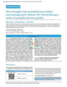
Nov 6, 2016 - optic neuropathy characterized by retinal ganglion cell. (RGC) apoptosis and ..... The previous study reported that ATR1 antagonist (losartan.
Effect of hyerptension on IOP, RGC and ET-1
窑Basic Research窑
Neuropathy optic glaucomatosa induced by systemic hypertension through activation endothelin-1 signaling pathway in central retinal artery in rats
Department of Ophthalmology, Faculty of Medicine, Brawijaya University, Malang 65145, Indonesia 2 Pharmacology Laboratory, Faculty of Medicine, Brawijaya University, Malang 65145, Indonesia Correspondence to: Prayitnaningsih Seskoati. Department of Ophthalmology, Faculty of Medicine, Brawijaya University, Malang 65145, East Java, Indonesia. seskoatip@ yahoo.com Received: 2016-03-01 Accepted: 2016-05-30 1
Abstract
· AIM:
To evaluate effect of hypertension on retinal
ganglion cell (RGC) apoptosis, intraocular pressure (IOP), and the activation of endothelin -1 (ET -1) signaling pathway in central retinal artery (CRA) in rats.
·METHODS: The experimental
study was performed on
20 male Sprague Dawley rats that were divided into control group, and hypertension groups. The hypertension was induced by subcutaneous deoxycorticoacetate (DOCA) 10 mg/kg twice a week and administered 0.9% NaCl solution daily for 2, 6, and 10wk. Blood pressure (BP) was measured using animal BP analyzer. IOP was measured by handheld tonometry. Retinal tissue preparations by paraffin blocks were made after enucleation. The expression of ET-1, eNOS, ET-1 receptor A (ETRA), ET-1 receptor B (ETRB), and phosphorylated myosin light chain kinase (MLCK), and caldesmon (CaD) in CRA and RGC apoptosis were evaluated through immunofluorescent staining method then observed using laser scanning confocal microscopy.
· RESULTS:
BP significantly increased in all of the
hypertension groups compared to control ( =0.001). Peak IOP elevation (7.78依4.14 mm Hg) and RGC apoptosis (576.15依33.28 Au) occurred on 2wk of hypertension. ET-1 expression (1238.6依55.1 Au) and eNOS expression (2814.2依 70.7 Au) were found highest in 2wk of hypertension, although the ratio of ET -1/eNOS decreased since 2wk. ETRA reached peak expression in 10wk of hypertension (1219.4 依6.3 Au), while ETRB significantly increased only in 2 weeks group (1069.2 依9.6 Au). The highest MLCK expression (1190.09依58.32 Au), CaD (1670.28依18.36 Au) were also found in 2wk of hypertension. 1568
· CONCLUSION:
Hypertension effects to activation of
ET-1 signaling pathway significantly in CRA, elevation of IOP, and RGC apoptosis. The highest value was achieved at 2wk, which is the development phase of hypertension.
·
KEYWORDS:
hypertension;
central
retinal
artery;
endothelin-1 signaling pathway; rats DOI:10.18240/ijo.2016.11.06 Prayitnaningsih S, Sujuti H, Effendi M, Abdullah A, Anandita NW, Yohana F, Permatasari N, Widodo MA. Neuropathy optic glaucomatosa induced by systemic hypertension through activation endothelin-1 signaling pathway in central retinal artery in rats. 2016;9(11):1568-1577
INTRODUCTION laucomatous optic neuropathy (GON) is a progressive optic neuropathy characterized by retinal ganglion cell (RGC) apoptosis and degeneration of its axons, that can result in permanent blindness. The relationship between hypertension and GON remains unclear. It has been reported that there is a positive correlation between hypertension and vascular autoregulation dysfunction in optic nerve [head normotension glaucoma (NTG)] while another study showed that there is the mechanical effect of hypertension on intraocular pressure (IOP) [primary open angle glaucoma (POAG)]. These two mechanisms support theory regarding ischemia in GON occurrence. On the other hand, some studies reported that there is no relationship between hypertension and GON[1-2]. Endothelin-1 (ET-1) is a strong vasoconstrictor agent which plays a major role in ischemia occurrence in optic nerve head. Elevated levels of ET-1 leads to hypertension and accumulation of ET-1 on high concentration causes organ failure in hypertension patients. Vascular autoregulation determines vascular endothelium function in perfusion, in which central retinal artery (CRA) is main nutrition source of the internal layer of the retina, whereas the imbalance between ET-1 and nitric oxide (NO) is the initial disruption in endothelial dysfunction. NO can act as a neurodegenerative or neuroprotective factor after ischemia depends on formed NO and nitric oxide synthase (NOS) isoform amount. NO, which formed from endothelial nitric oxide synthase (eNOS) and neuronal nitric oxide synthase
G
陨灶贼 允 韵责澡贼澡葬造皂燥造熏 灾燥造援 9熏 晕燥援 11熏 Nov.18, 圆园16 www. ijo. cn 栽藻造押8629原愿圆圆源缘员苑圆 8629 -82210956 耘皂葬蚤造押ijopress 岳员远猿援糟燥皂
(nNOS) on perivascular nitrergic neurons keep ocular blood circulation and prevents infarct[1-3]. Moreover, it was revealed that hypertension activates ET-1 signaling pathway in trabecular meshwork (TM). The increase of ET-1 and eNOS has reached their peak in the second week of hypertension related to ET-1 receptor A (ETRA) and ET-1 receptor B (ETRB) expression as ET-1 receptors. Other study results showed the possibility of hypertension effected on NTG occurrence and prolonged retinal arteriovenous passage (AVP) time[4-5]. The previous clinical study has shown that hypertension affects NTG occurrence. This study tries to reveal the effect of hypertension on ET-1 signaling pathway activation on CRA, IOP and RGC apoptosis. Effects of hypertension on eNOS expression and ET-1 expression in endothelial cells of CRA were examined in this study. ET-1 signaling pathway activation was determined by ETRA and ETRB expressions in smooth muscle of CRA, whereas ETRA is responsible in vasoconstriction and ETRB in vasodilatation. The roles of inositol triphosphate (IP3) and diacylglycerol (DAG) in CRA vasoconstriction were examined by measurement of phosphorylated myosin light chain kinase (MLCK) and caldesmon (CaD) expression. Deoxycorticoacetate (DOCA)-salt hypertensive model is an ideal observation model of kidney role in hypervolemia occurrence, this model is also related to ET-1 expression. There are some changes to DOCA-salt hypertension model in several phase of its development which occurs at second and sixth weeks. This study aimed to evaluate the effects of hypertension on ET-1 activity in CRA according to the hypertension duration. MATERIALS AND METHODS Animal Model Preparation This study was conducted from June to October 2014 at Pharmacology Laboratory, Faculty of Medicine and Central Laboratory of Biological Sciences, Brawijaya University. The experimental design was true experimental post only with control. Male Spraque Dawley rats 4-5 months old and 250-350 grams of weight were obtained from Animal Model Laboratory, Biomedical Center and Health Basic Technology (Jakarta, Indonesia). Twenty animal models were divided into four groups. The ethical clearance of this study was obtained from Health Research Ethics Committee of Faculty of Medicine, Brawijaya University. Deoxycorticoacetate -salt Hypertension Induction and Intraocular Pressure Measurement Rats were injected with DOCA 10 mg/kg dissolved in corn oil subcutaneously 2 times a week and had been administered 0.9% of NaCl solution for 2, 6 and 10wk. Rats were categorized as hypertensive if their BP were reached >150/90 mm Hg. The control group received corn oil administration 2 times a week subcutaneously and were given aqua bidest . Blood
pressure (BP) was measured by animal BP analyzer. Systolic blood pressure (SBP) value was calculated from the average of 3 lowest values from 5 SBP recorded values [6]. IOP was measured using hand-held non-contact tonometer after stabilization (ICare, USA). Central Retinal Artery and Retinal Tissue Sampling CRA and retinal tissue were taken by enucleation under the operating microscope (DECA 21, Inami Co. Ltd.). After being enucleated, they were directly put into bottles with 4% of paraformaldehyde. Eyeballs were cut transversely on the equator. After lenses were removed, anterior segments were divided into four quadrants, which allowed for easing in obtaining CRA and retinal tissues through paraffin blocking process[2]. Measurement of Endothelin-1, Endothelial Nitric Oxide Synthase, Endothelin -1 Receptor A/B Expressions and Phosphorylated Myosin Light Chain Kinase and Caldesmon Expression Assays Immunofluorescence (IF) staining was started with deparaffinization process. Primary antibodies used were anti-rat ET-1 antibody, eNOS-activated antibody, anti-rat ETRA antibody (Bioss, USA), anti-rat ETRB antibody (Bioss, USA), phospho-specific MLCK serine 19 antibody, anti CaD phosphor (Ser789), Smoothelin antibody (Bioss, USA), and von Willebrand factor (vWF). Secondary antibodies which used were fluorescein isothiocyanate (FITC) for eNOS, ETRB, and CaD while tetraethyl rhodamine isothiocyanate (TRITC) for ET-1, ETRA and MLCK. Apoptosis of RGC used annexin V and propidium iodide. Furthermore, slides were immediately observed with laser confocal microscope (LSCM) (Olympus fluo FV 10-ASW 1.7), with 400 × of magnification with 5 fields of view. Color intensity analysis was using the image of Olympus Image Binary Format was in the form of numerical data in Arbitrary units (Au). Statistical Analysis Data were analyzed using One-way ANOVA or Kruskal-Wallis test and Mann-Whitney test. All analysis were performed using SPSS 19.0 software for Windows. RESULTS Effect of Hypertension on the Increasing of Retinal Ganglion Cell Apoptosis Figure 1 shows that the most significant increase in apoptosis was found in 2-week group. Meanwhile, the 6-week and 10-week groups showed a decrease in apoptosis compared with 2-week group, although it still increased compared with the control group. Effect of Hypertension on the Intraocular Pressure The results of IOP measurement prior to treatment showed no significant differences in 4 groups ( =0.769), it indicates that initial IOP was homogeneous. Average of final IOP matched with the criteria of ocular hypertension was obtained in 2-week group, . equal to 21.42 依2.95 mm Hg. It is significant differences in delta IOP among the 4 groups with 1569
Effect of hyerptension on IOP, RGC and ET-1
Figure 1 Description of RGC undergoing apoptosis IF staining with Annexin V (green); RGC apoptosis increased in 2-week group (B), 6-week group (C) and 10-week group (D) compared with control group (A).
Figure 2 ET-1 expression (red) and CRA endothelial cell (green) The increase of cellular ET-1 expression is qualitatively visible in the 2-week group and 6-week group compared to the control group. While the 10-week group shows no difference with control. The pictures were obtained by IF staining observed under a confocal microscope with magnification 400伊. Cellular expression of ET-1 on endothelial CRA cells is shown by the arrow.
value of 0.026. Two-week group gave the highest delta IOP average, . 7.78依4.14 mm Hg (Table 1). Hypertension Effect on Expression of Endothelin-1 and Endothelial Nitric Oxide Synthase Expression of ET-1 in CRA endothelial cells significantly increased in 2-week group and 6-week group compared to control. Expression of ET-1 in 10-week group was lower than 2-week and 6-week group (Figure 2). ET-1 cellular expression in 10-week group was quite similar to ET-1 expression in control. eNOS expression on CRA endothelial cell in the control group and hypertension model group was significantly different. There is no significant difference of eNOS expression in 2-week group and 10-week group (Figure 3). There is a decrease in the ratio of cellular expression of ET-1 and eNOS activation since the second week although there is a slight increase in the 6th week (Figure 4). Effect of Hypertension Duration on Endothelin -1 Receptor A/B Expressions The duration of high ETRA 1570
Tabel 1 The differences of IOP measurement in 4 groups IOP
x ± s ; mm Hg
Hypertension duration (wk)
P
Control
2
6
10
Initial IOP
15.16±3.68
13.64±3.55
15.76±4.11
15.84±3.43
0.769
Final IOP
15.44±6.67
21.42±2.95
16.98±2.06
14.88±8.22
0.278
Delta IOP
0.28±3.59
7.78±4.14
1.22±3.38
-0.96±5.84
0.026
Table 2 The comparison of ETRA and ETRB expression in the CRA x ± s ; Au smooth muscle between control and hypertension groups Protein
Duration of hypertension
P
Control
2-week
6-week
10-week
ETRA
1075.2±9.6
1130.6±17.2
1200.1±6.4
1219.4±6.3
0.000
ETRB
938.7±11.6
1069.2±9.6
860.0±17.7
870.8±19.2
0.001
expression in CRA smooth muscle is longer than (Figures 4, 5). ETRA expression continues to increase the tenth week while ETRB expression increases only the second week and then decrease until the tenth (Table 2).
ETRB up to up to week
陨灶贼 允 韵责澡贼澡葬造皂燥造熏 灾燥造援 9熏 晕燥援 11熏 Nov.18, 圆园16 www. ijo. cn 栽藻造押8629原愿圆圆源缘员苑圆 8629 -82210956 耘皂葬蚤造押ijopress 岳员远猿援糟燥皂
Figure 3 eNOS expression (green) on CRA endothelial cells (red) The increase of cellular eNOS expression is qualitatively visible in the 2-week group, 6-week group, and 10-week group compared to the control group. The pictures were obtained by IF staining observed under a confocal microscope with magnification 400伊. Cellular expression of eNOS on endothelial CRA cells is shown by the arrow. Table 3 Comparison between duration of hypertension and MLCK and CaD activation in the ARS smooth x ± s ; Au muscles Protein
Duration of hypertension 2-week
MLCK
765.73±12.26
1190.09±58.32
875.69±9.53
779.77±8.52
0.001
CaD
882.04±9.37
1670.28±18.36
1273.10±8.81
1375.45±9.14
0.000
Figure 4 ET-1 and eNOS expression ratio on endothelial cell in the control group, 2 -week group, 6 -week group, and 10week group.
Effect of Hypertension on Cellular Expression of Phosphorylated Myosin Light Chain Kinase and Caldesmon Expression The limitation of CRA vasoconstriction observation with paraffin block method might cause evaluation of ET-1 activation is only up to its effect on CRA vasoconstriction. Observation of cellular expression of phosphorylated MLCK and CaD evaluated the role of IP3 and DAG in CRA vasoconstriction. The duration
6-week
10-week
P
Control
of CaD expression in CRA smooth muscle was longer than MLCK (Figure 7). All of the hypertension model groups showed the increasing in CaD, while MLCK significantly increased only up to the second week of hypertension duration which decreased afterward until the tenth week. Hypertension duration increased cellular expression of CaD in 2-week group, 6-week group, and 10-week group (Figure 8; Table 3). CaD showed highest cellular expression in 2-week group. There is a rise in mean cellular expression of MLCK and CaD in all groups with CaD expression rising being higher and longer than MLCK. DISCUSSION This research showed a significant increase in the final BP according to hypertension duration ( =0.001). Administration of DOCA-salt induced neurohumoral activation and increased blood volume. Intraneural ischemia has an important role in RGC apoptosis which occurs due to autoregulation dysfunction in CRA as nutrition main source of optic nerve head. Aldosterone induces superoxide formation through mineralocorticoid receptor which activates NADPH oxide and Rac1 in endothelial cells. The activation 1571
Effect of hyerptension on IOP, RGC and ET-1
Figure 5 Cellular expression of ETRA in CRA smooth muscle Cellular expression of ETRA recognized by TRITC staining in red color while the smooth muscle with FITC stains green. The increase of cellular ETRA expression is qualitatively visible in the 2-week, 6-week group, and 10-week group compared to the control group. The pictures are obtained by IF staining observed under a confocal microscope with magnification 400伊. Cellular expression of ETRA on endothelial CRA cells is shown by the arrow.
Figure 6 Cellular expression of ETRB in CRA smooth muscle Cellular expression of ETRB recognized by FITC staining in green color while the smooth muscle with TRITC stains red. The increase of cellular ETRB expression is qualitatively visible in the 2-week group compared with control, Reduction of ETRB cellular expression in 6-week group, and 10-week group. The pictures are obtained by IF staining observed under a confocal microscope with magnification 400伊. Cellular expression of ETRB on endothelial CRA cells is shown by the arrow.
of NADPH oxidase and Rac1 cause vascular damage by increasing the ROS formation. Increased levels of ROS induce NO synthase uncoupling and xanthine oxidase activation causing vascular endothelial dysfunction, apoptosis and inflammation[7-8]. Hypertension Effect on Retinal Ganglion Cells Apoptosis RGC apoptosis in GON occurred due to the presence of 1572
intraneural ischemia. It is caused by perfusion disturbance of optical nerve. Perfusion disturbance can occur when IOP suppress the blood supply (vascular disturbance). Long-term hypertension is indicated by increasing vascular resistance so it affects to low BP in the capillary bed. In addition, It can cause vascular endothelial dysfunction that can disrupt autoregulation of vascularisation and ischemia of optic nerve.
陨灶贼 允 韵责澡贼澡葬造皂燥造熏 灾燥造援 9熏 晕燥援 11熏 Nov.18, 圆园16 www. ijo. cn 栽藻造押8629原愿圆圆源缘员苑圆 8629 -82210956 耘皂葬蚤造押ijopress 岳员远猿援糟燥皂
Figure 7 Cellular expression of phosphorylated MLCK in CRA smooth muscle Cellular expression of phosphorylated MLCK (p-MLCK) recognized by TRITC staining in red color while the smooth muscle with FITC stains green. The increase of cellular p-MLCK expression is qualitatively visible in the 2-week group compared with control. Reduction of p-MLCK cellular expression in 6-week group, and 10-week group. The pictures were obtained by IF staining observed under a confocal microscope with magnification 400 伊 . Cellular expression of p-MLCK on endothelial CRA cells is shown by the arrow.
Figure 8 Cellular expression of phosphorylated CaD in CRA smooth muscle Cellular expression of CaD recognized by FITC staining in green color while the smooth muscle with TRITC stains red. The increase of cellular CaD expression is qualitatively visible in the 2-week group, 6-week group and 10-week group compared with control. The pictures were obtained by IF staining observed under a confocal microscope with magnification 400伊. Cellular expression of CaD on endothelial CRA cells is shown by the arrow.
Intraneural ischemia which would activate microglia and astrocytes. This condition also induces the releasing of TNF-琢, NO, ET-1, glutamate, serine, and sodium into retinal extracellular space which caused toxic effects in extracellular space. Toxic effect disturbed RGC mitochondria which lead apoptosis[1,9-13].
[14] These results also related with Nitta , using subcutaneous DOCA administration through osmotic minipump. A significant decrease in RGC amount was found in the peripheral retina in the second week and on the peripheral area or central retina was in the fourth week. In the 6th week, there was an increase of RGC amount in both of
1573
Effect of hyerptension on IOP, RGC and ET-1
peripheral area and central retina. There were no significant differences in the thickness of both of internal and external nuclear layers, also internal and external plexiform layers after the administration of aldosterone for 6wk compared with control group. This condition showed that severe neuronal losses only occurred in RGC layer. Cell death on retinal ganglion allegedly occurred due to renin-angiotensinaldosterone system (RAAS) system activation in brain and TM tissues which started from mineralocorticoid receptor (MR) activation. This activation led ET-1 signaling pathway which the interaction between ET-1 and ETRB would stimulate RGC apoptosis[14-15]. RGC apoptosis peak has increased in the 2nd week then followed by its decrease in the 6th and 10th week. Angiotensin 1-7 and Mas receptor followed by ETRB activation and increased production of NO explained this condition. Previous studies showed that RGC death occurred in two phases. Rapid phase occurred in the beginning of 3wk followed by slow phase occurred in a few months later. Mechanisms in rapid phase seemed to be related to the pressure, it was related to neuronal cells undergo apoptosis within 2 to 20h after the increasing of IOP. The process of secondary degeneration resulted in cell death. This condition related to this study results which the average of highest apoptosis occurs in initial 2wk (rapid phase) from DOCA-salt administration, then followed by average decreases of RGC apoptosis in the 6th and 10th week (slow phase)[11,14,16]. Hypertension Effect on Intraocular Pressure Hypertension effects of DOCA-salt model on TM were alleged due to the increased glucocorticoid receptor (GR). Sharif [17] mentioned that the effect of renin-angitensin (RAS) on TM was shown in angiotensin receptor 1 (ATR1) activity that mediated signaling for the accumulation of extracellular matrix and TGF-茁 on TM tissue that leads increase of IOP. [18] Stokes has found the high expression of GR mRNA and mineralocorticoid 11茁-HSD that interact with MR which has a role in the mechanism of ion and water transport, including the secretion of aqueous humor (AH). Also known that MR on TM is well-bonded with cortisol and aldosterone. Mineralocorticoid effects on eyes were mediated by epithelial sodium channels (EnaC), which involved in the increasing of AH formation and organize TM resistance through the regulation of TM cell volume[17-19] (Figure 9). The previous study reported that ATR1 antagonist (losartan and olmesartan medoxomil), angiotensin-converting enzyme (ACE) inhibitors (enalapril), and renin inhibitors (CGP38650, SRO43845 and Abbott-64 662) could decrease IOP by 15% -20% both in animal and human models. [11] Vaajanen reported that topical diminazene aceturate (DIZE) administration as ACE-2 activator could reduce IOP [ 20 ] in rabbit eyes . Research by Foureaux found that 1 mg/kg of DIZE daily oral administration for 1mo could 1574
Figure 9 Hypothesis of the involvement of RAS system and activation of ET -1 signaling pathway on IOP elevation in mice [17] This figure shows Ang-I, Ang-II, and Ang (1-7) production and their preferred receptor subtype Mas-1 and AT-1 receptors mediate signaling for activation of MLCK that responsible for raising IOP. Ang: Angiotensin; ATR1: Angiotensin receptor 1; IOP: Intraocular pressure; RAS: Renin-angitensin.
increase the ACE-2 expression in the retina. Hypotension effects of DIZE could be prevented by administration of Mas-receptor-selective antagonist A-779. This condition indicated DIZE has helped Ang (1-7) in Mas-receptor activation to decrease IOP. It was further stated that AH drainage from anterior camera oculi was stimulated by DIZE. RGC was maintained by DIZE through inhibition of caspase [21] 3 expressions. Meanwhile, Agarwal found that ACE inhibitor could induce the decreasing of IOP through its involvement in MMPs and cytokines modulation. Research [22] by Vaajanen found that the administration of enalaprilat dehydrates 1% (ACE inhibitor) single drop could reduce IOP by 20.3% after 3h of administration while losartan potassium 2% (ATR1 blocker) administration could decrease the IOP by 13.3%[11,20-23].
陨灶贼 允 韵责澡贼澡葬造皂燥造熏 灾燥造援 9熏 晕燥援 11熏 Nov.18, 圆园16 www. ijo. cn 栽藻造押8629原愿圆圆源缘员苑圆 8629 -82210956 耘皂葬蚤造押ijopress 岳员远猿援糟燥皂
Effect of Hypertension on Expression of Endothelin -1 and Endothelial Nitric Oxide Synthase The expression of ET-1 in CRA endothelial cells significantly increased on the second week (1238.6 依55.1 Au) compared to control (1069.2依22.6 Au). ET-1 expression started to decrease on the sixth week (897.7依14.0 Au) and it is similar with control on the tenth week (1111.9 依70.5 Au). eNOS expression significantly increased on second week (2814.2 依70.7 Au) and continue to increase (1935.9依22.1 Au on the sixth week and 2796.4依34.9 Au on the tenth week) compared to control (1049.8 依28.8 Au). Yamane 's [24] study revealed that DOCA-salt hypertension model has several developmental phases. In second until the sixth week of developmental phase, the activity of sympathic and humoral are balance indicated by the increasing of Vasopressin (VP)dan ET-1[6,24-25]. The increasing of ET-1 expression in this study suspected as the result of ET-1 localization in CRA endothelial cells occurs due to autoregulation dysfunction and mechanical compression. The previous study reports that endothelial cell will express the ET-1 as central baroreceptor-responsive and have a role in choroidal regulation against high systemic SBP. In this study, the administration of DOCA-salt causes ischemia in the CRA smooth muscle, probably by 1) involvement of aldosterone in increasing the activity of MR that increases NADPH oxidase and release of NO; 2) activation of sympathetic innervation in the choroid which increases IOP, destruction of blood retinal barrier (BRB) and release of ET-1. The imbalance of ET-1 and NO is the initial process of endothelial dysfunction in the CRA[26-27]. Toque 's [8] study supports the increasing of eNOS expression which described that DOCA-salt treatment for 6wk induces arginase (ARG) activity 1.5 times higher in the aorta and 2.6 times in mesenteric artery. ARG is a competitor for eNOS activity and regulator of NO production causing vascular dysfunction on DOCA-salt hypertension induction. Single nucleotide polymorphism is known affecting eNOS activation. There is positive correlation between hypertension and POAG. The decrease of ET-1/eNOS ration in the second week indicates the presence of compensatory mechanism by increasing eNOS to prevent further infarction. Hypertension causes damage to nerve fiber layer as the effect of compensatory hyperperfusion. Excessive NO accumulation can lead to peroxynitrite toxicity and generate apoptosis on RGC, therefore, we need further study in longer duration of hypertension[28]. The Effect of Hypertension on Endothelin -1 Receptor A/B Expressions in Central Retiral Atrery Smooth Muscle The effect of ET-1 expression is mediated by ETRA and ETRB expression. ETRA is commonly found in smooth muscle cells while ETRB is mostly found in other tissues such as brain and some epithelial cells. These receptors induce smooth muscle contraction, including smooth muscle of
CRA. ETRA expression significantly increased in 2-week group (1130.6依17.2 Au), 6-week group (1200.1依6.4 Au), and 10-week group (1219.4 依6.3 Au) compared to control (1075.2依9.6 Au). ETRB expression significantly increased on the second week of hypertension (1069.2依9.6 Au) compared to control (938.7依11.6 Au). It started to decrease on the sixth week of hypertension (860.0 依17.7 Au) and continuously decreased until the tenth week of hypertension (870.8依19.2 Au). Mature ET-1 activates ETRA and ETRB through autocrine and paracrine signaling which depend on its concentration. Oxidative stress and excessive vascular superoxide anion (O2-) production induce the increasing of ETRA and ETRB expressions through NADPH oxidase pathway on the second week of hypertension leading to endothelial dysfunction. Accumulation of O2- inhibits endothelin-converting enzyme (ECE) activation and ET-1 synthesis. Jimenez 's[29] study showed that ROS has a complex role in ET-1 synthesis. Production of O2-, NO, and hydrogen peroxide determine ET-1 level. Internalized ETRA is then released into the pericentriolar compartment and recycled. The recycled ETRA appear in plasma membrane without ligand whereas internalized ETRB is separated and degraded in lysosome causing longer vasoconstriction effect by ETRA than ETRB. Protein scaffolding such as Tip60 and HDAC7 are also responsible in life span. ET-1 affinity is stronger when it bound with ETRA than with ETRB. The expression of ETRA in smooth muscle is associated with hypertension duration. Disturbance of ETRB elimination also affects ET-1 elimination. The accumulation of ET-1 activates more ETRA causing continues vasoconstriction[30]. Effect of Hypertension on Phosphorylated Myosin Light Chain Kinase and Caldesmon Expression Actin-myosin interaction in molecular level is controlled by 2 isoforms of myosin II. These isoforms activities are determined by phosphorylation of posttranslational myosin regulatory light chain 9 (Myl9) through antagonist activity of MLCK and myosin light chain (MLC) phosphatase (MYP). Post-translation modification of Myl9 is triggered by RhoA/ROCK signaling pathway enhanced by MLC phosphorylation through Myosin Light Chain Phosphorylation (MLCP) inhibition on porcine retinal artery [23]. MLCK activation significantly increased on CRA smooth muscle in 2-week group (1190.09依58.32 Au) and 6-week group (875.69依9.53 Au) compared to control group, while there is no significant difference between 10-week group (779.77依8.52 Au) and control (765.73依12.26 Au). [31] show that DOCA-salt hypertension Study of Licht induction with subcutaneous injection of 50 mg pellet for 21d causes arteriolar vasoconstriction on retina. It also increases expression of Myl9 and phosphorylated Myl9 level in smooth muscle cells of retinal vascular. This condition is related to ET-1 which induced by activator protein-1 (AP-1) as a transcription factor of cellular contractility, vascular smooth 1575
Effect of hyerptension on IOP, RGC and ET-1
muscle migration, proliferation during neointimal formation, and endothelial cell migration and development. ET-1 also stimulated ERK activity and further AP-1 activities. Myl9 upregulation is a response of vascular smooth muscle on stress[32]. Phosphorylated CaD expression in CRA smooth muscle cells significantly increased in all hypertension model groups compared to control (882.04依9.73 Au). It decreased significantly on the sixth week of hypertension (1273.10 依8.81 Au) and increased again on the tenth week (1375.45 依9.14 Au) but lower than the second week (1670.28依18.36 Au). Duration of CaD activation is longer than MLCK activation [33]. These results support the theory that hypertension increases CRA vasoconstriction significantly. Interaction of ET-1 with ETRA receptor activates phospholipase C (PLC-茁) and increases expression of IP3 and DAG. IP3 stimulates Ca2+ to be released from the sarcoplasmic reticulum (SR). ET-1 stimulates Ca2+ entering Ca2+ channels which allow it binds to CaD. Ca2+ and CaD complex activates MLCK, MLC-P causing actin-myosin interaction in smooth muscle contraction. Diacylglycerol (DAG) activates PKC which then induces actin-binding protein calponin (CAP) phosphorylation also protein kinase cascades involve Raf, MAPK kinase (MEK), and MAPK (ERK1/2). Protein kinase cascades activation increases myofilament sensitivity to Ca2+[15]. Ca2+ and CaD complex formation is not required for MLCK activation through IP3 pathway while CaD activation is Ca2+-independent which might cause more requirement of time in CaD activation than MLCK. The control group in this study was one group treated similarly with 10-week group. The control group should be treated similarly with 2-week group and 6-week group. CRA contractility could not be observed due to paraffinated samples. The limitation of this study is there was only one control group, followed for 10wk. The control group should be also in 2 and 6wk to exclude age factor. Another limitation was paraffin blocks preparation which cause TM contractility could not be assessed directly. Hypertension of DOCA-salt model affects significantly on the activation of ET-1 signaling pathway in CRA endothelial cells and smooth muscle cells, IOP and RGC apoptosis elevation. The peak of ET-1 activation signaling pathway occurs in the development phase of hypertension. ACKNOWLEDGEMENTS Foundation: Directorate General of Higher Education (DGHE), National Education Ministry Republic of Indonesia. Conflicts of Interest: Prayitnaningsih S, None; Sujuti H, None; Effendi M, None; Abdullah A, None; Anandita NW, None; Yohana F, None; Permatasari N, None; Widodo MA, None. 1576
REFERENCES 1 Agarwal R, Gupta SK, Agarwal P, Saxena R, Agrawal SS. Current concepts in the pathophysiology of glaucoma.
2009;57
(4):257-266. 2 Tektas OY, Lutjen-Drecoll E. Structural changes of the trabecular meshwork in different kinds of glaucoma. 769-775.
2009;88 (4):
3 Toda N, Nakanishi-Toda M. Nitric oxide: ocular blood flow, glaucoma, and diabetic retinopathy.
2007;26(3):205-238.
4 Plange N, Kaup M, Remky A, Arend KO. Prolonged retinal arteriovenous passage time is correlated to ocular perfusion pressure in normal tension glaucoma.
2008;246(8):1147-1152.
5 Prayitnaningsih S, Sujuti H, Abdullah Hamid A, Permatasari N, Aris Widodo M. The effect of hypertension on intraocular pressure and apoptosis of retinal ganglion cell through ET-1 signaling pathway activation in trabecular meshwork of hypertension rat model.
2015;
93(S255). 6 Prahalathan P, Kumar S, Raja B. Effect of morin, a flavonoid against DOCA-salt hypertensive dependent study.
2012;
2(6):443-448. 7 Iyer A, Chan V, Brown L. The DOCA-Salt hypertensive rat as a model of cardiovaskular oxidative and inflammatory stress.
2010;6
(4):291-297. 8 Toque HA, Nunes KP, Rojas M, Bhatta A, Yao L, Xu Z, Romero MJ, Webb RC, Caldwell RB, Caldwell RW. Arginase 1 mediates increased blood pressure and contribute to vascular endothelial dysfunction in deoxycorticosterone acetate-salt hypertension.
2013;4:219.
9 Harris A, Rechtman E, Siesky B, Jonescu-Cuypers C, McCranor L, Garzozi HJ. The role of optic nerve blood flow in the pathogenesis of glaucoma.
2005;18(3):345-353.
10 Hayreh SS. Pathophysiology of glaucomatous optic neuropathy: role of optic nerve head vascular insufficiency.
2008;2
(1):6-17. 11 Vaajanen A, Vapaatalo H, Kautiainen H, Oksala O. Angiotensin (1-7) reduces intraocular pressure in the normotensive rabbit eye. 2008;49(6):2557-2562. 12 American Academy of Ophthalmology Staff. . San Franscisco: American Academy of Ophthalmology; 2011:1-100. 13 Nickells RW. From ocular hypertension to ganglion cell death: a theoretical sequence of events leading to glaucoma. 2007;42(2):278-287. 14 Nitta E, Hirooka K, Tenkumo K, Fujita T, Nishiyama A, Nakamura T, Itano T, Shiraga F. Aldosterone: a mediator of retinal ganglion cell death and the potential role in the pathogenesis in normal-tension glaucoma. 2013;4:e711. 15 Mazzuca MQ, Khalil RA. Vascular endothelin receptor type B: structure, function and dysregulation in vascular disease. 2012;84(2):147-162. 16 Minton AZ, Phatak NR, Stankowska DL, He S, Ma HY, Mueller BH, Jiang M, Luedtke R, Yang S, Brownlee C, Krishnamoorthy RR. Endothelin B receptors contribute to retinal ganglion cell loss in a rat model of glaucoma.
2012;7(8):e43199.
17 Sharif NA. Novel potential treatment modalities for ocular hypertension : focus on angiotensin and bradykinin system axes. 2015;31(3):131-145. 18 Stokes J, Noble J, Brett L, Phillips C, Seck JR, O'Brien C, Andrew R. Distribution of glucocorticoid and mineralocorticoid receptors and
陨灶贼 允 韵责澡贼澡葬造皂燥造熏 灾燥造援 9熏 晕燥援 11熏 Nov.18, 圆园16 www. ijo. cn 栽藻造押8629原愿圆圆源缘员苑圆 8629 -82210956 耘皂葬蚤造押ijopress 岳员远猿援糟燥皂 11β-Hydroxysteroid dehydrogenases in human and rat ocular tissues. 2000;41:1629-1638.
during isometric exercise in healthy humans. 2003;44(2):728-733.
19 May CA. Could mineralocorticoids play a role in the pathophysiology of
27 Flammer J, Konieczka K, Flammer AJ. The primary vascular dysregulation
open angle glaucoma?
syndrome: implications for eye diseases.
2012;2012:196418.
2013;4(1):14.
20 Foureaux G, Nogueira JC, Nogueira BS, Fulgencio GO, Menezes GB,
28 Kang JH, Wiggs JL, Rosner BA, Haines J, Abdrabou W, Pasquale LR.
Fernandes SO, Cardoso VN, Fernandes RS, Oliveira GP, Franca JR, Faraco
Endothelial nitric oxide synthase gene variants and primary open-angle
AA, Raizada MK, Ferreira AJ. Antiglaucomatous effects of the activation of
glaucoma: interactions with hypertension, alcohol, and cigarette smoking.
intrinsic angiotensin-converting enzyme 2.
2011;129(6):773-780.
2013;54(6):4296-4306.
29 Jimenez R, Lopez-Sepulveda R, Kadmiri M, Romero M, Vera R,
21 Agarwal R, Krasilnikova AV, Raja IS, Agarwal P, Mohd Ismail N.
Sanchez M, Vargas F, O'Valle F, Zarzuelo A, Duenas M, Santos-Buelga C,
Mechanisms of angiotensin converting enzyme inhibitor-induced IOP reduction in normotensive rats. 2014;730:8-13.
Duarte J. Polyphenols restore endothelial function in DOCA-salt hypertension: role of endothelin-1 and NADPH oxidase.
22 Vaajanen A, Kalesnykas G, Vapaatalo H, Uusitalo H. The expression of mas-receptor of the renin-angiotensin system in the human eye. 2015;253(7):1053-1059.
2007;43(3):462-473. 30 Prasanna G, Narayan S, Krishnamoorthy RR, Yorio T. Eyeing endothelins: a cellular perspective.
2003;253(1-2):71-88.
23 Inoue T, Tanihara H. Rho-associated kinase inhibitors: A novel
31 Licht AH, Nubel T, Feldner A, Jurisch-Yaksi N, Marcello M,
glaucoma therapy.
Demicheva E, Hu JH, Hartenstein B, Augustin HG, Hecker M, Angel P,
2013;37:1-12.
24 Yemane H, Busauskas M, Burris SK, Knuepfer MM. Neurohumoral
Korff T, Schorpp-Kistner M. Junb regulates arterial contraction capacity,
mechanisms in deoxycorticosterone acetate (DOCA)-salt hypertension in
cellular contractility and motility via its target
rats.
2010;120(7):2307-2318.
2010;95(1):51-55.
in mice.
25 Grobe JL, Buehrer BA, Hilzendeger AM, Liu X, Davis DR, Xu D,
32 Shehadeh LA, Webster KA, Hare JM, Vazquez-Padron RI. Dynamic
Sigmund CD. Angiotensinergic signaling in the brain mediates metabolic
regulation of vascular myosin light chain (MYL9) with injury and aging.
effects of deoxycorticosterone (DOCA)-salt in C57 mice.
2011;6(10):e25855.
2011;57(3):600-607.
33 Lin JJ, Li Y, Eppinga RD, Wang Q, Jin JP. Chapter 1: roles of
26 Fuchsjager-Mayrl G, Luksch A, Malec M, Polska E, Wolzt M,
caldesmon in cell motility and actin cytoskeleton remodeling.
Schmetterer L. Role of endothelin-1 in choroidal blood flow regulation
2009;274:1-68.
1577