Jul 31, 1984 - growth in LLC-MK2 cells. The HAV antigen was isolated from CsCI gradients and consisted primarily of the ...... 35:572-574. 4. Emini, E. A., B. A. ...
Vol. 52. No. 2
JOURNAL OF VIROLOGY, Nov. 1984, p. 465-473
0022-538X/84/110465-09$02.00/0 Copyright C) 1984, American Society for Microbiology
Neutralizing Monoclonal Antibodies to Hepatitis A Virus: Partial Localization of a Neutralizing Antigenic Site JOSEPH V. HUGHES,* LINDA W. STANTON, JOANNE E. TOMASSINI, WILLIAM J. LONG, AND EDWARD M. SCOLNICK Department of Virology and Cell Biology, Merck Sharp & Dohme Research Laibor-ator-ies, West Poinit, Pennsylvania 19486 Received 13 April 1984/Accepted 31 July 1984
BALB/c mice were immunized with purified preparations of hepatitis A virus (HAV) isolated after 21 days of growth in LLC-MK2 cells. The HAV antigen was isolated from CsCI gradients and consisted primarily of the following three proteins as analyzed after sodium dodecyl sulfate-polyacrylamide gel electrophoresis and Coomassie blue staining: VP-1 at 33,000 daltons, VP-2 at 29,000 to 30,000 daltons, and VP-3 at 27,000 daltons. The spleen cells isolated from two BALB/c mice, immunized with two inoculations of HAV, were fused with SP 2/0 myeloma cells and grown in hypoxanthine-aminopterin-thymidine medium. Of 270 hybridomas initially screened, 72 were positive for binding HAV by a noncompetitive radioimmunoassay. All 72 were tested for the ability to neutralize the infectivity of HAV in an in vitro cell culture assay that was adapted for microtiter plates and that used detergent-treated virus for improved neutralization sensitivity and newborn cynomolgus monkey kidney cells for rapid growth. Eighteen hybridomas were positive for neutralization; 16 remained stable. Of the 16, 9 were able to compete with labeled polyclonal serum for binding to HAV. The nine competing hybridomas could be separated into two groups which appear to be directed towards two different sites on HAV and could complement each other in the competitive radioimmunoassay against polyclonal sera. Of the original 16 neutralizing hybridomas, 4 were subcloned through two cycles of limit dilutions. All four monoclonal antibodies retained their original neutralizing and competitive properties; three were immunoglobulin G2a, and one was immunoglobulin Gl. All four monoclonal antibodies readily precipitate whole '25I-labeled HAV but are not able to recognize the disrupted proteins of the virus (as tested by immune precipitations of heat- and detergent-disrupted virions or Western blot analyses). However, the heterobifunctional cross-linking reagent toluene-2,4-diisocyanate was used to cross-link purified Fab fragments of two different monoclonal antibodies (2D2 and 6A5) to HAV before disruption. This reagent demonstrated a specific reaction of the monoclonal antibodies to the VP-1 of HAV, suggesting this major surface protein contains at least one of the major neutralization sites for HAV.
of the virus and would provide immunological probes to localize the neutralization epitopes of HAV. This study has also made use of a recently reported procedure for quantitating the infectious titer of HAV in cell culture by a radioimmunofocus assay (11, 12) that allows one to quantitate infectious centers for this noncytopathic virus. In the present study, adaptations in the cell line used, culture vessel size, and detergent treatment of viral inoculum have increased, respectively, the speed and versatility of the assay and the sensitivity of HAV to neutralization. MATERIALS AND METHODS Growth and purification of HAV. LLC-MK2 cells were infected with the CR 326 strain of HAV at a multiplicity of 0.1 to 0.5 and maintained at 35°C in Eagle minimal essential medium-Earle salts (Flow Laboratories) with 0.2% fetal calf serum-2 mM glutamine for 21 to 28 days. The infected cells were then washed and scraped in phosphate-buffered saline (PBS) and pelleted. The cells were suspended in lysis buffer (10 mM Tris-hydrochloride [pH 7.5]. 10 mM NaCl, 1.5 mM MgCl2, 1% Nonidet P-40) and subjected to two cycles of sonication for 20 s and incubation on ice for 10 min. Cell debris was removed by centrifugation at 10,000 x g for 20 min. The supernatant was removed, and sodium Sarkosyl (SLS) was added to a concentration of 0.5%. After a 30-min incubation at 37°C and a brief sonication, the viral supernatant was pelleted through a 20% sucrose shelf in 0.1% SLS in TNE (10 mM Tris-hydrochloride [pH 7.4], 150 mM NaCl. 1.0 mM EDTA) for 20 h at 100,000 x g. After centrifugation, the
Hepatitis A virus (HAV) has only recently been characterized in sufficient detail to classify it as a picornavirus (7). Several investigators have attempted to characterize the structural proteins of this virus by surface labeling techniques (3, 6, 19, 20). However, the study of HAV and its structural components has been complicated by the slow, low-yielding, nonlytic growth of the virus in cell culture systems and by low yields from infected mammalian livers. In particular, the structural proteins which contain the viral neutralization epitopes have not been described. Only one other study has reported on neutralizing monoclonal antibodies to HAV without any characterization of the viral components that are recognized (15). As with other picornaviruses, including poliovirus, rhinoviruses, and mengovirus (13, 14, 18), VP-1 of HAV appears to be the major surface component of the virion (2, 6). VP-2, VP-3, and VP-4 are apparently less exposed (2, 6). However, it remains to be determined whether VP-1 contains any neutralization epitopes for HAV as was recently demonstrated to be the case for poliovirus (4, 5, 16). Although isolated polypeptides have demonstrated that VP-1 can induce low but significant titers of neutralizing antibodies for poliovirus (2) and foot-and-mouth disease virus (9), such an approach for HAV has been extremely difficult due to the low amounts of purified proteins obtained from cell culture or infected livers. We, therefore, attempted to produce a number of monoclonal antibodies that would neutralize the infectivity *
Corresponding author.
465
466
J. VIROL.
HUGHES ET AL.
viral pellet was resuspended in 0.5% SLS in TNE, sonicated, and incubated for 30 min at 37°C. Virus was separated on 25 to 45% preformed linear cesium chloride gradients by centrifugation for 60 h. Fractions were collected and assayed for refractive index and HAV antigen by radioimmunoassay (RIA). The major viral peaks were pooled, diluted 20-fold in TNE with 0.1% SLS, and pelleted. Quantitation and neutralization of HAV. The quantitation and neutralization of infectious HAV was determined in a cell culture assay similar to that described by Lemon et al. (11, 12), with a number of modifications. Newborn cynomolgus monkey kidney cells were used for all of these assays and were set up from frozen stock cultures. To visualize the viral antigen, the agarose-medium plugs were gently removed by aspiration, and the cells were gently washed three times with PBS and fixed with acetone. After air drying, 1251_ labeled antibody to HAV (HAVAB kit, Abbott Laboratories) was added to each well (0.2 to 0.4 ml/well in a 12-well plate) and incubated for 4 h with rocking at 35°C. The 125iantiserum was aspirated off, and the monolayers were washed five times with PBS. After air drying, the plates were exposed to Kodak XAR-5 film with a DuPont intensifying screen at -70°C for 1 to 3 days. The sensitivity of neutralization was increased when the stock HAV cultures were first treated with detergent. SLS was added to 0. 1%, and the viral stock was incubated for 60 min at 37°C. The virus was then dialyzed extensively against PBS for 14 h and filter sterilized. Radioactive labeling of HAV. To label HAV with 1251, 5 to 20 Fig of purified HAV was incubated for 60 s with 50 pug of chloramine-T dissolved in sodium phosphate (0.5 M; pH 7.5) and 1 mCi of [125I]iodine. Sodium bisulfite (40 ,ug in 0.1 ml) was added to end the reaction. Nal (300 ,ul of stock [10 mg/ ml]) was added. The 125I-labeled HAV was separated from the unreacted [1251]iodine by chromatography on a Sephadex G-100 column. SDS-PAGE of HAV. For sodium dodecyl sulfate-poly-
acrylamide gel electrophoresis (SDS-PAGE) of HAV, viral preparations, immune precipitations, and cross-linked antibody-virus mixes were analyzed by the discontinuous gel system of Laemmli (10), with the following modifications: a separating layer of 15% acrylamide with 3.5 M urea and a 5% stacking layer with 3.5 M urea were utilized. Production of hybridomas. Adult BALB/c mice were injected once intraperitoneally with 5 jig of purified HAV suspended in complete Freund adjuvant (0.5 ml/mouse). Mice were bled, and sera were prepared 18 days after the viral inoculation. Mice that were positive for anti-HAV antibodies by both RIA and neutralization assays were inoculated once more, 3 days before the cell fusion, by injection of 3.5 p.g of purified HAV in the tail vein (in aqueous solution; 0.2 ml/mouse). The spleen cells from immunized animals were washed, pelleted, counted, and fused to SP 2/0 myeloma cells at a ratio of 10:1. A polyethylene glycol (average molecular size, 1,000 daltons [1 Kd]; Aldrich Chemical Co., Inc.) mix at 35% with dimethyl sulfoxide at 5% was added, and the fusion proceeded for a total exposure time of 6 min in a standard manner. Culture wells which contained cells after 12 days at a confluency of 15% or greater (usually 40 to 80%) were tested for production of antibodies to HAV by the noncompetitive RIA and neutralization assays. Purification of monoclonal antibodies from tissue culture medium. The monoclonal antibodies used in cross-linking experiments, immune precipitations of labeled HAV, and other experiments to identify the neutralizing sites on the virus were purified from tissue culture fluid taken from growing cultures of the hybridoma cells for each individual antibody as described by Emini et al. (4). Normally the tissue culture fluid was filtered through a Whatman 4M filter paper, a glass wool-containing column, and a column containing Sepharose 6B (Pharmacia Fine Chemicals, Inc.) and finally was chromatographed on a protein A column. To obtain Fab fragments of the individual monoclonal antibod-
0 x
E
0.
Fraction Number FIG. 1. Separation of HAV on CsCl gradients. HAV was isolated after 21 days of growth by lysis and sonication of LLC-MK22 cells and then pelleted through a 20% sucrose shelf. The viral pellet was solubilized and centrifuged to equilibrium on a 25 to 45% CsCl gradient. Fractions were collected, and samples were analyzed for the refractive index and for viral antigen by RIA.
VOL. 52, 1984
MONOCLONAL ANTIBODIES TO HAV S td
66K-
*
9 2 K-
6 O K30.
20K
14 K
FIG. 2. Separation of purified HAV by SDS-PAGE. Both peaks of HAV antigen as separated on CsCl gradients (Fig. 1) were resolved by SDS-PAGE with a 15% separating layer and a 5% stacking layer; both layers contained 3.5 M urea. Approximately 8.0 ,ug of HAV was loaded for each peak. The gels were stained with Coomassie blue upon completion. Numbers on the left are molecular sizes in thousands (K). Std, Standard.
ies, 10 to 15 mg of the purified antibody was lyophilized and dissolved in 100 mM sodium phosphate-10 mM cysteine (pH 7.2, 2 ml/20 mg of antibody). Papain (Sigma Chemical Co.) was added in a ratio of 1:100 for enzyme to antibody by weight. The reaction was incubated overnight for 16 h at 37°C and was terminated with the addition of iodoacetamide (Sigma) to a final concentration of 30 mM. The digested antibody was then chromatographed over a Sephadex G-75 column and a protein A-Sepharose column. The Fab fragments excluded from the protein A column were pooled, dialyzed against distilled water, and lyophilized in 200-,ug portions. Noncompetitive RIA to detect antibodies to HAV. Polystyrene beads (Precision Plastic Ball Co.) were coated with goat anti-mouse immunoglobulin G (heavy and light chains) antiserum (Cappel Laboratories) at a dilution of 1:400. For the assay, 0.01 to 0.03 ml of each serum or monoclonal tissue culture fluid was diluted to 0.2 ml with PBS containing 1% bovine serum albumin and added to a well in a plastic 103
5X
I04
104
467
reaction tray. One bead coated with goat anti-mouse immunoglobulin G was added per well and incubated with the test serum for 2 h at 37°C. The beads were washed three times with 5 ml of water; 0.2 ml of HAV (prepared as described above and diluted to 50 ng/ml in PBS with bovine serum albumin) was added and incubated overnight (16 h) at room temperature. The beads were again washed three times with 5 ml of water per wash. 125I-antibody to HAV, from the Abbott HAVAB kit, was added (0.2 ml) and incubated for 2 h at 37°C. The beads were washed as described above and counted in a gamma counter for 1 min/sample. Positive controls included mouse and marmoset sera from HAVimmunized animals; negative controls were either PBS, medium alone, or preimmunization serum. A sample was scored as positive if the counts per minute were 5 standard deviations greater than the mean of the negative controls. Modified competition assay for antibodies to HAV. For this procedure, some of the reagents and the general procedures that are used in the Abbott Laboratories kit for measuring antibodies to HAV were used with a few modifications. The competitive ratio of test sample to labeled serum was reduced from the normal HAVAB ratio of 1:20 to 1:1. Briefly, tissue culture medium (from hybridoma cells) or serum was added (normally 100 ,u) to the wells of a 20-well microtiter plate. If less than 100 ptl was used, then PBS containing 1 mg of bovine serum albumin per ml was added to bring the volume up to 100 RI1. One hundred microliters of 125I-labeled human antibody to HAV and a polystyrene bead, to which HAV was bound, was then added to each well. After an overnight (16-h) incubation at room temperature, the test serum or medium and competing labeled antibodies were aspirated off, and the beads were washed three times with 5 ml of distilled water. The beads were counted in a gamma counter for 1 min. Immune precipitations. 125I-labeled HAV (usually 0.01 ml/ reaction; 50,000 to 100,000 cpm) was first preabsorbed for 30 min at 4°C against staphylococcus A cells (0.05 ml/reaction; PANSORBIN from Calbiochem-Behring) in 0.25 ml of IP buffer (20 mM Tris-hydrochloride [pH 7.4], 5 mM MgCl2, 100 mM NaCl, 1% Triton X-100, 0.5% deoxycholate, 50 mg of bovine serum albumin per ml). For some experiments, 125I-labeled HAV was first disrupted by the addition of SDS and 2-mercaptoethanol to 0.2 and 1%, respectively, and boiling for 3 min. The staphylococcus A cells were removed by centrifugation, and 2 to 40 p,l of a specific serum or hybridoma culture fluid was added, along with 5 R1 of a normal rabbit serum. After a 2-h incubation at 4°C, 50 p.1 of 5X 10⁵
105
5X iO 6
H AV
H AV
Chimp Post Serum
FIG. 3. Neutralization assay for HAV. Untreated stock preparations of HAV were incubated at 35°C for 60 min in the presence or absence of postimmune serum from a chimpanzee. The virus or virus-antibody mixture was absorbed to newborn cynomolgus monkey kidney cells for 4 h at 35°C, before the addition of medium containing 0.5% agarose and incubation for 7 days at 35°C. The agarose was then removed, the cells were fixed and stained with the addition of 125I-labeled antibody to HAV, and the viral antigen was visualized as described in the text. The addition of preimmune serum did not result in any significant reduction of the viral titer.
468
HUGHES ET AL.
A B
A
IX
!03
D
C
E
F
M
5XI4
.
J. VIROL.
G
HT
J
'* *. SI
4
*.l z. w
..
X IO4
: s .s
B
A
B
C
D
E
F
G
H
taking samples at various times, usually less than 15 min, and incubating them at 0°C for 10 min to solidify the TDI. TDI was twice removed by pelleting at 2,000 x g at 0°C. The final supernatant was added to either I251-labeled HAV, with 50,000 to 70,000 cpm per reaction, or 35S-labeled HAV, with 3,000 to 7,000 cpm per reaction, for 30 min at room temperature. Saturated tribasic phosphate buffer was added to raise the pH to 9.6 for 10 min at room temperature. Saturated monobasic phosphate was then added to lower the pH to 7.0. The samples were dialyzed against distilled water overnight with three changes, lyophilized to dryness, and run on an SDS-polyacrylamide gel.
NO ANTIBODY
POST SERA
RESULTS
Preparation of HAV for immunization. For the immunization of BALB/c mice, HAV was cultured for 21 to 28 days in
20
50
the
continuous
was
found
cell
line
in the cell
LLC-MK2.
medium,
Since
even
little
viral
after this
antigen
prolonged
200
growth period, the viral antigen was isolated from cells lysed in hypotonic buffer with Nonidet P-40. The cell supernatant
500
after
UN INFECTED
'wL'.z.
FIG. 4. (A) Microneutralization assay. HAV, at three different dilutions, was preincubated with the following before its addition to newborn cynomolgus monkey kidney cells in 96-well microtiter plates: no serum added (lane A), 10 ,ul of postimmune serum from five BALB/c mice (numbered 1 to 5) immunized with HAV (lanes B to F). 10 .1l of preimmune serum from mouse no. 1 matched with serum from lane B (lane G), 5 p1 of serum from an HAV-immunized chimpanzee (lane H). and uninfected cells (lanes I and J). After preincubation, the virus-serum mix was added to the cells for a 4-h absorption. The cells were briefly washed, media was added, and the mixture was incubated for 5 days at 35°C. After being washed, the cells were fixed and stained with 25l-labeled antibody, and the viral antigen was visualized as described in the text. (B) Neutralization of HAV after detergent treatment. Stock HAV was incubated in the absence (lanes A to D) or presence (lanes E to H) of 0.1% SLS for 60 min at 35°C and dialyzed against distilled water before its use in this titration-neutralization assay. The two viral preparations were diluted for 10-fold dilutions (i.e., lanes A and E are 101 dilutions, lanes B and F are 102 dilutions, etc.) and then incubated with various dilutions of postimmune serum from a chimpanzee (from 1:20 to 1:500) before addition to newborn cynomolgus monkey kidney cells. After a 4-h absorption and a 5-day growth period, the viral antigen was visualized after the addition of '25I-labeled antibody to HAV.
staphylococcus A cells was added, and the incubation continued for 60 min at 4°C with mixing at 15-min intervals. The samples were then pelleted by centrifugation and washed twice in BW buffer (10 mM sodium phosphate [pH 8.0], 100 mM NaCl, 1 mM EDTA, 1% Triton X-100, 0.5% deoxycholate, 0.1% SDS). The final pellet was dissolved in sample buffer for polyacrylamide gels, boiled for 3 min, and then centrifuged for 5 min. The supernatant was resolved by SDSPAGE. Cross-linking monoclonal antibodies to HAV. Neutralizing monoclonal antibodies directed toward HAV were crosslinked to the virus by the use of the heterobifunctional crosslinker toluene-2,4-diisocyanate (TDI), with minor adaptations of the procedure described by Emini et al. (4). Fab fragments of a monoclonal antibody, at a final concentration of 0.5 tkg/pLl in sodium phosphate buffer (10 mM; pH 7.2) were incubated with TDI (1.1 p.l/200 [l of Fab) at either 4°C or at room temperature. The reaction was terminated by
lysis and low-speed centrifugation (to
remove
nuclei
and cell debris) was centrifuged over a 20% sucrose shelf to remove the majority of the soluble cellular proteins. The
TABLE 1. Antibody assay of hybridoma fluids' Noncompetitive assay
Tissue culture fluid or serum
1G9 2F3 3B4 4A11 4C9 4E5 4E10 4F7 4F8 4F11
5C7" SC11 SDS
5D9O
SD10 SE5 5G10 6F3 4C12
4D2 4F3 5B2
5D3 6B1 6D3 6H3
Control
HAV neutral-
bound
cpm
Reaction
ization reaction
3,893 27,222 16,668 29,031 24.918 23,194 7,065 21,623 14,378 23,552 22,770 16,093 23,330 14,740 23,995 22,160 21,581 27,712 19,279 8,635 25,772 23,182 1,865 2,057 989
+ + + + + + + + + + +
+ + + + + + + + + + +
586 714
Mouse postimmune no. 1 Mouse preimmune no. 1
8,254
Chimpanzee postimmune
5,214
Competitive assay
reaction + + + +
+ +
+
ND + ND
+
+
+ + + + + + + + + +
+ + + + + + -
+/+/-
ND ND ND
ND ND ND ND ND
+
+
+
+
+
+
+
ND ND ND
506
no. 1 " The noncompetitive assay (see text) used 30 pLl of hybridoma tissue culture medium, control medium, or 5 ,u1 of serum; the neutralization assay (see text) was as described in the legend to Fig. 4: and the competitive assay used 100 ,ul of hybridoma tissue culture medium. Symbols: +, strong reaction; +/-. marginal reaction: and -, negative reaction. ND, Not determined. " Two hybridomas initially selected later lost all activity to HAV.
MONOCLONAL ANTIBODIES TO HAV
VOL. 52, 1984
A
B
469
D
C
1001-"
c
801-
0
0 0
-00--
.I
A.&A
-
'~
-
0
/
60 c
40
-
_
_
20 _ I
Li of *1 20 40 60 80 ^1 of e2 80 60 40 20
20 40 60 80 80 60 40 20
I
1
20 40 60 80 80 60 40 20
1
1
1
20 40 60 80 80 60 40 20
FIG. 5. Competition RIA for antibodies to HAV. The tissue culture fluids from various hybridomas were tested for the ability to inhibit the binding of human polyclonal sera to HAV either individually or in combination with another hybridoma culture fluid, as described in the text. In each panel, the microliters of culture fluids added to the competitive assay for the no. 1 fluid added alone (0), no. 2 added alone (0), or for both added together (A) is indicated. (A) No. 1, 4A11; no. 2, 2F3; (B) no. 1, 4F11; no. 2, SC11; (C) no. 1, 4A11; no. 2, 4F11; and (D) no. 1, 4A11; no. 2, 3B4. Normal tissue culture media and all those hybridomas listed in Table 1 as noncompetitive demonstrated less than 10% inhibition in this assay.
solubilized pellet was then centrifuged to equilibrium on a 25 to 45% cesium chloride gradient, which typically resulted in a separation of the viral antigen into two distinct species
(Fig. 1). Both peak I and peak II were relatively pure as demonstrated by SDS-PAGE and Coomassie blue staining (Fig. 2). The three major proteins in the peak I material were 33 Kd (VP-1), 29 Kd (VP-2), and 27 Kd (VP-3), with one minor band at 30 Kd, which may be related to VP-2. The major proteins in peak II typically includes the 33-, 30-, 29-, and 27-Kd proteins, although the relative proportions of the 30- and 29-Kd species are typically the reverse of those in peak I. A number of higher-molecular-weight proteins were also seen in peak II in contrast to peak I, which are presumably soluble proteins or minor contaminants. In separate experiments, it was demonstrated that the infectivity of peak I is 3 to 4 log1o higher than that of the material in peak II. In addition, when the infected cell cultures were labeled by the addition of [3H]uridine to the growth medium, peak I contained the vast majority of labeled RNA, with little radioactive material associated with peak II. Neutralization assay for HAV. A neutralization assay was set up to measure neutralizing activity against HAV, a virus that is both nonlytic and slow growing in cell culture. For this assay, HAV was grown in newborn cynomolgus monkey kidney cells, in which the viral antigen can be readily detected after 5 to 7 days of growth with either fluorescent or 125I-labeled antibody to HAV. When the virally infected cell cultures were grown under media containing 0.5% agarose for 7 days, foci of labeled viral antigen could be detected (Fig. 3). This assay readily allowed the quantitation of infectious virions and could measure a neutralization titer when the virus was incubated with various serum dilutions before infection. A number of sera from HAV-immunized marmosets or chimpanzees and naturally infected humans were tested in this assay, resulting in neutralization over a wide range of dilutions, similar to that demonstrated by the chimpanzee postimmune serum in Fig. 3. Preimmune serum had no significant effect on the growth of HAV.
A variation of this technique was utilized to measure HAV neutralization for one of the first screenings of the hybridoma cell culture fluids. Newborn cynomolgus monkey kidney cells were grown to confluency in 96-well microtiter plates; three different dilutions of HAV were preincubated with serum (mice, chimpanzee, or marmoset) or hybridoma fluids before adsorption to the cells. No agarose was included in the growth medium for the 5-day growth period. After 5 days, the cells were fixed with acetone and labeled with 125I. antibody to HAV; the entire plate was exposed to X-ray film. Although HAV growth could be readily detected above background, this assay allowed a quick assessment of protection for a number of samples from sera of immunized animals (Fig. 4a) and from hybridoma fluids. Separate experiments with this technique also demonstrated that pretreatment of HAV used in the neutralization assay with detergent (0.1% SLS) resulted in an increased sensitivity of HAV to immune serum (Fig. 4b). Thus, in all future screening assays, the HAV stock was pretreated to provide a more sensitive measure of neutralizing ability. Preimmune serum
TABLE 2. Properties of subcloned hybridomasa Original hybridoma
Cloned hybridoma
2F3 5D5 3B4
2D2
5D10
1B9 3E1 6A5
Reaction in:
Subtype
Noncompetitive RIA
IgG2a IgGl IgG2a IgG2a
+ + + +
Competitive RIA
HAV neutral-
+
+ + + +
+ +
ization
a The original hybridomas that were selected (Table 1) were subcloned with two cycles of limit dilutions. The final monoclones, termed cloned hybridomas, were subtyped for the immunoglobulin type. Each was also assayed in the noncompetitive and competitive RIAs and neutralization assays to establish that they had retained their original properties (Table 1). See footnote b of Table 1 for an explanation of symbols.
J. VIROL.
HUGHES ET AL.
470
A
B
C
D
E
1, ' 5X
105
105
FIG. 6. Neutralization assay for monoclonal antibodies. Detergent-treated HAV stock was diluted and incubated with no additions (A), a 1:200 dilution of preimmune chimpanzee serum (B), a 1:200 dilution of postimmune chimpanzee serum (C), 20 p1 of monoclonal 2D2 tissue culture fluid (D), or 20 p.1 of monoclonal 6A5 before absorption and growth on newborn cynomolgus monkey kidney cells as described in the legend to Fig. 3 (E). Normal tissue culture fluids did not have any effect on the appearance of HAV antigen. The monoclonal antibodies 3E1 and 1B9 also demonstrated similar levels of neutralization of HAV (data not shown).
did not decrease the growth of HAV in this assay (data not shown). Hybridoma production and initial screening. BALB/c mice, immunized with a single dose of 5 pug of purified peak I HAV, were positive after 18 days for the presence of antibodies to HAV by the noncompetitive and competitive RIAs and a neutralization assay. Two mice were then reinjected 3 days before cell fusion, 25 days after the first injection with 3.5 p.g of HAV. The immunized spleen cells from two BALB/c mice were fused with SP 2/0 myeloma cells and plated in six 96-well plates; hybridomas were selected by growth in hypoxanthine-aminopterin-thymidine medium. Twelve days after fusion, all cultures containing growing cells at a confluency ranging from 15 to 80% were screened for antibodies to HAV by a noncompetitive RIA. Of the 270 cultures that were screened, 72 were positive for antibodies in the culture media that bound HAV (the results from a few are listed in Table 1). A number of cultures had very strong reactions for binding HAV (e.g., 2F3, 4A11, etc.); others demonstrated weaker or marginal activities (e.g., 1G9, 4E10, and 4D2), yet these were readily differentiated from the negative cultures (e.g., 6D3 and 6H3). The quality of the reaction was not related to the confluency of the cells. All 72 of the positive cultures were then tested in the neutralization assay with these first hybridoma culture fluids in the 96-well plate assay (described in the legend to Fig. 4). Eighteen were positive for neutralizing activity against HAV (Table 1); 16 remained stable and still maintained this neutralizing effect, even after further culturing, freezing, and unfreezing of these hybridomas. Most of the hybrids that demonstrated a strong positive binding for HAV also neutralized the virus. However, a few exceptions were noted: some strong binding hybridoma fluids (4C12, 4F3, and 5B2) did not effectively neutralize HAV, and some (1G9 and 4E10) that demonstrated weaker binding still neutralized the viral infectivity. Competitive antibody assays. Initially, a noncompetitive RIA was used to select from the first hybridoma culture fluids, as it was not expected that any anti-HAV monoclonal antibodies produced in the selected hybridomas would be capable of competing effectively against anti-HAV polyclonal serum for binding to the virus. However, at a later
stage when the tissue culture fluids from the 16 stable neutralizing hybridomas were assayed in a competitive RIA, 9 were positive for competition against the polyclonal serum (Table 1). As only the 16 neutralizing hybridomas were saved from this fusion, it was not possible to determine whether this high percentage (56%) of competing hybridomas was based on the preselection of neutralizing activity. Mixtures of each of the nine positive hybridoma fluids with all possible combinations of two fluids were assayed for competition against polyclonal serum for binding to HAV. These mixture experiments resulted in separating the hybridomas into two groups. When any of the antibodies from group 1 (4E5, 2F3, 5D10, and 4F11) was combined with any of the antibodies from group 2 (4A11, 3B4, 5C11, 1G9, and 4E10), the competition with polyclonal serum was enhanced to various degrees (Fig. 5A, B, and C). A combination of any of the hybridomas within a group (e.g., 4A11 combined with 3B4) would not result in any enhanced competition against the polyclonal serum (Fig. 5D). Subcloning of anti-HAV hybridomas. Four of the neutralizing hybridomas were subcloned through two successive clonings by limit dilutions. For each subcloning, each culture that reached 20% confluency after limit dilution was tested for activity in the competitive and noncompetitive RIAs and the neutralization assay. The resultant monoclones (Table 2)
A B C
D
E
F G
H
UK
L
92KK 66K 43K
30 K --
.. VP-2 ~~~~~~~~~~~VP P3
2 OK ---
4K
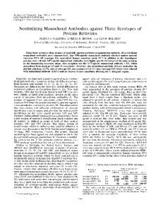
FIG. 7. Immune precipitationoflabeled HAV. 125I-labeled HAV was reacted with various levels of different sera or monoclonal antibodies, and staphylococcus A cells were added to precipitate specific antibody-antigen complexes. As described in the text, the immune precipitates were then analyzed by SDS-PAGE. After electrophoresis the gel was fixed, dried, and exposed to X-ray film. The additions to the labeled HAV (40,000 cpm/reaction) were as follows: 10 RI of preimmune chimpanzee serum (A), 2 ,ul of postimmune chimpanzee serum (B), 10 R.I of postimmune chimpanzee serum (C), 2 ,ul of postimmune mouse no. 1 serum (D), 2 p.1 of postimmune mouse no. 2 serum (E), 10 p.1 of culture medium (F), 2 p.1 of 6A5 culture fluids (G), 2 p.1 of 2D2 culture fluids (H), 10 p1 of 2D2 (I), 2 p.1 of 3E1 culture medium (J), 10 p.l of 3E1 culture medium (K), and 10 p1I of 1B9 culture medium (L). Numbers on the left are molecular sizes in thousands (K). The normal migration of the viral structural proteins is shown on the right.
VOL. 52, 1984
were inoculated into mice to produce ascites fluid which was then tested to determine the subtype of the monoclonal antibody. The final monoclonal antibodies, isolated from ascites fluid or from antibody purified by protein A column chromatography, retained their original activities in the two RIAs. The purified antibodies from these four monoclones were also tested in the original neutralization assay (with cells grown under 0.5% agarose) and were as effective as polyclonal immune serum in inactivating viral infectivity (two are demonstrated in Fig. 6). Immune precipitations with monoclonal antibodies. To characterize the target of the neutralizing monoclonal antibodies, these antibodies were first tested for their ability to bind and precipitate 125I-labeled HAV. For these experiments, peak I material was labeled with 1251 by the chloramine-T method. Typically, when this reaction was stopped after 60 s or less, VP-1 was much more highly labeled than VP-3 was, with little or no 125I present on the VP-2 protein (Fig. 7). Small amounts of either the unpurified hybridoma culture medium or purified antibodies from protein A columns were able to readily precipitate the labeled HAV in a manner similar to polyclonal serum from immunized animals (Fig. 7). The controls included preimmune serum from animals and the medium used for the growth of the hybridomas, which resulted in little or no background reaction. When this labeled viral preparation was disrupted with SDS-,-mercaptoethanol and boiled for 3 min before the immune precipitations, there was no apparent reaction of the monoclonal antibodies to the disrupted, labeled proteins of HAV. Even large microgram quantities of the monoclonal antibodies failed to react with the disrupted proteins (data not shown). However, the postimmune serum from HAVimmunized animals was also unable to demonstrate any consistent activity towards disrupted proteins. Purified HAV was also separated on gels and then transferred to nitrocellulose paper for reaction with pre- and postimmunized animal sera and monoclonal antibodies. Although the immune sera from marmosets and chimpanzees reacted with all of the viral proteins, as detected by either [125I]protein A- or horseradish peroxidase-labeled second antibody, none of the four monoclonal antibodies was capable of reacting with the viral proteins (data not shown). Cross-linking neutralizing monoclonal antibodies to HAV. Since the monoclonal antibodies did not bind or recognize the viral structural proteins in a denatured state, the monoclonal antibodies were covalently cross-linked to the whole virus before disruption. For these experiments, HAV was labeled with ['251]iodine by the chloramine-T reaction for 3 to 4 min, which resulted in extensive labeling of VP-1 and VP-3 and a small amount of labeling of VP-2 (Fig. 8, which contrasts to the rapid labeling of HAV in Fig. 7). Fab fragments of the neutralizing monoclonal antibodies, purified from protein A chromatography and subsequent papain digestion, were prepared and reacted at either 4°C or room temperature with the heterobifunctional cross-linking reagent TDI. Fab fragments were separated from TDI by centrifugation and then reacted with the 125I-labeled virus. A change in pH effected the second cross-linking of the Fab fragment to the proteins of HAV. After pH neutralization and dialysis to remove excess salts, the HAV proteins were separated by SDS-PAGE. The cross-linking of the monoclone 2D2 Fab fragments resulted primarily in the linking of antibody to VP-1, with a concomitant loss of the '25I-labeled material from the region in which VP-1 migrates. Whether 2D2 was first reacted with TDI at 4°C or at room temperature, this antibody readily recognized VP-1 and formed large aggregates, most of which remained at the top of the gel or
MONOCLONAL ANTIBODIES TO HAV
471
stacking layer (Fig. 8). Longer incubation of Fab fragments with TDI, particularly at room temperature, apparently resulted in more bound cross-linker being added to the Fab fragment, which resulted in a reaction with not only VP-1 but also VP-2 and VP-3. Both 2D2 and 6A5 were cross-linked to labeled HAV, and both demonstrated a more specific reaction to VP-1 (data for 6A5 match that for 2D2). As a control for these cross-linking experiments, Fab fragments from a monoclonal antibody specific for rhinovirus were used under the same cross-linking conditions. No specific reaction was seen to any of the structural proteins of HAV, even when the TDI was added at room temperature for up to 10 min (data not shown). DISCUSSION The only other study that has reported on the production of monoclonal antibodies to HAV (15) utilized a competitive assay to select the initial hybridomas, which may explain the rather low percentage (less than 1%) of positive hybrids identified. In contrast, the RIA used in the present study was designed to identify any mouse immunoglobulin that could bind HAV. The high ratio (72 of 270 tested) of positive
A
B
C
D E F VP Om
_* .
AVP-2 VP-3
FIG. 8. Cross-linking of Fab fragments of 2D2 to HAV. The purified Fab fragments of the monoclonal antibody 2D2 were incubated with the cross-linking reagent TDI at room temperature for 5 min (A), 10 min (B), or 15 min (C), or at 4°C for 5 min (D) or 10 min (E); a sample of 2D2-Fab was also incubated at room temperature for 15 min in the absence of TDI (F). Unreacted TDI was removed by solidification, and pelleting was done as described in the text. The various Fab preparations (with or without TDI) were then added to 125I-labeled HAV (60,000 cpm) and incubated for 30 min at room temperature. The pH was raised to 9.6 to complete the crosslinking for 10 min. The pH was then lowered to 7.0, and the samples were dialyzed to remove excess phosphate and then analyzed on polyacrylamide gels. Upon completion, the gels were fixed, dried, and exposed to X-ray film. The HAV preparation for this experiment was labeled with 1251 longer than normal (see the legend to Fig. 7) to obtain more equal labeling of the structural proteins. The normal migration of the viral structural proteins is shown on the right.
472
HUGHES ET AL.
cultures in the noncompetitive RIA suggests that this assay could allow the selection of a more diverse set of monoclonal antibodies. A wide variance was certainly demonstrated in the reaction of the initial hybridoma culture fluids for binding HAV in this assay (Table 1). The variety in the response was not related to the confluency of the hybridomas and remained at consistent levels for the first few weeks that the cultures were maintained and assayed. In addition, when some of the original hybridomas were then tested for competition for binding to HAV against labeled human anti-HAV antiserum, a number of different activities were identified. Some of the hybridoma cultures were incapable of competing, whereas two groups of hybridomas were clearly identified that could compete and could complement with the heterologous group. Such complimentary activity suggests that these hybridoma antibodies are directed to two different sites on the virion structure, and these two compete directly or indirectly with polyclonal serum for binding. Antisera to these two sites are apparently well represented in labeled human immune sera since the hybridoma fluids compete so readily for binding (Fig. 5). The number of such major sites that are readily recognized in polyclonal sera and the nature of the competition will be further explored in future studies with the more well-characterized monoclonal antibodies (from the subcloned hybridomas). The present study also examined a number of parameters of an in vitro assay for measuring the growth of HAV and its neutralization by various antibody preparations. Although previous studies have described the measurement of focal areas of viral antigen after a 2-week growth in cells (11, 12), this length of time seemed prohibitive as an assay to screen hybridomas rapidly. Accordingly, newborn cynomolgus monkey kidney cells were evaluated for the growth of HAV under agarose; they were well suited for producing focal areas of antigen after only 7 days of growth (Fig. 3) and resulted in titers that were consistent with other methods of quantitating HAV. By adapting this assay to 96-well microtiter plates, the incubation time could be reduced to 5 days, allowing a more rapid evaluation of growth and neutralizing activity. The pretreatment of HAV with detergent also increased the sensitivity of the virus to neutralization. This increased sensitivity was detected for several different stock preparations and strains of virus, was independent of the monoclonal or polyclonal nature of the antibody, and suggests that HAV may be present normally as an aggregate of several virions. All of the neutralizing monoclonal antibodies that were selected demonstrated a very specific and high affinity for the hepatitis virion as measured by both types of RIAs and the immune precipitations of '25I-labeled HAV (Fig. 7). The reactivity of these monoclonal antibodies (6A5, 2D2, 1B9, and 3E1) appears to be as specific as that seen with polyclonal serum from immunized animals or naturally infected humans. However, when the virion structure was disrupted, it was more difficult to demonstrate any recognition of the viral structure proteins by either the polyclonal or monoclonal antibodies. When labeled virus was disrupted by the addition of SDS-,B-mercaptoethanol and subsequent boiling, there was little or no reaction detected between any monoclonal or immune polyclonal serum and the viral proteins in the immune precipitation assay. Even after separation of the disrupted virus by gel electrophoresis and subsequent transfer to nitrocellulose membranes, no specific reactions were detected for the monoclonal antibodies. All 16 of the original neutralizing hybridomas (presumably partially cloned) and
J. VIROL.
the four monoclonal antibodies were negative in all of the assays to measure a specific reaction to the disrupted viral components.
The lack of reaction of the neutralizing monoclonal antibodies to the disrupted viral proteins is consistent with the results from other studies on monoclonal antibodies to poliovirus. Although a number of investigators have produced numerous neutralizing monoclonal antibodies to all three subtypes of poliovirus (4, 8, 17), only one monoclonal antibody has been described that binds to isolated denatured VP-1 (1). Apparently, the structural conformation of the proteins within the virion, in particular that recognized by the neutralizing antibodies, is drastically altered when the proteins are separated away from the whole virion structure. Therefore, it was only through cross-linking experiments that a specific reaction could be demonstrated for a specific HAV protein. Emini et al. (4) had previously demonstrated that the TDI cross-linking reagent could be utilized to detect a specific reaction of a neutralizing monoclonal antibody to the VP-1 of poliovirus. Following their procedure closely, we prepared Fab fragments from the monoclonal antibodies 6A5 and 2D2 to prevent nonspecific binding by the Fc portion of the antibody. The Fab fragments which were reacted with TDI at room temperature demonstrated reaction with not only VP-1, so that almost all of the VP-1 was cross-linked to highmolecular-weight aggregates at the top of the gel, but also to some extent with VP-2 and VP-3. However, when TDI was initially added at 4°C, the reaction was much more specific to VP-1, with very little of the VP-2 or VP-3 bound into large aggregates. Apparently, the lower temperature (4°C) leads to fewer molecules of TDI bound per antibody, which results in a more specific cross-linking of only the antibody-combining site to the viral antigenic site. Two other proteins that are detected in preparations of radiolabeled HAV (at 25 and 22 Kd) also appear to cross-link somewhat in this reaction. These proteins are at very low levels on a molar basis, as they are not readily detected when the viral preparations are stained. It is presently not clear whether these peptides are cellular contaminants that could be closely associated with the viral surface (and are nonspecifically cross-linked to the Fab fragment) or breakdown products of the viral structural protein. Further experiments will be needed to more fully characterize this apparently nonspecific reaction. Both 6A5 and 2D2 demonstrated very similar reactions in the cross-linking experiments, with similar temperature dependencies and kinetics for reaction to HAV, suggesting that the neutralization site recognized is on VP-1. Neither 3E1 nor 1B9 has yet been examined for a specific reaction by cross-linking. That at least one of the neutralization epitopes for HAV is present on VP-1 is not too surprising, given that it appears to be the major surface component of this virus. The rather extensive radiolabeling of VP-1 in contrast to VP2, VP-3, or VP-4 (Fig. 7) has been previously described by a number of other investigators for HAV (3, 6). The identification of VP-1 as the major surface protein and now as containing at least one neutralization epitope is consistent with the results for a number of other picornaviruses, including poliovirus, rhinovirus, and foot-and-mouth disease virus (1, 2, 4, 5, 13, 14, 16, 18). Presumably, VP-1 of HAV could be the basis for a subunit vaccine, if the polypeptide in part or whole retains some ability to induce a protective response to the whole virus. Since these subcloned hybridomas (Table 2) have retained the ability to both compete with immune serum for binding
MONOCLONAL ANTIBODIES TO HAV
VOL. 52, 1984
and also neutralize the infectivity of HAV, these could be ideal immunodiagnostic reagents. Indeed, the use of monoclonal antibodies (after labeling) in a competitive assay would be useful to measure not only an anti-HAV response but also the neutralizing activity present in a particular test sample. Presumably, since the monoclonal antibodies would recognize only the sites for neutralization on the virus, a competition assay would measure the neutralizing antibodies present in a polyclonal sera that are directed to the same site.
9.
10. 11.
ACKNOWLEDGMENTS We acknowledge the many helpful discussions with P. Provost and E. Emini. The strain of HAV, CR326 (developed for growth in LLC-MK2 cells by P. Provost and his colleagues), and the immune sera from humans, marmosets, and chimpanzees were also generously provided by P. Provost. We also gratefully acknowledge technical assistance from P. Giesa, F. Banker, A. Schlabach, D. Williams, L. Callahan, A. Palumbo, R. Lynch, W. Hurni, and W. Miller. LITERATURE CITED 1. Blondel, B., 0. Akacem, R. Crainic, P. Couillin, and F. Horodniceanu. 1983. Detection by monoclonal antibodies of an antigenic determinant critical for poliovirus neutralization present on VP1 and on heat-inactivated virions. Virology 126:707-710. 2. Chow, M., and D. Baltimore. 1982. Isolated poliovirus capsid protein VP-1 induces a neutralizing response in rats. Proc. Natl. Acad. Sci. U.S.A. 79:7518-7521. 3. Coulepis, A. G., S. A. Locarnini, and I. D. Gust. 1980. lodination of hepatitis A virus reveals a fourth structural polypeptide. J. Virol. 35:572-574. 4. Emini, E. A., B. A. Jameson, A. J. Lewis, G. R. Larsen, and E. Wimmer. 1982. Poliovirus neutralization epitopes: analysis and localization with neutralizing monoclonal antibodies. J. Virol.
43:997-1005. 5. Emini, E. A., B. A. Jameson, and E. Wimmer. 1983. Priming for and induction of anti-poliovirus neutralizing antibodies by synthetic peptides. Nature (London) 304:699-703. 6. Gerlich, W. H., and G. G. Frosner. 1983. Topology and immunoreactivity of capsid proteins in hepatitis A virus. Med. Microbiol. Immunol. 172:101-106. 7. Gust, I. D., A. G. Coulepis, S. M. Feinstone, S. A. Locarnini, Y. Moritsugu, R. Najera, and G. Siegl. 1983. Taxonomic classification of hepatitis A virus. Intervirology 20:1-7. 8. Icenogle, J., S. F. Gilbert, J. Grieves, J. Anderegg, and R.
12. 13. 14.
15.
16.
17.
18.
19. 20.
473
Rueckert. 1981. A neutralizing monoclonal antibody against poliovirus and its reaction with related antigens. Virology 115:211-215. Kaaden, 0. R., K. H. Adam, and K. Strohmaier. 1977. Induction of neutralizing antibodies and immunity in vaccinated guinea pigs by cyanogen bromide-peptides of VP-3 of foot and mouth disease viruses. J. Gen. Virol. 34:397-400. Laemmli, U. K. 1970. Cleavage of structural proteins during the assembly of the head of bacteriophage T4. Nature (London) 277:680-685. Lemon, S. M., and L. N. Binn. 1983. Serum neutralizing antibody response to hepatitis A virus. J. Infect. Dis. 148:10331039. Lemon, S. M., L. N. Binn, and R. H. Marchwicki. 1983. Radioimmunofocus assay for quantitation of hepatitis A virus in cell cultures. J. Clin. Microbiol. 17:834-839. Lonberg-Holm, K., and B. E. Butterworth. 1976. Investigation of the structure of polio and human rhinovirions through the use of selective chemical reactivity. Virology 71:207-216. Lund, G. A., B. R. Ziola, A. Salmi, and D. G. Scraba. 1977. Structure of mengovirion. V. Distribution of the capsid polypeptides with respect to the surface of the virus particle. Virology 78:35-44. MacGregor, A., M. Kornitschuk, J. G. R. Hurrell, N. I. Lehmann, A. G. Coulepis, S. A. Locarnini, and I. D. Gust. 1983. Monoclonal antibodies against hepatitis A virus. J. Clin. Microbiol. 18:1237-1243. Minor, P. D., G. C. Schild, J. Bootman, D. M. A. Evans, M. Ferguson, P. Reeve, M. Spitz, G. Spanway, A. J. Cann, R. Hauptmann, L. D. Clarke, R. C. Mountford, and J. W. Almond. 1983. Location and primary structure of a major antigenic site for poliovirus neutralization. Nature (London) 301:674-679. Osterhaus, A. D. M. E., A. L. vanWezel, B. van Steenis, G. A. Drost, and T. G. Hazendonk. 1981. Monoclonal antibodies to polioviruses: production of specific monoclonal antibodies to the sabin vaccine strains. Intervirology 16:214-218. Putnak, J. R., and B. A. Phillips. 1982. Poliovirus empty capsid morphogenesis: evidence for conformational differences between self- and extract-assembled empty capsids. J. Virol. 41:792-800. Siegl, G., G. G. Frosner, V. Gauss-Muller, J. D. Tratschin, and F. Deinhardt. 1981. The physiochemical properties of infectious hepatitis A virus. J. Gen. Virol. 57:331-341. Tratschin, J. D., G. Siegl, G. G. Frosner, and F. Deinhardt. 1981. Characterization and classification of virus particles associated with hepatitis A. III. Structural proteins. J. Virol. 38:151156.