Available online at www.sciencedirect.com
ScienceDirect Physics Procedia 69 (2015) 87 – 91
10 World Conference on Neutron Radiography 5-10 October 2014
Neutron Radiography Facility at IBR-2 High Flux Pulsed Reactor: First Results D.P. Kozlenkoa*, S.E. Kichanova, E.V. Lukina, A.V. Rutkauskasa, G.D. Bokuchavaa, B.N. Savenkoa, A.V. Pakhnevichb , A.Yu. Rozanovb a
Frank Laboratory of Neutron Physics, Joint Institute for Nuclear Research, 141980, Dubna, Russia b Paleontological Institute of the Russian Academy of Sciences, 117997, Moscow, Russia
Abstract A neutron radiography and tomography facility have been developed recently at the IBR-2 high flux pulsed reactor. The facility is operated with the CCD-camera based detector having maximal field of view of 20x20 cm, and the L/D ratio can be varied in the range 200 – 2000. The first results of the radiography and tomography experiments with industrial materials and products, paleontological and geophysical objects, meteorites, are presented. © Published by Elsevier B.V.B.V. This is an open access article under the CC BY-NC-ND license © 2015 2015The TheAuthors. Authors. Published by Elsevier (http://creativecommons.org/licenses/by-nc-nd/4.0/). Selection and peer-review under responsibility of Paul Scherrer Institut. Selection and peer-review under responsibility of Paul Scherrer Institut Keywords: neutron radiography; neutron tomography; meteorites; paleontology
1. Introduction The neutron radiography and tomography methods are the powerful tool of non-destructive analysis, which demonstrates importance in industrial and scientific research [Anderson et al. (2009) and Domanus (1992)]. The fundamental difference in nature of neutrons interaction with matter compared to X-rays provides additional benefits to neutron methods, including sensitivity to light elements, notable difference in contrast between neighboring elements or isotopes, high penetration effect through metals or heavy elements. All these features make neutron radiography and tomography highly demanded tools with growing range of applications in industry,
* Corresponding author: Tel.: +7-49621-63783 E-mail address:
[email protected]
1875-3892 © 2015 The Authors. Published by Elsevier B.V. This is an open access article under the CC BY-NC-ND license (http://creativecommons.org/licenses/by-nc-nd/4.0/). Selection and peer-review under responsibility of Paul Scherrer Institut doi:10.1016/j.phpro.2015.07.012
88
D.P. Kozlenko et al. / Physics Procedia 69 (2015) 87 – 91
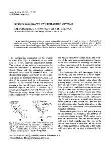
geophysics [Perfect et al. (2014)], paleontology, archeology and other various fields, included culture heritage investigations [Middleton et al. (2005)]. As result of above-mentioned advantages of neutron radiography and tomography, the extensive activities in a neutron radiography facilities development on a research reactors and spallation neutron sources [Lehmann and Ridikas (2014)] are sighted. The IBR-2M high flux pulsed reactor is one of the most powerful pulsed neutron sources in the world with the average power 2 MW, power per neutron pulse 1850 MW and neutron flux in pulse of 5⋅1015 n/cm2/s. The pulsed operation regime of the IBR-2 reactor with 5 Hz frequency and long pulse duration for thermal neutrons of 350 μs makes it attractive not only for traditional neutron imaging applications but in particular also for development of modern energy-selective techniques used time-of-flight methods [Lehmann et al. (2009) and Kockelmann et al. (2007)]. In recent years the neutron radiography and tomography facility at the IBR-2 reactor has been developed. Here, the design and main parameters of this facility and a short overview of the first obtained results are presented. 2. The design and main parameters of the neutron radiography and tomography facility The new neutron radiography and tomography facility is placed on the 14th beamline of the IBR-2 high flux pulsed reactor. The layout of this neutron facility is shown in Fig. 1a. a
b
Fig. 1. (a) The layout of a 14th beamline of IBR-2 reactor. The collimator system with evacuated tube, geometry of concrete biological shielding and beamstopper place are shown. (b) The light-tight box of a scintillator-based detector. The additional lead shielding of a CCD camera are on top of box.
The neutron beam is formed by means of a collimator system, which consists of four cylindrical divergent inserts of boron-contained polyethylene disks and steel rings for construction rigidity. The collimator holes expand in diameter from initial 5.0 to 23.7 cm. The incident beam aperture can be varied in 1.0 – 5.0 mm limits. The collimator system is placed in evacuated tubes to avoid air scattering. The basic parameters of neutron radiography and tomography facility on IBR-2 reactor are listed in Table 1. The neutron flux on sample place in the present configuration is I ~ 5.5(2)x106 n/cm2/s and have been measured by gold foils activation method. The IBR-2 reactor provides a thermal neutron beam with wavelengths ranged from ~0.2 to 8 Å and a spectral distribution maximum of ~1.8 Å. The scintillator-based detector system is illustrated in Fig. 1b. As scintillation screen, the 6LiF/ZnS scintillator of 0.2 mm thickness manufactured by RC TRITEC Ltd (Switzerland) is used. The camera box was produced by Neutron Optics (Grenoble, France). The light is reflected out of the beam by a mirror and focused on CCD chip by an optical lens Nikon 50 mm 1:1.4D AF-Nikkor. The sample manipulator system is based on HUBER goniometer with x-, y- translation and z- rotation stages. The minimumal rotational step is 0.02 deg.
D.P. Kozlenko et al. / Physics Procedia 69 (2015) 87 – 91
Table 1. The main parameters of the neutron radiography station on the IBR-2 reactor. Parameter L/D ratio
200-1000
Aperture diameter D
10 – 50 mm
Distance between input aperture and sample L
10 m
Field of view (FOV)
20x20 cm2
CCD camera type
VIDEOSCAN-11002-2001
Active pixels
4008x2672
Pixel size (µm)
9x9
CCD chip area (mm)
36x24
Digitization Scintillator screen specifics
12 Bits 6
LiF/ZnS scintillator
The imaging data are subtracted by the dark current image and are divided to the incident neutron beam by means of an ImageJ software [Schneider et al. (2012)]. The tomographic reconstruction is performed by an HPITRE program [Chen et al. (2012)]. The VGStudio MAX 2.2 software of Volume Graphics (Heidelberg, Germany) is used for the visualization and analysis of reconstructed 3D data. The evaluated spatial resolution for the field-of-view 20x20 cm2 is about 300 µm. 3. The first radiography and tomography results obtained on new radiography facility 3.1. Neutron radiography measurements The photography and neutron radiography of a large metal casing manometer are presented in Fig. 2a. The inner brass ring-shaped membrane and steel gears mechanisms are visible and well distinguishable. The photography and neutron radiography of a steel - zirconium bimetallic adapter of RBMK reactor fabricated by cold welding are shown in Fig.2b. The sharp contrast between different metal parts was observed. The internal threads shape is clearly visible. a b
Fig. 2. (a) The photography and neutron radiography of the metal casing manometer. (b) The photography and neutron radiography of the steelZr bimetallic adapter.
3.2. The neutron tomography studies of the inner structure of meteorites The inner structure of iron-stone meteorites studies by means the neutron tomography have been performed. We studied Seymchan [van Niekerk et al. (2007)] and Marjalahti [Ryder (1984)] meteorite. The both meteorites are pallasites and generally contain olivine grains, metallic Fe–Ni alloy and small amount of other oxides [Buseck and Goldstein (1969)].
89
90
D.P. Kozlenko et al. / Physics Procedia 69 (2015) 87 – 91
The tomography experiments have been performed with rotation step of 0.5o and total number of measured projections was 360. The exposure time for one projection was 10 s and resulting measurements lasted for 4 h. The obtained 3D models of metal alloy matrix of investigated meteorites are presented in Fig. 3a-b. The distinguishable inner compactions in metal matter of both meteorites have been observed. These compactions can correspond with some density or composition fluctuations in iron-nickel alloy. a
b
Fig. 3. (a) The extracted 3D model of metal Fe-Ni distribution of the Seymchan meteorite after the tomographic reconstruction. (b) The tomography 3D model of metal Fe-Ni alloy distribution of the Marjalahti meteorite. The red inclusions correspond to denser part in alloy basis.
3.3. The neutron tomography studies of the paleontology objects a
b
Fig. 4. (a) The photography and reconstructed 3D model of inner structure of a fossil cone Protosequoia sp. (b) The photography and tomography reconstructed model of stromatolites colony from Petuhovskoe Lake (Russia). The red regions indicate the stromatalites presence in rocks.
The neutron tomography studies of fossil cone Protosequoia sp. dated from Cretaceous period have been performed. The reconstructed 3D models of inner composition like as cones leafs and its crushed stem are shown in Fig. 4a. Stromatolites are layered structures which formed in shallow water by the trapping, binding and cementation of sedimentary grains Cyanobacteria. The fossil stromatolites provide information about life behavior on the Earth
D.P. Kozlenko et al. / Physics Procedia 69 (2015) 87 – 91
more than 3.5 billion years ago. We studied the stromatalite colony in CaCO3 gritstone rock from alkaline Petuhovskoe Lake (Altai reg., Russia). The 3D model of stromatalites colony distribution into rocks is shown in Fig. 4b. 4. Conclusions In this paper, the newly developed neutron radiography and tomography facility at a high flux pulsed reactor IBR-2 is described. The obtained technical parameters are suitable for a wide range of applications, as demonstrated in the given review of the performed studies of industrial, paleontological and meteorite objects. Further developments will be focused on the improvement of the technical parameters and implementation of energy selective time-of-flight based techniques. Acknowledgements This work has been supported by IAEA research contract 17217and RFBR grant 14-22-01001-ofi_m. The authors would like to acknowledge A.A.Kustov (JINR) for assistance with technical drawings of the facility, A.V.Belushkin (JINR), A.M.Balagurov (JINR), E.Lehmann (PSI, Switzerland), N.Karjilov (HZB, Germany), I.A.Bobrikov (JINR) for useful discussions, E.A. Krasavin (JINR) for kindly lending the meteorite objects and O.S.Samylina, A.B.Sokolova, L.V.Zaitseva (PIN RAS) for providing a paleontological objects. References Anderson, I.S., McGreevy, R.L., Bilheux, H.Z. (Eds.), 2009. Neutron Imaging and Applications: A Reference for the Imaging Community. Springer, New York, pp. 341. Buseck, P.R., Goldstein, J.I. 1969. Olivine compositions and cooling rates of pallasitic meteorites. Geological Society of America Bulletin. 80, 2141-2158. Chen, R.C., Dreossi, D., Mancini, L., Menk, R., Rigon, L., Xiao, T.Q., Longo, R. 2012. PITRE: software for phase-sensitive X-ray image processing and tomography reconstruction. Journal Of Synchrotron Radiation 19, 836-845. Domanus, J. C. (Ed.), 1992, Practical neutron radiography. Kluwer academic publishers, Netherlands, Dordrecht, pp. 269. Dragunov, Yu. G., Tretiyakov, I. T., Lopatkin, A.V., et. al., 2012. Modernization of the IBR-2 pulsed research reactor. Atomic Energy 113, 2938. Lehmann, E.H., Frei, G., Vontobel, P., Josic, L., Kardjilov, N., Hilger, A., Kockelmann, W., Steuwer, A. 2009. The energy-selective option in neutron imaging. Nuclear Instruments and Methods in Physics Research A 603, 429–438. Lehmann, E.H., Ridikas, D., 2014. “Status of neutron activities in a worldwide context”, 10th World Conference on Neutron Radiography. Grindelwald, Switzerland, p. 29. Kockelmann, W., Frei, G., Lehmann, E.H., Vontobel, P., Santisteban, J.R., 2007. Energy-selective neutron transmission imaging at a pulsed source. Nuclear Instruments and Methods in Physics Research A 578, 421-434. Middleton, A., Tum, J., Lang, J., (Ed.), 2005. Radiography of Cultural Material. 2nd edition, Routledge, pp. 208. Perfect, E., Cheng, C.-L., Kang, M., Bilheux, H.Z., Lamanna, J.M., Gragg, M.J., Wright, D.M.. 2014. Neutron imaging of hydrogen-rich fluids in geomaterials and engineered porous media: A review. Earth-Science Reviews 129, 120-135. Ryder, G., 1984. Minor elements in Marjalahti olivine. Meteoritics 79-83. Schneider, C.A., Rasband, W.S., Eliceiri, K.W. 2012. NIH Image to ImageJ: 25 years of image analysis. Nature Methods 9, 671–675. van Niekerk, D., Greenwood, R. C., Franchi, I. A., Scott, E. R. D., Keil, K. 2007. “Seymchan: A Main Group Pallasite - Not an Iron Meteorite”, 70th Annual Meteoritical Society Meeting, Tucson, Arizona, USA, 42, p. 5196.
91