32 ICTTD-3 ON TIC R E T T E L NEWS
ICKT D KS A N
F SO E S EA DIS E N B OR
FEBRUARY 2007 S C I P TRO HE T N KI C O ST E V FROM THE EDITOR LI
THE GLOBAL IMPORTANCE OF TICKS From the Coordinator Frans Jongejan Ticks transmit a greater variety of pathogenic micro-organisms than any other arthropod vector group, and are among the most important vectors of diseases affecting humans and animals. Although the ICTTD project and this Newsletter is focussed on ticks and tick-borne diseases affecting livestock production in the tropics, tick-borne diseases have gained enormously in notoriety in other parts of the world, creating a global problem. For instance, more than 1100 human cases of Crimean-Congo haemorrhagic fever have been reported in a continuing series of outbreaks which started in Turkey in 2002 constituting the largest epidemic of this disease since it was first recognized in 1944. Another striking example is the fact that acaricides for companion animals have become the major component of the market portfolio of the global animal health industry in response to the heavy toll that companion animals, in particular dogs, pay to tick-borne diseases. One of the aims of the Integrated
To the homepage of ICTTD
Consortium on Ticks and Tickborne Diseases (ICTTD-3) project is to gain a better understanding of tickhost-pathogen interactions under changing climatic conditions. The complex vector-pathogen-host interrelationship is also studied using genomics tools that have now become available. Ultimately, factors that determine the co-evolution of pathogens and ticks in wildlife and domestic livestock will have to be identified. The ICTTD project will hold its third annual meeting between 24 and 28 September 2007 in Zanzibar, Tanzania. During the first two or three days of the meeting, the theme of the meeting will be: Co-evolution of Pathogens, Hosts and Vector ticks. As a result of the previous annual meeting, held in September last year in South Africa, 10 comprehensive reviews have been submitted to “Trends in Parasitology” to be considered for inclusion into a special issue of this leading parasitology journal. The special issue, to be presented at the forthcoming annual meeting in Tanzania, will be accompanied by a centrefold poster entitled, “Genomes, Ticks and Pathogens”, which will illustrate key features of pathogen genomes and genomics tools for ticks, such as RNA interference. 1
Gerrit Uilenberg The Plos Biology Open Access journal has started a new series highlighting fundamental challenges in biology. The first one is: Levin S.A. 2006. Fundamental questions in biology. PLoS Biol., 4 (9): e300 (1471-1472). (E-mail:
[email protected]). This is an interesting discussion (which deserves to be read in full) on changes in the discipline of biology in universities. It is to a large extent relevant to the medical/veterinary field (including the discipline of ticks and tick-borne diseases); just replace the word “biology” by “medical/veterinary science” (although I don’t want to imply that such science is equivalent to basic science.) See page 2
I 1 1 4 5 14 15 20 24 25 28 28 31 31 32 33 35 36
N
S
I
D
E
From the editor From the coordinator - General - Ticks - Tick-borne diseases - Theilerioses - Babesioses - Diseases by Rickettsiales - Anaplasmoses - Ehrlichioses - Cowdrioses - Rickettsiosis - Dermatophilosis - Other diseases - Letters to the editor - Announcements Addresses of the editors
From page 1 There has been increasing specialisation, and with the development of new tools more scientists from outside biology departments have become involved. While allowing rapid progress on specific problems, these often have little interest in the broader scope of biology. Existing departments of biology have split and split, creating tensions, for funding; administrators have tended to accept that areas that require or attract less funding are outdated and dispensable. But after having acquired so much information in genomics, it is now time to identify and address fundamental questions in biology, cutting across disciplines. All this was already evident over 20 years ago, when Harry Hoogstraal in his Presidential Address at the 60th Annual Meeting of the American Society of Parasitologists in 1985 (J. Parasitol., 71: p. 686) said: “Two scientists, Joan and John, were strolling on the beach one sunny day. Suddenly something scurried in the sand beside their feet. “What’s that?” asked John. “Why, don’t you know,” Joan replied, “that’s a sandcrab … you’ve been studying its synapses for four years.” And he went on to say essentially what Prof. Levin is saying today, and talked about the necessity of preserving the identity of the Society as a group dedicated to explaining holistic parasitism. Although it is not new, it is worthwhile, as far as our field is concerned, to attract the attention of some of the specialised scientists in laboratories to the fact that the ultimate aim of their research in for instance molecular science or immunology is to help improve the control of ticks and tickborne diseases of tropical livestock, and ultimately animal productivity and human food security. It will avoid suggesting that PCR is a practical way of diagnosing acute clinical anaplasmosis (historical!). On another level, this also brings to mind what the Director of CIRADEMVT during most of my time at that institute (Georges Tacher) used to say, when a young scientist came bubbling with enthousiasm with some new bright idea: 1° What can it be used for? 2° How much does it cost? 3° Who is going to pay for it?
In the section Letters to the Editor there is a reply to Nick McHardy’s letters in issue n° 30. In fact it is only in part a reaction to those letters and concerns mainly immunisation against ECF. From this letter and those in n° 30, a common view can be distilled, which I wholeheartedly support: Donor organisations and governments should not only finance research that might end in improved solutions in the future, but also help to apply existing cost/benefit-positive methods for the control of diseases with an economical impact. This includes financing stabilate production with good quality control for the existing infection and treatment method of immunisation against ECF. There seems to be only a limited amount of stabilate left; who will make it, who will finance that? It also includes financing practical research on improvements in imperfect existing control methods.
N.Y. Acad. Sci., 2006, 1081: 552 pp. ISBN 1-57331-637-7. Relevant papers are presented in the newsletter under the various sections. See also under ANNOUNCEMENTS. The proceedings are dedicated to Jim C. Williams, who was president of the American Society for Tropical Veterinary Medicine, now the STVM, from 1989 to 1993 and was instrumental in planning and organising the first STVM Biennial meeting in Puerto Rico in 1991: Katherine M. Kocan. 2006. Dedication. Jim C. Williams. Ann. N.Y. Acad. Sci., 1081: xvii-xviii.
Another new co-editor is Ulrike Seitzer in Germany; she will share the field of babesioses (except those caused by B. bigemina and B. bovis) with Frank Katzer, and particularly use her experience acquired in the field of the babesioses of small ruminants.
The Annals of the New York Academy of Science have also published (in 2005 and 2006) other volumes of interest to many workers on tick-borne diseases, as they contain the proceedings of the 4th International Conference on Rickettsiae and Rickettsial Diseases, which was held in June 2005 in Spain. The reference is: K.E. Hechemy, J.A. Oteo, D.A. Raoult, D.J. Silverman & J.R. Blanco (Eds). Rickettsioses: From genome to proteome, pathobiology, and rickettsiae as an international threat. Ann. N.Y. Acad. Sci., 2005, 1063: 474 pp. ISBN 1-57331-601-6, and, with the same editors: Century of rickettsiology: Emerging, reemerging rickettsioses, molecular diagnostics, and emerging veterinary rickettsioses. Ann. N.Y. Acad. Sci., 2006, 1078: 626 pp. ISBN 1-57331-639-3. Most of the papers are not of relevance for tropical livestock, but some are and these are presented in this issue of the newsletter, or if not obtained in time in the next one. I have to remind once more the authors who would like their papers presented in the newsletter, to send me or the relevant co-editor, a pdf or “hard” reprint of the paper, as some are missed, in spite of all efforts.
The proceedings of the 8th STVM biennial meeting held in Hanoi (Vietnam) in June/July 2005 have now been published as vol. 1081 of the Annals of the New York Academy of Sciences. There are 77 papers. The full reference is: E.F. Blouin & J.-C. Maillard. Impact of emerging zoonotic diseases on animal health: 8th biennial conference of the Society for Tropical Veterinary Medicine. Ann.
A new section is created for this issue: “Diseases caused by Rickettsiales”. This includes anaplasmoses, ehrlichioses and cowdriosis, as well as to a limited extent infections by species of the genus Rickettsia (zoonoses which are not normally of direct importance to tropical livestock). The section may from time to time appear in future issues, depending on publications presented.
Time continues to go by. This time John Molloy, coordinator and editor for babesiosis caused by B. bigemina and B. bovis, retires from his position at the Queensland Department of Primary Industries and Fisheries. Fortunately he will continue to share his experience with us as editor in this field, but Louise Jackson will take over as coordinator. Thank you very much John for all the time you have put in this task and for doing it so well. Louise may sometimes delegate references to others than the “official” co-editors, and in that case the name of the reviewer is given in full, not just as initials (e.g. Bert de Vos).
2
To the Index
Received from Prof. Reuben Kaufman: Jack Gregson (17 June 1910 – 29 October 2006) John Douglas (Jack) Gregson passed away peacefully at his home in Kamloops, British Columbia, Canada. He is survived by his wife, Barbara, their five children, nine grandchildren and four great grandchildren. Jack was born near Red Deer Alberta. He developed an early passion for butterflies and, as a teenager, had amassed an impressive collection. He earned his B.A. at the University of British Columbia (1934), and his M.Sc. in Medical Entomology from the University of Alberta (1936), with a thesis entitled: “A Preliminary Study of Tick and Host in Relation to Western Canadian Tick-borne Diseaeses”. It’s a wonderful historical document, not only because of its broad scientific scope, but also for some exquisite diagrams, hand-drawn and coloured by Jack. Following his M.Sc., Jack took up a position in the Veterinary and Medical Entomology labs of the Canada Department of Agriculture (now Agriculture and Agri-Food Canada) in Kamloops, where he spent his whole career, serving as the lab’s Director from 1944 to his retirement in 1971. His interest in ticks was very broad, including feeding dynamics, host immunity (publishing a bare 3 years after Trager’s classical paper of 19391), taxonomy, natural history, morphology/histology and tick paralysis. It was his interest in tick paralysis associated with the Rocky Mountain wood tick, Dermacentor andersoni, which led him to an observation in 1967 that helped solve a long-time puzzle of tick physiology. Although it had been known for at least two decades that ticks concentrate their blood meal by excretion of excess fluid, the route of this excretion remained an enigma, because most ticks do not excrete urine during and immediately after the feeding period as do blood-sucking insects. It was his observations on tick mouthparts attached to everted hamster cheek pouches that led him to propose salivation as the mechanism of blood meal concentration2. This hypothesis was confirmed experimentally by Roger Tatchell, also in 19673 which, in turn, formed the foundation of new research directions in tick physiology.
Jack was one of those remarkably talented and self-reliant people who seem to be able to do everything. In addition to building their house in the 1940s, and landscaping their extensive grounds, he was also a keen naturalist, photographer and artist. Some years ago their property was designated a “Heritage Garden site” in Kamloops, and it is, perhaps, a taste of what paradise might be like; for years it has been a favoured venue where newly married couples are formally photographed. In 1936 he established the Kamloops Outdoor Club, and in 1970 the Kamloops Naturalist Club. The many alpine trips that he led in remote areas of British Columbia and beyond served as inspiration for his landscape oil paintings, some of which have been displayed in the Vancouver and Kamloops Art Galleries. Jack has been a ‘local hero’ in Kamloops. He spearheaded the development of the McArthur Island Waterway Park in 1980, and a butterfly garden in that park in 1994. The bicycle path there is named “The Jack Gregson Trail”, and he was named a freeman of the city of Kamloops in 1990. Among numerous local and Canadian provincial awards he received over the years, in 2000, Jack was awarded an Honourary Doctor of Letters degree from the University College of the Cariboo. Fittingly, a tick from Eastern Canada has been named after him: Ixodes gregsoni4. I feel most privileged to have known Jack since my PhD years at the University of British Columbia, when he was an inspiration to my research program on tick salivary gland physiology. I am honoured to have been welcomed in Jack and Barbara’s home frequently since then, and to have been blessed with the warmest expressions of their hospitality. It is appropriate that Jack should have the last word here. His tireless efforts in environmental advocacy over the years should remind us all of our responsibility, as biologists, to the preservation of our natural heritage, wherever we live in the world. Once, when a local group claimed the right to develop a ski village because they “owned” the land, Jack composed the following poem, which is reprinted here from the Proceedings of the 4th International Conference on Ticks and Tick-borne Pathogens (TTP 4)5, with kind permission of Springer Science and Business Media. 3
To the Index
1
2
3
4
5
Trager, W. (1939). Acquired immunity to ticks. J. Parasitol. 25, 57-81. Gregson, J.D. (1967) Observations on the movement of fluids in the vicinity of the mouthparts of naturally feeding Dermacentor andersoni Stiles. Parasitology 57, 1-8. Tatchell, R.J. (1967) Salivary secretion in the cattle tick as a means of water elimination. Nature 213, 940941. Lindquist, E.E., Wu, K.W. and Redner. J.H. (1999). A new species of the tick genus Ixodes (Acari: Ixodidae) parasitic on mustelids (Mammalia: Carnivora) in Canada. Canadian Entomologist 131, 151170. Jongejan, F. & Kaufman, W. Reuben (Eds) Ticks and Tick-Borne Pathogens, Kluwer Academic, Dordrecht/Boston/London 2003.
Territorial Claims (J.D. Gregson) Said a cricket to an ice-bug as they sat on Mt. Paul’s slide, “Don’t you love my rocky talus - it’s the topmost of my pride”. “But yours it’s not”, the ancient bug reprovingly replied, “Three hundred million years I’ve lived, and you have just arrived.” Said an eagle to a ground-squirrel as it soared o’er peaks sublime, “Be careful how you dig the earth and spoil this land of mine.” But the rodent queried rightly, as the best he could define “You birds were not around at all when mammals had their time.” The moral of this issue is, as far as I can see, This land belongs to none of us, not even you and me! We’re all just lucky tenants on an earth that came to be. Signed: Grylloblatta, the Ice-bug
GENERAL Presentations of ICTTD-3 Working Group on Zoonoses The ICTTD-3 data base on ticks and tick-borne zoonoses in the tropics and sub-tropics and the working group on (sub-)tropical tick-borne zoonoses have been presented in a poster at the Zoonosis Congress in Argentina (May 2006) and in an oral presentation at the Annual Meeting of the European College of Veterinary Public Health in Lyon (France) (Dec. 2006). In both cases the authors were: D. De Meneghi, T. Avsic-Zupanc, A. Bouattour, A. Estrada-Peña; A. Gueye, M. Labruna, A. Lakos and F. Jongejan. The respective titles were, respectively: “A data base on ticks and tick-borne zoonoses in the tropics and sub-tropics” and “The Integrated Consortium on Ticks and Tick-borne Diseases (ICTTD-3): Development of a data base on ticks and tick-borne zoonoses in the tropics and sub-tropics”. More on climate change and tickborne diseases In the previous issue (p. 8) I thought it interesting that Ostfeld et al., 2006, in PLoS Biology, 4 (6) did not find the abundance of deer of predictive value for the risk of Lyme disease. Interesting, because this is in contrast to the generally held opinion that the increase in deer as principal hosts of adult Ixodes vectors of Lyme borreliosis is to a great extent responsible for the increase in the disease. Sarah Randolph drew my attention to a reaction she and others sent after reading that paper, and that was published on September the 6th, 2006 online, together with a reponse from Ostfeld et al. These letters are unfortunately easily overlooked as they are not part of any of the issues of the journal. Go to http://biology.plosjournals.org, type “Ostfeld” in the search PLoS Biology window, select the paper from the list, then choose “Read Other Responses” (under CONTRIBUTE on the right). What it amounts to is that Randolph and co-authors of the letter believe that the conclusions of Ostfeld et al. may be due to flawed statistical procedures and cannot be accepted as reliable until the data are re-analysed using
“standard” practices. In their response Ostfeld and co-authors maintain that their statistical procedures did show that deer abundance had no effect on temporal variation in Lyme disease risk. Recent publications Bowman D.D. 2006. Successful and currently ongoing parasite eradication programs. Vet. Parasitol., 139: 293307. (E-mail:
[email protected]) (The summary in this newsletter is limited to ticks and TBDs. Among the successful programmes the eradication of Boophilus ticks, and Texas cattle fever (babesiosis) which they transmit, is of course given as a great success story, and rightly so. As the author correctly states, the eradication programme stops at the USA border and constant surveillance is required to prevent reintroduction. Among ongoing eradication programmes mentioned, there is the Caribbean Amblyomma Programme (CAP), which was intended to eradicate the tropical bont tick from the western hemisphere, and the diseases it transmits or is associated with, heartwater and (not mentioned in the paper) severe dermatophilosis. As we now know, CAP has had to be abandoned (NL n° 30), and the paper does not even mention the heavily infested French Antilles. One category is not mentioned in the paper, and that is unsuccessful eradication programmes. CAP can now be ranged into that category, and another unsuccessful programme I can think of was the attempt to eradicate the tick Boophilus microplus from a relatively small north-eastern corner of New South Wales (Australia), in spite of greatly increased knowledge of the tick and better acaricides than were available during the eradication of Boophilus ticks in the USA. There is more to eradication than scientific knowledge!) (GU) Raoult D., Fournier P.-E., Eremeeva M., Graves S., Kelly P.J., Oteo J.A., Sekeyova Z., Tamura A., Tarasevich I. & Zhang L. 2005. Naming of rickettsiae and rickettsial diseses. Ann. N.Y. Acad. Sci., 1063: 1-12. (E-mail:
[email protected]) (A very useful review of the rules of the Bacterial Code and the impact of molecular techniques on the taxonomy and phylogeny of the Rickettsiales, 4
To the Index
which are bacteria. The rules of the Bacterial Code are quite different from those of the Zoological Code, much more rigid. For example, any new name (after the more or less arbitrary Approved List of 1980 was published) has to be validated by publication in the Int. J. Syst.Bacteriol., now Int. J. Syst. Evol. Microbiol.), and as the authors point out, any species named after the Approved List was established has to fulfil certain criteria in order to be validated, criteria which are not always attained for the Rickettsiales, as they include for instance its isolation in pure culture. The advent of genome sequencing has been of great value. There is also a discussion about the names of rickettsial diseases. It is preferable that each disease has a single name, in order to avoid confusion. But for some several names exist, for instance the disease caused by R. conorii is called Mediterranean spotted fever, Marseilles fever or boutonneuse fever Rocky Mountain spotted fever, caused by R. rickettsii occurs also in South America; and there are many other examples. There is a list of the validated species and subspecies of the genus Rickettsia, The list is becoming longer every year!) (GU) Uilenberg G. & Goff W.L. 2006. Polyphasic taxonomy. Ann. N.Y. Acad. Sci., 1081: 492-497. (E-mail:
[email protected]) (This is a follow-up to earlier reactions to recent taxonomic papers, many concerning tick-borne organisms and ticks. Authors’ abstract: “Several organisms from a number of prokaryotic and eukaryotic groups have presented problems for systematists for a long time. Both phenotypic and genotyîc methods for sorting out these relationships have been employed. There are limitations with each method when taken alone. Since the purpose of systematics is to determine the correct genealogical relationships among biological organisms, it is necessary to use all available means to arrive at consensus associations, and polyphasic taxonomy, which takes into consideration both methods, is a rational approach. In this short article, we provide a number of examples where polyphasic taxonomy is serving as the means of arriving at the desired consensus.”) (GU)
TICKS THPbase: call for support In the last ICTTD meeting, the members of the Action group 1 (THPbase) agreed to strongly recommend the updating of the current version of the tick data set. While this database now contains a huge number of records, it is necessary to keep it as much updated as possible, and to incorporate records yet unnoticed by the compilers. The compilation work is a huge task and the help of all possible supporters of this task is needed. The database needs records with adequate coordinate references, that are commonly overlooked when the report is published. Local volunteers willing to cooperate in finding the right coordinates would be of great help too, also when the same name applies to more than one village or town, as is the case sometimes. The potential contributors wishing their tick collections to be included in the database should contact Agustin Estrada-Peña (
[email protected]) to obtain a sample of the computer file to be used. We hope this important step in the objectives of the Concerted Action will be greatly enhanced with the help of the readers of the Newsletter.
the immunopathology of their interaction with the host, their control, modern techniques used in taxonomy, and more. This is a very useful book for Latin America, and its usefulness would be even more enhanced if it were to be translated into Spanish. One additional remark: The three authors given above are in fact editors, apart from being authors, because many others have collaborated in writing the various chapters. Writing about translation into Spanish: The 2003 book on ticks of the neotropical region, written in English by Guglielmone and others (NL n° 25, p. 6), has also appeared in Spanish since, with the same authors, under the title: “Las garrapatas (Acari: Ixodida:) de la región zoogeográfica neotropical. Ediciones INTA.” Mediterranean region Prof. Daniele De Meneghi of Turin sent me a book on the ticks of Italy which was published in 2005. A considerable number of collaborators have been involved in its preparation and it has been edited by Drs. G. Cringoli, A. Iori, L. Rinaldi, V. Veneziano and C. Genchi. The title is “Zecche”. ISBN 88-89132-05-1. Published by the University of Naples “Federico II”, 263 pages. This is the 6th book in the
New books
series “Mappe Parasitologiche” of which Dr. Cringoli is the series editor. The authors are to be congratulated for doing a thorough job and for gathering the scattered information on the ticks of Italy and the pathogens which they transmit. There are many and mostly excellent photos, drawings, keys, diagrams, and maps. Humoristic drawings enliven the book. As a considerable bonus an interactive CD-ROM, prepared by Drs. A. Iori, A. Di Giuglio and S. De Felici, comes with the book, and makes it easy to find quickly anything one is looking for in particular. These comments are by necessity too short to do fully justice to the value of this volume. The title is “Zecche” and indeed most of the emphasis is on the ticks themselves, tick-borne infections are dealt with in a much more succinct way. All tick taxonomists may not always agree with the systematics of ticks used, but I have to leave it to them to judge. Of course, in a publication of this scope it is unavoidable that a few mistakes and omissions occur. I haven’t gone through every detail, and will mention for instance only that there does not appear to be any reason for considering ticks as reservoir and vectors of Haemobartonella spp., and that these are no longer considered as belonging to the Rickettsiales but to the Mycoplasmatales. The book is logically in Italian, and although an English version of the CD-ROM is planned, it is not available in the specimen I have.
Neotropical region The ICTTD-supported publication on neotropical ticks which was mentioned in newsletter n° 28 (p. 39) has now been published:
Protocol for surveys and collection of ticks
Barros-Battesti D.M., Arzua M. & Bechara G.H. 2006. Carrapatos de importância médico-veterinária da região neotropical: Um guia ilustrado para identificação de espécies. (Ticks of medical-veterinary importance of the neotropical region: An illustrated guide to the identification of species.) 223 pp. Instituto Butantan, São Paulo. ISBN no. 85-99909-01-0. (In Portuguese.)
ICTTD has of course no ‘official’ protocol for tick surveys, but a summary of the protocol set up by Ivan Horak and Fred Potgieter for South Africa and sent by Ivan to several participants in ICTTD-3, is a useful guide for others. A more detailed version of the protocol can be obtained from Ivan:
In addition to keys to the families, genera and species, there are chapters on biology of ticks, their medical and veterinary importance, including their role as vectors of various pathogens, 5
To the Index
SURVEYS TO ESTABLISH THE LOCAL, PROVINCIAL OR NATIONAL DISTRIBUTION OF TICKS Sample sites Small scale farmers. All regions should provide a list of dip-tanks or villages by district, and approximate figures on cattle or stock density. By assigning numbers to the dip-tanks or villages, tables of random numbers can be used to identify those that must be sampled. Commercial farmers. Farms that lie in the centre of quarter or half-degree latitude and longitude squares, or that lie where the lines of latitude and longitude intersect, can be selected for survey purposes. This will ensure a gridlike pattern of collection sites. Target animals The animals to be sampled should be chosen to yield maximum information. Only untreated animals should be sampled, as acaricide use would interfere with the results. Sampling procedure. Each team should be provided with printed work sheets. Upon arrival at the sampling locality, geo-reference and other information for the site should be recorded. Sample size and target animals Five healthy, but visibly tick-infested cattle approximately 1 year old, and if possible each animal belonging to a different owner at each dip-tank site, or village. Whenever available five healthy adult goats (or sheep), preferably with visible tick infestations, and if possible each animal belonging to a different owner at each dip-tank site, or village. Five healthy dogs, preferably with visible tick infestations, at each dip-tank or village. Sample size free-living ticks a) Free-living ticks are collected from three 100 m long drag-samples. These drags must not be performed in the immediate vicinity of the dip-tank because of the possible effect that residual dip-wash dripping from cattle may have on freeliving ticks. b) Whenever possible fowl tampans are collected from two poultry coops or roosting places in the vicinity of the dip-tank or at the village.
METHODS
(ii) Free-living adult ticks
Please note: All labels used to identify any tick collection must be written in lead pencil or be computer-printed. These labels must be small enough to fit into the vials containing alcohol and collected ticks. Do not use a ballpoint pen on labels as the ink will dissolve in the alcohol. Do not write on the outside of the vials as spilt alcohol will dissolve the writing. Do not write on the lids of vials as these may become separated from the vials containing the ticks.
(a) Drag-sampling Many questing adult ixodid ticks can be collected from the vegetation by drag-sampling as described above. (b) Vegetation sampling Adult ticks can be collected by hand from the tips and stems of grass within a specified measured area or alongside a measured length of road or a path, or during a specific period of time, say 15 or 20 minutes. Use a separate vial and label for the ticks recovered from each of the collection sites. (c) Sampling hen-coops Fowl tampans can be collected from under the bark of any wooden structure in the hen-coop. Tampans are seldom found in hen-coops made only from stone or brick. Use a separate vial and label for the ticks recovered from each of the hen-coops.
Collection of Free-Living Ticks (i) Free-living immature ticks Ten 1 000 mm x 100 mm flannel strips, with an 80 mm x 4 mm diameter metal rod sewn into their loose ends as a weight, are attached adjacent to one another on a 1 200 mmlong wooden spar by means of Velcro tape (a wooden broom stick is suitable). Each collection is made by an operator walking at a normal pace, pulling this spar, with the flannel strips attached, by means of a string or twine harness attached to its ends, for a distance of 100 m over the vegetation. At the completion of each drag the wooden spar with the flannel strips attached to it is suspended about 1.5 metre above the ground on upright stands. If no stands are available the spar can be suspended between an open car door and the top of the dashboard, or between two trees, or across a corner where two fences meet. The flannel strips are individually detached from the spar, always working from one end of the spar and not from both ends towards the middle. The ticks are removed from the flannel strips by means of fine-tipped forceps and placed in vials containing 70 % ethyl alcohol. Each cleaned flannel strip is then re-attached to the wooden spar in readiness for the next drag. Ideally at least three drags, at a distance of at least 50 metres apart from each other are performed. Use a separate vial and label for the ticks collected from each of the three drags. Drags are not done over dew-laden grass early in the morning or over grass after rain, as this wets the flannel strips and decreases their efficacy. 6
To the Index
Tick Collection from Host Animals Total tick collections are not always possible for practical reasons. Instead, six clearly defined sites on each host animal can be selected because of their importance as feeding sites for the different species and developmental stages of the more common cattle ticks. The ticks on these sites are collected either by hand or with forceps. If Hyalomma species are present, forceps and gloves should be used, as they may be infected with CCHF virus. If there are large numbers of ticks present in any of these sites it is not necessary to collect more than ten of them as the surveys are not aimed at determining the number of ticks present, but rather at what species are present at a particular locality. If two or more tick species are observed in the same sample site on an animal, for example around the anus, make sure that you collect ticks of both or all species. The sites are as follows: (1) Ear: Both surfaces of a single ear. This site is important for all stages of Rhipicephalus appendiculatus and immature Rhipicephalus (Boophilus) decoloratus. The external ear canal can be included, but care must be taken not to injure the animal. This site is important for the immature stages of Rhipicephalus evertsi evertsi and Otobius megnini.
(2) Neck: Includes the lateral surfaces, the dewlap and the mane. Only one side of the neck of each bovine is sampled. This is an important site for all stages of development of R. (Boophilus) decoloratus and R. (Boophilus) microplus. (3) Leg: Includes the axilla, the leg (from elbow to fetlock) and foot (below fetlock). Only one foreleg of each animal is sampled. This site is important for the feeding of all stages of R. (Boophilus) decoloratus, all stages of development of Amblyomma hebraeum and adult Hyalomma spp. (4) Tail: Includes the tail and tail brush, and is important for the feeding of Rhipicephalus simus, Hyalomma truncatum and A. hebraeum adults. (5) Upper perineum: Extends from the base of the tail, and includes the area around the anus, to about 10 cm below the anus, and is important for the feeding of R. evertsi evertsi, A. hebraeum and Hyalomma marginatum rufipes adults. (6) Lower perineum: Extending from below the upper perineum to the base of the scrotum, or udder is an important feeding site for adult A. hebraeum and all stages of development of R. (Boophilus) decoloratus and R. (Boophilus) microplus. All the ticks collected from a single animal can be stored in 70% alcohol in a single vial with a label. A similar procedure can be followed with goats and sheep. Dogs must be properly restrained, and a muzzle should be used. Ticks can be collected from the whole dog by palpating the animal for engorging ticks and collecting these, followed by working through the hair-coat, paying particular attention to the ears, face and neck. All the ticks collected from a single dog can be stored in 70% alcohol in a single vial with a label. Transporting vials All the hard work can be undone if the vials in which ticks have been stored before transport to the laboratory are not regularly checked for evaporation and topped up if required. If possible deliver the vials to the laboratory at which the ticks will be examined personally, or have somebody
reliable do it. If they are to be posted or couriered make sure that they are securely packaged and padded to avoid breakage. Secure the lids of the vials, particularly if they are to be transported by air, because the reduced pressure at altitude may cause the lids to pop. Identification and Counting All the ticks that have been collected are identified and counted at a laboratory by means of a stereoscopic microscope. Any ticks that cannot be identi-
fied should be placed in a separate vial with a label and stored for later detailed examination or for consultation with an expert. WORK SHEETS Examples of work sheets that have been used in tick surveys conducted in the Eastern Cape Province, South Africa, and in southern Mozambique are attached. Work sheet 1 is used to enter the data identifying the collection
WORK SHEET 1 Tick Survey Date Province District Municipality Name of dip-tank, village, or farm Geographic coordinates Altitude Name of stock owner Veld-type Type of farming Dipping program Tick-borne diseases
................................... ................................... ................................... ................................... ................................... ................................... ................................... ................................... ................................... ................................... ................................... ...................................
WORK SHEET 2 Tick Survey Computer-printed labels to be placed in vials containing ticks and 70% ethyl alcohol Examples Municipality: O.R. Tambo; District: Mbizana; Ntlamvukazi Dip-Tank Calf 1, ORT, Mbizana, Tlamvukazi
Calf 2, ORT, Mbizana, Tlamvukazi
Calf 4, ORT, Mbizana, Tlamvukazi
Calf 5, ORT, Mbizana, Tlamvukazi
Goat 1, ORT, Mbizana, Tlamvukazi
Goat 2, ORT, Mbizana, Tlamvukazi
Goat 4, ORT, Mbizana, Tlamvukazi
Goat 5, ORT, Mbizana,Tlamvukazi
Sheep 1, ORT, Mbizana, Tlamvukazi
Sheep 2, ORT, Mbizana, Tlamvukazi
Sheep 4, ORT, Mbizana, Tlamvukazi
Sheep 5, ORT, Mbizana,Tlamvukazi
Dog 1, ORT, Mbizana, Tlamvukazi
Dog 2, ORT, Mbizana, Tlamvukazi
Dog 4, ORT, Mbizana, Tlamvukazi
Dog 5, ORT, Mbizana, Tlamvukazi
Hen-coop 1, ORT, Mbizana, Tlamvukazi
Hen-coop 2, ORT, Mbizana, Tlamvukazi
Drag 1, ORT, Mbizana, Tlamvukazi
Drag 2, ORT, Mbizana, Tlamvukazi
7
To the Index
Calf 2, ORT, Mbizana, Tlamvukazi
Goat 3, ORT, Mbizana, Tlamvukazi
Sheep 3, ORT, Mbizana, Tlamvukazi
Dog 3, ORT, Mbizana, Tlamvukazi
Drag 3, ORT, Mbizana, Tlamvukazi
site, as well as any relevant data concerning the animals that are sampled, acaricide treatment, tick-borne diseases etc. Work sheet 2 consists of computer pre-printed labels to be placed in the vials with the ticks collected from each drag or from each animal. At the laboratory the tick identifications and numbers of ticks collected from each animal or drag or hen-coop can be entered on Work sheet 3. These worksheets can be adapted to your own needs. Alan Walker’s contribution Arrieta M.C., Lekiw B.K. & Kaufman W.R. 2006. Antimicrobial activity in the egg wax of the African cattle tick Amblyomma hebraeum. Exp. Appl. Acarol., 39: 297-313. (E-mail:
[email protected]) (Ticks, like other arthropods investigated, have a variety of antimicrobial peptides, usually described as defensins, that are part of the innate immune system vital to their survival. Eggs of ticks are specially vulnerable to microorganisms because they are rich, concentrated source of food, exposed to a huge variety of microorganisms in moist warm environments. Indeed, other workers have speculated that the distribution of some tick species may be limited by susceptibility of the eggs to pathogenic microorganisms, along with their susceptibility to dessication. The egg wax of tick eggs is secreted by Genés’ organ which becomes active during oviposition and is extruded between the capitulum and the main body of the tick for action. There is very little information on how this organ works or on it protective function against microorganisms. The authors made extracts of eggs with chloroform/methanol and tested them against a variety of bacteria. They found that the growth of Escherichia coli, Serratia marcescens and S.epidermidis was inhibited. Various data are given to further describe the nature of this inhibition and on the activity of Genés’ organ in this tick species.) Azizi S. & Yakhchali M. 2006. Transitory lameness in sheep due to Hyalomma spp. infestation in Urmia, Iran. Small Ruminant Res., 63: 262264. (E-mail:
[email protected]) (A sample of 457 sheep were examined because of lameness and this revealed heavy tick infestation,usually
WORK SHEET 3 Tick Survey Tick identifications and tick counts for individual animals Municipality: O.R. Tambo; District: Mbizana; Ntlamvukazi Dip-Tank Tick Counts, Date:……………………... Animal
No.
Species and numbers of ticks collected:
Calf Calf Calf Calf Calf TOTAL
1 2 3 4 5
................................................. ................................................. ................................................. ................................................. ................................................. .................................................
Goat Goat Goat Goat Goat TOTAL
1 2 3 4 5
................................................. ................................................. ................................................. ................................................. ................................................. .................................................
Dog Dog Dog Dog Dog TOTAL
1 2 3 4 5
................................................. ................................................. ................................................. ................................................. ................................................. .................................................
Hencoop Hencoop TOTAL
1 2
................................................. ................................................. .................................................
Drag Drag Drag TOTAL
1 2 3
................................................. ................................................. ................................................. .................................................
of only one aggregate of ticks on each limb. Lameness of a non-weight bearing sort was more prevalent in lambs than adults. The ticks apparently causing this lameness were Hyalomma anatolicum anatolicum and H. asiaticum asiaticum. Heavy infestation of the hosts with these ticks could be the main cause of lameness which is likely to be caused by the inflammatory reaction of the sheep to the ticks.)
period Haemaphysalis punctata was the commonest tick, whilst Ixodes ricinus was commoner during 2003-2004. Tick collection rates in areas with moderate to high tick density were positively correlated with temperature during actual sampling and the rainfall seven days previously. More ticks were found at medium than high altitudes, in forested rather than meadow vegetation.)
Barandika J.F., Berriatua E., Barral M., Juste R.A., Anda P. & García-Pérez. 2006. Risk factors associated with ixodid tick species distribution in the Basque region in Spain. Med. Vet. Entomol., 20: 177-188. (E-mail:
[email protected]) (Blanket drags on vegetation during two one year studies 11 years apart, monthly, in forty zones in this region of Spain were made, totalling 162 thousand ticks collected. In the earlier
Buresova V., Franta Z. & Kopacek P. 2006 Chryseobacterium indologenes pathogenicity to the soft tick Ornithodoros moubata and the hard tick Ixodes ricinus. J. Invertebr. Pathol., 93: 96-104. (E-mail:
[email protected]) (This bacterium was found in the guts of 65% of the soft ticks moribund laboratory colony. Artificially feeding on blood contaminated with C. indologenes was lethal to all ticks at concen-
8
To the Index
trations about 106 bacteria/ml. compared to a similar dose applied to the hard tick Ixodes ricinus which did not cause significant mortality. Examination of the guts found the bacteria multiplying exponentially in the soft tick but cleared from the hard tick within one day. Haemocytes from both soft and hard ticks had phagocytic activity against the bacteria. These results are an encouraging indication that this bacterium could be used for comparative studies of innate immunity in ticks.) Estrada-Peña A. 2006. Prediction of habitat suitability for ticks. Ann.N.Y.Acad.Sci., 1078: 275-284. (E-mail:
[email protected]). (This paper is published as a contribution to the 4th international conference on rickettsiae and rickettsial diseases held in Spain in 2005 and Dr. EstradaPeña tells us that it is intended as a short summary of his personal views on the current opportunities in predictive mapping of distributions of ticks. Topics covered are the concept of climate space or bioclimatic envelope approach to modelling which has its foundations in ecological niche theory. This is contrasted with the effect of climate broadly defined at continental scales as the prime determinant of tick distribution. However, there are many minor variations in distribution patterns of a single species within such a broad scale and this may be due to rapid evolutionary adaptation of subpopulations to local climates, host availablity and possibly livestock management. Some of the methods available are considered, including the Gower similarity approach, neural networks, generalized linear and similar statistical models. Whatever methods are used they should be fully demonstrable and open, proprietary black boxes as mapping methods should be avoided. These general points are illustrated with maps of distribution of some tick species in the Mediterranean basin and in New York State.) Estrada-Peña A., Venzal J.M. & Sanchez Acedo C. 2006. The tick Ixodes ricinus: distribution and climate preferences in the western Palaearctic. Med.Vet.Entomol., 20: 189-197. (Email:
[email protected]) (The authors describe in detail methods for classifying ecological regions in respect of the factors that determine the distribution of ticks (with Ixodes
ricinus in Europe being the example used). The data from which the classification of these regions is derived are from satellite, primarily monthly records of normalized difference vegetation index. The ecological regions identified total 10 and the tick subpopulations can be regarded as clades. The distribution of I. ricinus from numerous published sources is fitted on this distribution of clades as a means of identifying ecological regions most favourable to the ticks. In addition this approach is considered an important step in identifying the effects of different responses to environmental conditions of the various sub-populations of
the tick that will occur in such a large area.) Guglielmone A.A., Beati L., BarrosBattesti D.M., Labruna M.B., Nava S., Venzai J.M., Mangold A.J., Szabo M.P.J., Martins J.R., González-Acuña D. & Estrada-Peña A. 2006. Ticks on humans in South America. Exp. Appl. Acarol., 40: 83-100. (E-mail
[email protected]) (Twenty-eight species of ticks have been found on humans in South America comprising 21 species of Amblyomma, one of Boophilus, two of Dermacentor, two of Haemaphysalis, one of Ixodes and one Rhipicephalus
Drawing of an Amblyomma tigrinum female, a tick which is occasionally found on man in South America. Its specific epithet is derived from the Latin 'tigris' meaning 'tiger', which refers to the tiger stripe-like coloration of the scutum of this species. This picture is reprinted from Aragão H de B, Fonseca F da. 1961. Notas de Ixodologia. VIII. Lista e chave para os representantes da fauna ixodológica brasileira. Mem. Inst. Oswaldo Cruz; 59(2):115-29. 9
To the Index
species. Some of these ticks are of restricted distribution such as that of Amblyomma triste at 21 sites in Uraguay. The most widespread tick infesting humans in S.America is A.cajennense, followed by A.ovale. Amblyomma species are clearly the most important potential threat to humans as vectors of pathogens in this continent, in contrast to Ixodes species on other continents.) Ketchum H., Strey O.F. & Longnecker M.T. 2006. Mating success of two geographically distinct populations of Gulf Coast ticks, Amblyomma maculatum Koch. Vet. Parasitol., 140: 143147. (E-mail:
[email protected]) (The Gulf Coast tick is a potential vector of Ehrlichia ruminantium that is spreading in the southern states of the USA. There are various sub-populations with different phenologies etc. and this study was to find if they are reproductively compatible. Crosses were made in the four ways possible using matings together with feeding on cattle, and the immature instars were raised on birds. The crosses were compared using the conversion efficiency index, which is a measure of the rate at which females convert engorgement mass into eggs, and also the eclosion rate of the eggs was recorded. The crosses between different sites had slightly lower reproductive performance than the same site crosses. Thus this potentially interbreeding and widespread population of ticks is considered a potentially greater threat to livestock industry than it already is if it became infected with the agent of heartwater.) Koch K.R. & Burg J.G. 2006. Relative abundance and survival of the tick Amblyomma americanum collected from sunlit and shaded habitats. Med. Vet. Entomol., 20: 173-176. (E-mail:
[email protected]) (This paper on the lone star tick of America is an interesting comparison with those on distribution of Ixodes ricinus and other ticks in Europe because it is a fairly mobile hunter tick in contrast to the relatively sessile questing ticks. In addition the females are larger and more resistant to dessication than the males. The authors studied the relative distribution of the sexes in sunlit and shaded areas in a habitat in Kansas and found that females are commoner in
sunlit areas and males (as well as nymphs) in shaded areas. This has obvious significance for small scale studies of habitat preferences on this and possibly similar hunter species within the Amblyomma and Hyalomma genera.) Zeman P. & Lynen G. 2006. Evaluation of four modelling techniques to predict the potential distribution of ticks using indigenous cattle infestations as calibration data. Exp. Appl. Acarol., 39: 163-176. (E-mail:
[email protected]) The authors used a dataset of 809 sites in Tanzania sampled during 1998 to 2001 (Lynen G., Bakuname C., Mtui P. & Sanka P. 2000 Tick and Tick Borne Disease Survey in the northern regions in Tanzania. Proceedings Tanzanian Veterinary Association Conference, 19, 76-85). At these sites 10 to 20 sentinel cattle were sampled for ticks. The data for Rhipicephalus appendiculatus only were used for this study because they are already well understood, thus suitable for testing models of distribution. The data set for distribution of this tick was divided into two equal sub-sets, one used for the model fitting and the other to test the fit of the models. A wide range of environmental variables was derived from various satellite derived sources with particular emphasis on temperature and humidity. The models were all probability based, using published versions of linear discriminant analysis, quadratic discriminant analysis, generalized regression, and finally a modification by Dr. Zeman of the published methods for weights of evidence. The latter is described fully in an appendix, it has the advantage of dealing best with spatially imprecise data as might be obtained from mobile herds of cattle, in contrast to data from vegetation dragging for questing ticks. The performance of all these models was good and there was an increase in performance in the order of the models as above. A detailed discussion of the merits of the models is given in terms of their reality, precision, parsimony and practicality. This up to date, detailed contribution to geographical information systems on tick borne disease will be important reading for all working on this topic.) Zhou J.L., Ueda M., Umemiya R., Battsetseg B., Boldbaatar, B. Xuan X.A. & Fujisaki K. 2006. A secreted cystatin from the tick Haemaphysalis 10
To the Index
longicornis and its distinct expression patterns in relation to innate immunity. Insect Biochem. Mol. Biol., 36: 527535. (E-mail:
[email protected]) (Proteins that inhibit cysteine protease have been identified as cystatins and are widely distributed in nature. They have been proposed as part of the immune system of arthropods but this has not been confirmed for ticks. The authors cloned a gene encoding cystatin and designated it as Hlcyst-2 (H. longicornis cystatin-2). Its full-length cDNA is 569 bp, and it encodes a putative 133 amino acid protein with an obvious signal peptide. Sequence analysis demonstrated its homology with other cystatins. It was produced as recombinant protein and its inhibitory activity against papain, cathepsin L, and cathepsin B was studied using fluorescent substrates. This cystatin was expressed in the tick’s gut and haemocytes and feeding induced expression in the gut. Ticks challenged with LPS or Babesia gibsoni were found using real-time PCR expressed Hlcyst-2 more than the saline controls. The authors concluded that this cystatin Hlcyst-2 is involved in innate immunity of this species of tick.) Control Bahiense T.C., Kamp E.K. & Bittencourt V.R.E.P. 2006. Compatibility of the fungus Metarhizium anisopliae and deltamethrin to control a resistant strain of Boophilus microplus tick. Vet. Parasitol., 141: 319-324. (E-mail:
[email protected]) The aim of the study was to increase efficacy of both chemical and botanical pesticide by studying their compatibility in order to integrate their use. A further aim was to reduce loss of efficacy of the entomopathogens (presumably loss of virulence) and slow down rate of acquisition of resistance to chemical pesticides. Ticks were raised on cattle and fungus was cultured on rice. Ticks were tested by immersion of free unfed larvae in deltamethrin at concentrations of 0.39, 0.78, 1.56, 3.12 and 6.12 ppm, and concentrations of M. anisopliae at 105, 106, 107 and 108 conidia ml-1. The mortality of larvae in deltamethrin was 7 to 36.5% for increasing concentrations. For increasing concentrations of M. anisopliae, the mortality rates were 10 to 96.9%. These rates were proportional to the concentrations used for both types of
pesticide, and they resulted in higher larval mortalility rates than those obtained with the non-associated concentrations. The authors conclude that these two very different types of pesticide are compatible in normal use.) Pereira J.R. & Famadas K.M. 2006. The efficiency of extracts of Dahlstedtia pentaphylla on Boophilus microplus in artificially infested bovines.Vet. Parasitol., 142: 192-195. (E-mail:
[email protected]) (Roots of this legume were cultivated then air dried, pulverized and the potential botanical acaricide extracted with 100% ethanol at a ratio of 1 kg of roots to 3 litres of ethanol. Dilutions of the standard extract were made with water at 1: 10 and 1:20. These were sprayed onto the skin of 30 cattle with various stages of laboratory infestation with B.microplus. The survival of ticks on the cattle was counted and the reproductive performance of the engorged female ticks was estimated. The highest acaricidal efficacy (76%) was against ticks on the cattle, 3 days after application of the extract at 1 : 10. There was no inhibition of egg laying or larval hatching.) So far Alan Walker’s review Peter Willadsen’s contribution Ciprandi A., Kobe de Oliveira S., Masuda A., Horn F. & Termignoni, C. 2006. Boophilus microplus: its saliva contains microphilin, a small thrombin inhibitor. Exp. Parasitol., 114: 40-46. (E-mail:
[email protected]) (The control of host blood coagulation and haemostasis continues to be of interest to several research groups. Microphilin, whose purification and characterization is described in this paper, is the smallest thrombin inhibitor identified so far, with a molecular mass of only 1770 Da. B. microplus also contains a much larger thrombin inhibitor, BmAP of 60kDa. The authors suggest the two may act cooperatively.) Engracia Filho J.R., Bechara G.H. & Teodoro R.L. 2006. Dermal mast cell counts in F2 Holstein x Gir crossbred cattle artificially infested with the tick Boophilus microplus (Acari: Ixodidae). Ann. N.Y. Acad. Sci., 1081: 476-478. (E-mail:
[email protected])
(This paper studies the role of dermal mast cells on resistance to B. microplus in F2 Holstein x Gir crossbred cattle artificially infested with ticks. Dermal mast cells were counted both before and after infestation. The cattle showed moderate to high tick resistance and, on average, a doubling in dermal mast cell numbers after infestation. The number of cattle studied (148) ensured the significance of the result. The authors suggest the resistance acquired by these crossbred cattle was probably associated with this increase in mast cell number. In the 1970s it was shown that mast cell degranulation seemed to be greater in tick resistant Bos taurus compared with non-resistant cattle. This paper adds significantly to that observation.) Garg R., Juncadella I.J., Ramamoorthi N., Ashish Anathanarayanan S.K., Thomas V., Rincón M., Krueger J.K., Fikrig E., Yengo C.M. & Anguita, J. 2006. Cutting edge: CD4 is the receptor for the tick saliva immunosuppressor, Salp15. J. Immunol., 177: 6579-6583. (E-mail:
[email protected]) (The Ixodes scapularis salivary protein Salp15 may be responsible for immunomodulation of the host and protection of Borrelia burgdorferi against antibody-mediated killing. It inhibits CD4+ T cell activation. This paper shows Salp15 binds specifically to CD4 in both lymphocytes and in non-lymphocytic cells expressing CD4. It shows further that early signalling events in T cell activation are inhibited leading, downstream, to a reduction in IL-2 production. This is the most detailed study to date on the way one tick may inhibit a host immune response.) Pedra J.H.F., Narasimhan S., Deponte K., Marcantonio N., Kantor F.S. & Kikrig, E. 2006. Disruption of the salivary protein 14 in Ixodes scapularis nymphs and impact on pathogen acquisition. Am. J. Trop. Med. Hyg., 75: 677-682. (E-mail:
[email protected]) (Salp14 was isolated from an I. scapularis salivary gland cDNA library through screening with serum from tick immune rabbits. It is an anticoagulant and reduction of salp14 expression in adults by RNAi led to reduced engorgement weights. It occurs as a family of at least 30 paralogs in adult I. scapularis. In this paper, expression 11
To the Index
of the salp14 family and the paralog salp9pac was suppressed in nymphs using RNAi. Unlike adults, it was found that reduction in salp9pac did not affect nymphal feeding. Reduction in salp14 expression did not affect the acquisition of Anaplasma phagocytophilum or Borrelia burgdorferi by ticks feeding for 72 hours. The authors suggest that the positive result of gene suppression in adults and the absence of an effect in nymphs may suggest the two stages have different feeding mechanisms.) Pruett J.H., Untalan P.M. & Davey, R.B. 2006. Identification and partial purification of serologically defined Boophilus microplus larval antigens by natural ectoparasite exposure. Vet. Parasitol., 140: 148-157. (E-mail:
[email protected]) (In this paper, the authors use bovine antiserum raised by repeated infestation with larval B. microplus to identify larval antigens and purify them by conventional means. Tick infestations on cattle were terminated each time after 4 days of larval exposure, presumably to bias the antibody response towards larval antigens. They describe in particular a 19kDa protein that was allergenic and could be used in an ELISA assay to indicate previous tick exposure. The paper focuses on one of a number of potentially antigen-containing fractions, making this the first in a series of studies of stage-specific antigens. The paper also notes that pre-infestation sera react with a number of bands on a Western blot of larval extract. This kind of observation has been made and reported for many years. It is usually tentatively attributed, as in this paper, to cross-reactivity due to exposure to other, unidentified, parasites. In reality, the source and importance of such “pre-exposure” antibodies remains elusive.) Titus R.G., Bishop J.V. & Mejia J.S. 2006. The immunomodulatory factors of arthropod saliva and the potential for these factors to serve as vaccine targets to prevent pathogen transmission. Parasite Immunol., 28: 131-141. (E-mail:
[email protected]) (This brief review brings together three ideas: the fact that the saliva of most if not all blood-feeding arthropods contains immunomodulatory or immunosuppressive factors; the fact that saliva
can enhance the infectivity of pathogens; and the idea that arthropodtransmitted pathogens might be controlled by vaccination with the appropriate factors from vector saliva. Many of the factors that have been identified come from ticks, making this review a useful starting point for this literature. The examples of the effects of vaccination or natural exposure used in this review however are for Leishmania transmission. Several papers showing the potential for tick-transmitted diseases have been reviewed in the ICTTD newsletter before.) So far Peter Willadsen’s review David Kemp’s contribution Control Davey R.B., George J.E. & Miller R.J. 2006. Comparison of the reproductive biology between acaricide-resistant and acaricide-susceptible Rhipicephalus (Boophilus) microplus (Acari: Ixodidae). Vet. Parasitol., 139: 211-220. (Email:
[email protected].) (It is important to know the biological fitness of acaricide resistant strains of ticks. This might indicate how long a resistant strain would survive in the absence of acaricide selection pressure and if the acaricide of concern can be brought back into use. There is very little information on biological fitness of resistant ticks and in this paper, biological fitness of three acaricide resistant strains of Rhipicephalus (Boophilus) microplus is assessed. The strains were resistant to the organophosphate, coumaphos (OP), the pyrethroid, permethrin (P) or the formamidine, amitraz (F). The biological fitness criteria were, female preoviposition period, oviposition period, number of eggs laid, egg incubation period and egg hatchability. Compared to a susceptible strain, the resistant strains showed only small reductions in the most important criteria, number of eggs laid and egg hatchability. The O strain was more affected than the P or F strains. Anyone planning similar experiments should realise that fitness is not easy to measure. For example, the engorgement weight of the resistant strains was less than of the susceptible strain. While this is important information, it is known that lighter ticks lay fewer eggs, so ticks to be tested for fitness were selected within similar weight
range for all strains. Another important preliminary to the planning of such experiments is to read this paper before proceeding. It is necessary to repeatedly treat the tick strains with the selected acaricides to try and obtain homozygous resistant populations. The results of these experiments suggest that it would be unlikely that the resistant populations would rapidly die out after removal of acaricide selection pressure. Possibly, this might not apply to the OP population since they showed some loss of biological fitness. However there are other fitness criteria that need to be tested, for example, mating success where resistant and susceptible populations are on the same animal. It would be interesting to know the biological fitness of other P strains resistant to the more commonly used cypermethrin and deltamethrin. The P strain used here had a resistance factor of 39.4. This is lower than the resistance factor of other P strains. A minor typographical error; it should be larval eclosion from the egg, not enclosed from the egg.) Gutierrez J. A., Zhao X., Kemper C.J., Plummer P.R., Bauer S.M., Hutchens D. E., Smith C.K. & White W.H. 2006. An in vivo rodent model for identifying and characterizing acaricides. J. Med. Entomol., 43: 526-532. (E-mail:
[email protected]) (Developing new acaricides is expensive. Larval immersion tests and other in vitro methods can be used to screen for many thousands of potential candidates but taking suitable candidates further often involves using cattle, particularly when checking efficacy against Boophilus microplus which are more or less specific to cattle and certainly do not complete their feeding cycle on suitable laboratory hosts. Applying new test acaricides to cattle needs relative large amounts of the new chemical. These problems are discussed and in this paper an alternative in vivo test with laboratory rats has been developed using four well known acaricides, fipronil, ivermectin, permethrin and chlorpyriphos. For permethrin, the rat model was also compared with this acaricide used to treat ticks on cattle. The LC50 was determined in the rat model as well as engorgement weight and time to engorgement. In the rat model, a flexible container 12
To the Index
was glued to the back and a small volume of acaricide applied in ethanol (fipronil, ivermectin, permethrin and chlorpyriphos) and a commercial permethrin formulation was also used diluted in water. Ten nymphs of Amblyomma americanum were inserted into each container. The order of potency in the results for mortality was fipronil > ivermectin > permethrin = chloryriphos. The potency for reduction in engorgement weight was fipronil > ivermectin > chlorpyriphos > permethrin. The concentration to reach half maximum adult tick mortality (EC50) with permethrin was 0.28 mg/cm2 for acaricide applied in ethanol to an enclosed area on cattle. This was close to the EC50 of 0.035 mg/cm2 applied to nymphal ticks on rats. The EC50 for commercial (aqueous) permethrin, when used to treat whole area of cattle infested with adult ticks, was 0.015 mg/cm2. This shows that a much lower quantity of a chemical needed on rats can give a good predictive value of its potential on cattle.) Lima A.C., Sasaki S.D. & Tanaka A.S. 2006. Bmcystatin, a cysteine proteinase inhibitor characterized from the tick Boophilus microplus. Biophys. Biochem. Res.Commun., 347: 44-50. (E-mail:
[email protected]) (Selected clones from a Rhipicephalus (Boophilus) micropus cDNA library of the fat body were sequenced. One protein with 98 amino acids had 70% amino acid identity to type 1 cystatin from Ixodes scapularis and was named Bmcystatin. The Bmcystatin gene was cloned and expressed in Escherichia coli. The recombinant Bmcystatin was purified and shown to have a molecular mass of 11 kDa. The recombinant Bmcystatin was shown to be a Cl cysteine peptidase inhibitor and an inhibitor of vitellin degrading cysteine endopeptidase. RT-PCR confirmed the expression of Bmcystatin in the fat body and ovary of the tick. In addition, protein bands from extracts of salivary glands, ovary and fat body were found using anti-Bmcystatin antibody in Western blots. The authors suggest a number of potential functions of these Bmcystatins. In the ovary cystatin could have a role in regulating endogenous proteolysis during embryogenesis. In the salivary glands the Bmcystatin could act as a defence protein or a regulator of immune responses by the host. In the gut they might
protect against harmful injected factors. There is potential for Bmcystatin to be tested in a vaccine against the tick, as a B. microplus seriene protein inhibitor already shows promise. This paper is an excellent example of how new technologies are adding to our knowledge of tick biochemistry.) (GU: See also the paper by Zhou et al. on the cystatine of Haemaphysalis longicornis, above in Alan’s contribution.)
of treatment would be correlated with resistance but this was not the case. Amitraz resistance remains perplexing. It is worth noting that the larvae were immersed in amitraz for 72 hours, rather than in amitraz impregnated paper and this might be a better diagnostic test. More needs to be done.) So far David Kemp’s review Other recent publications
Rodriguez-Vivas R. I., RodriguezArevalo F., Alonso-Diaz M.A., Fragoso-Sanchez. H., Santamaria V.M. & Rosario-Cruz. R. 2006. Prevalence and potential risk factors for amitraz resistance in Boophilus microplus ticks in cattle farms in the State of Yucatan, Mexico. Prev. Med. Med., 75: 280286. (E-mail:
[email protected]) (There have been a number of recent studies on “risk factors” associated with emergence of acaricide resistance in the tick Boophilus microplus. Resistance to amitraz is of particular importance because it is a low cost, highly effective acaricide with no residual effect, so it causes minor residue problems. However diagnosis of amitraz resistance has always been difficult. For example, it has been difficult to determine a discriminating dose that would distinguish resistant from susceptible tick populations. In fact, in some tests, the highest amitraz concentrations can give less mortality than lower concentrations. This is the reason for the different tests that have been used and in general, these tests are probably underestimates of the percentage of resistant ticks in the population. It is sometimes difficult to reconcile the results of these tests with actual resistance in the field. In this paper the authors use a reasonable compromise. The discriminating dose was set at 0.00025% in a larval immersion test which killed 100% of susceptible ticks. Any survivors were considered to be amitraz resistant. The results from field samples from the Yucatan, Mexico, showed 80.6% had no amitraz resistance, 17.4% had low to moderate resistance and 2% had strong resistance. There was no correlation between amitraz resistance and the parameters tested, which included, frequency of dipping/spraying, stocking density, use of amitraz at the recommended concentration, amitraz resistance on neighbouring farms, etc. It is generally expected that frequency
Alonso Diaz M.A., Rodriguez Vivas R.I., Fragoso Sanchez H. & Rosario Cruz R. 2006. (Resistance of Boophilus microplus to ixodicides.) Arch. Med. Vet., 38: 105-113. (In Spanish.) (In Mexico. By title only.) Da Rocha C.M.B.M., de Oliveira P.R., Leite R.C., Cardoso D.L., Calic S.B. & Furlong J. 2006. (Perception of dairy farmers from Passos county, MG, Brazil, xoncerning the tick Boophilus microplus (Acari: Ixodidae), 2001.) Ciencia Rural, 36: 1235-1242. (In Portuguese.) (Most of the 25 dairy farm owners interviewed have a college degree and more of 6 years of experience in dairy farming. They had no proper knowledge on the biology of the tick nor on the toxicological risks of acaricide application and the tick control measures they used favoured resistance.to acaricides.) (GU. Based on authors’ abstract.)
Ghosh S., Azhahianambi P. & de la Fuente J. 2006. Control of ticks of ruminants, with special emphasis on livestock farming systems in India: present and future possibilities for integrated control – a review. Exp. Appl. Acarol., 40: 49-66. (E-mail:
[email protected]) (An extensive and important review of what goes on in this field in India. The paper consists of several reviews: Reviews of livestock farming systems in India, tick species on livestock in India and their control, and of TBDs and their economic impact on livestock in India, reportedly estimated from US$ 500 to 800 million annually. The paper ends with a review of integrated methods for the control of ticks and tick-borne diseases, possible future developments in this field, research in this field in India and possible future application in India.) (GU: It is stated that the existence of Babesia bovis in India needs further confirmation, which surprises me, but perhaps because I have never worked in India. For example Gautam states: “Babesia bigemina is the main species prevalent in the bovines [in India]”, but also: “The occurrence of B. bovis (B. argentina) in buffaloes … and in cattle … has also been recorded”, and he quotes 4 references, 2 for each host species. (Gautam O.P. 1980. Babesiosis in India. In: O.P. Gautam, R.D. Sharma & S. Dhar (eds.) Haemoprotozoan diseases of domestic animals. Proceedings of a Seminar), pp. 115-131.) One minor spelling
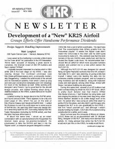
Massive tick infestation by Boophilus microplus and Hyalomma anatolicum on a cross-bred cow in India. Photograph by Vanlalhmuaka and S. Ghosh. 13
To the Index
error: Theileria lestocardi instead of T. lestoquardi.) Guglielmone A.A. 2006. (Vectors of spotted fevers in Argentina.) Abstract of Round Table presentation. Proc. Congreso de Zoonosis, La Plata, Argentina, 10-12 May 2006, in: Acta Bioquímica Clinica Latinoamericana (ABCL) (Suppl. 3). (In Spanish.) and Guglielmone A.A. & Nava S. 2006. (Amblyomma ticks parasitising humans in Argentina.) Abstract of Round Table presentation. Proc. Congreso de Zoonosis, La Plata, Argentina, 10-12 May 2006, in: Acta Bioquímica Clinica Latinoamericana (ABCL) (Suppl. 3. (In Spanish.) (Ten species have been found on human beings in Argentina: A. aureolatum, A. brasiliense, A. cajennense, A. coelebs, A. neumanni, A. ovale, A. parvum, A. pseudoparvum, A. tigrinum & A. triste, with records of A. cajennense, A. neumanni and A. parvum being the most frequent. As in other American countries, A. cajennense appears to constitute the greatest risk for humans, both by its occurrence in populated areas and its vector capacity of the most lethal agent of human rickettsiosis, R. rickettsi. It has also been found infected with R. honei (causal agent of Flinders rickettsiosis), and R. amblyommii (of as yet unknown pathogenicity). But such species as A. neumanni and A. parvum should also be considered as potentially dangerous. Preliminary research results indicate that A. neumanni may be infected with R. belli and R. amblyommii and A. parvum with a rickettsia related to R. parkeri. All three species are common in northern Argentina, and relatively frequent on humans. Several other tick species have been found infected in Argentina or other countries with Rickettsia spp. of the SF group. The dog tick, Rhipicephalus sanguineus, is only found sporadically on human beings in Argentina, but is known to be vector of R. conorii and R. massiliae, and has been found infected with R. rickettsii.) (GU.) (See also the more extensive paper in Alan’s contribution above: Guglielmone et al. in Exp. Appl. Acarol.) López M.E., Flores J., Mendoza P., Vázquez V., Liébano E., Bravo A., Herrera D., Godínes E., Vargas P. & Zamudio F. 2006. Use of Bacillus
thuringiensis toxin as an alternative method of control against Haemonchus contortus. Ann. N.Y. Acad. Sci., 1081: 347-354. (E-mail:
[email protected]) (GU: The paper is not about ticks. But if B. thuringiensis toxin can be manufactered economically, it might be a way to get it inside ticks by injecting it into the host! Seems worth a followup.) Nava S., Mangold A.J. & Guglielmone A.A. 2006. The natural hosts for larvae and nymphs of Amblyomma neumanni and Amblyomma parvum (Acari: Ixodidae). Exp. Appl. Acarol., 40: 123131. (E-mail:
[email protected]) (A. neumanni and A. parvum are two 3-host neotropical tick species that have adapted to introduced domestic livestock. Although preliminary experiments to transmit heartwater with A. neumanni have given negative results (Rev. Elev. Méd. Vét. Pays Trop., 1985, 38: 34-42), it is an experimental vector of bovine anaplasmosis (Folia Parasitol., 1995, 42: 75); the role of A. parvum as a disease vector is unknown. All 3 stages of A. neumanni were found on cattle and horses, and no larvae and nymphs were found on other animals. Cattle can sustain the complete cycle of this species, which was presumably a parasite of endemic Artiodactyls before domestic ruminants were introduced. This tick is therefore quite suitable as a vector of anaplasmosis, which is transmitted transstadially by ticks. On the other hand, no larvae or nymphs of A. parvum were found on domestic animals and virtually all immatures were found on a medium-sized rodent of the Fam. Caviidae, while adults are common on cattle and goats.) (GU) Stachurski F., Bouyer J. & Bouyer F. 2005. (Actually this last issue of vol. 58 was published in 2006.) Lutte contre les ectoparasites des bovins par pédiluve: méthode innovante utilisée en zone périurbaine subhumide du Burkina Faso. (Innovative method to control cattle ectoparasites in suburban areas of the subhumid zone of Burkina Faso: the footbath.) Revue Elev. Méd. Vét. Pays Trop., 58: 221-228. (In French.) (E-mail:
[email protected]) (The first author, F. Stachurski, has studied the infestation process of cattle by the tick Amblyomma variegatum, 14
To the Index
the most harmful tick species in the area, and armed with the results of these painstaking studies has developed an original control method: Most of the ticks first attach temporarily on the feet and only reach their predilection sites when the animals lie down at night (Stachurski. 2000. Med. Vet. Entomol., 14: 391-399). Treating only the lower extremity of the legs, by having the animals pass every 2 to 3 days in a footbath containing an acaricide, at night before they regain their night quarters, eliminates most of these ticks, not only in theory, but also in reality, as shown in the present paper and another one in press. The necessary frequency of the application depends on the residual effect of the acaricide used. This paper extends the method also to the control of the local riverine species of tsetse flies (Glossina tachinoides and G. palpalis gambiensis), not only because many also tend to attack the legs of cattle, but also because the acaricide (to which the flies are far more susceptible than the tick).reaches other parts of the body by splashing, diffusion, contact, etc. The construction of the footbath is shown in detail. It requires an initial investment, but daily exploitation is cheap, and the method controls both tick and tsetse.) (GU. This is a good example of how a non-conformist approach can result in a novel, practical control measure.)
TICK-BORNE DISEASES Recent publications Kivaria F.M. 2006. Estimated direct economic costs associated with tickborne diseases on cattle in Tanzania. Trop. Anim. Health Prod., 38: 291299. (E-mail:
[email protected]) (It is impossible to arrive at accurate estimations of the economic impact of TBDs, and this is fully realised by the author. As he says, estimates are confounded by the heterogeneous nature of cattle production systems and other complexities, the unreliability of reporting, as well as the impracticality of gathering data that present all possible factors. In the estimated 17.7 x 10⁶ cattle in
the country, the major TBDs (East Coast fever, anaplasmosis, heartwater and babesiosis) are estimated to be responsible annually for a total loss of US$ 364 x 106. This figure includes the cost of disease and tick control, but not that of other TBDs nor other effects of tick infestation.) Although it is more than 6 times higher than an earlier estimate (1999) which arrived at a total cost of ticks and TBDs in Tanzania of US$ 55 x 106, it is still thought likely to be an underestimation.) (GU. For full details of the methodology, please read the paper. Without necessarily accepting any estimate, I agree completely with the last sentence of the author: “Thus, despite the limitations of the estimates presented here, it is evident that tick-borne diseases inflict substantial economic losses on cattle production and resource use in Tanzania.”) Okuthe O.S. & Buyu G.E. 2006. Prevalence and incidence of tick-borne diseases in smallholder farming systems in the western-Kenya highlands. Vet. Parasitol., 141: 307-312. (E-mail:
[email protected] or
[email protected]) (Serology using the ELISA for theileriosis (meant is presumably T. parva infection), anaplasmosis (A. marginale) and babesiosis (only B. bovis is mentioned, but what about B. bigemina?) was effected on 408 cattle in the rural area and 192 in the periurban area in the rainy season (September) and 114 and 46 respectively in the dry season (February). Suspect clinical cases of these TBDs were furthermore monitored during almost 2 years by blood smears, and in case of suspicion of theileriosis also by lymph-node biopsy. Seroprevalence in the rural area was 60% for theileriosis, 50% for anaplasmosis and 37% for babesiosis, these figures were 73, 32 and 27,5% in the peri-urban area, respectively. The incidence of laboratory-confirmed cases of clinical TBD was 4.9, 4 .6 and 0.4% for theileriosis, anaplasmosis and babesiosis, respectively, in the rural area, and as high as 39.1, 32.1 and 1.5%, in the peri-urban area. The differences in prevalence and incidence between the rural and periurban areas may be due to differences in tick control and management) (GU. Such surveys are quite useful, to give an idea of the TBD problems in certain areas. Unfortunately, the cattle
breeds involved are not discussed, one might perhaps expect more improved and therefore more susceptible cattle in peri-urban areas [it is only in the Conclusions that the authors mention smallholder dairy farming].)
THEILERIOSIS Effect of the carrier state or chronic theileriosis. In NL n° 27 of August 2005, I wrote that Dr. Mohamed Darghouth in Tunisia drew the attention to the fact that the Theileria annulata carrier state is a very important factor of production loss. I added that previously this had not really been taken into account. While Prof. Darghout is probably right, what I added is wrong. In fact, I recently came across the summary of a paper of almost 18 years ago which shows that negative effects of chronic T. annulata infection had been studied well before: Michael S.A., El Refaii A.H., McHardy N. & Rae D.G. 1989. Effect of treatment of chronic theileriosis with buparvaquone on milk yields. Trop. Anim. Health Prod., 21 : 218222. Chronic theileriosis of Friesian cattle in Egypt was the cause of severely depressed milk yields, as treatment with buparvaquone was followed by a doubling of the milk yield. It should be added that chronic theileriosis as described in the paper is different from the carrier state, but it is perhaps difficult to distinguish clearly between the two. I would be very grateful for documented or published contributions to the subject, including on other TBDs. Recent publications Vaccination Darghouth M.A., Boulter N.R., Gharbi M., Sassi L., Tait A. & Hall R. 2006. Vaccination of calves with an attenuated cell line of Theileria annulata and the sporozoite antigen SPAG-1 produces a synergistic effect. Vet. Parasitol., 142: 54-62. (E-mail:
[email protected]). (Although cell line vaccines for T. annulata have been available for many 15
To the Index
years, the down-sides of live pathogen vaccines have meant that the take-up has, with a few exceptions, been limited. A synthetic vaccine would have many advantages, but only one T. annulata recombinant protein, the immuno-dominant sporozoite specific antigen SPAG-1, has shown any success in vaccination trials, and even this was partial. SPAG-1 induces antibodies that block sporozoite entry, but cell lines are probably more efficacious in inducing protection because T. annulata proteins expressed at the schizont stage activate cellular immune mechanisms. The Tunisian cell line used in this paper has previously been shown to protect against an LD50 dose of heterologous sporozoites. However in this paper male Holstein calves were challenged with an LD100. Four groups of seven animals were immunised with: cell line alone; SPAG-1 alone; cell line followed by SPAG-1; and a control group received PBS. All seven controls and six out of seven calves which received SPAG-1 alone had severe responses and were euthanased. The cell line immunised animals fared better with four out of seven calves requiring euthanasia. However all of the animals that received both cell line and SPAG-1 survived. In terms of clinical and parasitological parameters, the combination of both cell line and SPAG-1 markedly increased the time to first appearance of schizonts when compared to the treatments separately. These results suggest that a synthetic vaccine would be optimal if it included both sporozoite and schizont antigens. Interestingly, the combined treatment resulted in lower anti-SPAG-1 antibodies than in the animals that received SPAG-1 alone, confirming the importance of non-humoral protective pathways. The authors speculate that maybe cell line vaccination changes the cytokine environment which militates against an optimal antibody response e.g. it is believed that a Th1 type response involving interferon-γ is necessary for protection against the schizont stage, whereas a Th2 type response may be more important for antibody induction, and the two pathways can be mutually inhibitory.) (LG) Epidemiology, transmission, distribution Aktas M., Altay K. & Dumanli N. 2006. PCR-based detection of
Theileria ovis in Rhipicephalus bursa adult ticks. Vet. Parasitol., 140: 259263. (E-mail:
[email protected]) (The authors collected 420 R. bursa ticks from 95 sheep and 55 goats in the Elazig province of eastern Turkey in the spring of 2004. They chose 192 of these to assess their infection rate of Theileria; therefore, they dissected salivary glands of these ticks and extracted DNA from them. The DNA was used in PCRs to amplify a 520 bp ssu rRNA gene fragment to confirm the presence of Theileria. Sequence analysis of some of the PCR products revealed that the Theileria species present was T. ovis. The infection rate of the ticks was 19.27% and no significant difference between infection rates in male and female ticks was observed. The authors conclude that R. bursa may play an important role as a natural vector for T. ovis.) (FK) Ali A.M., Bakheit M.A., Mukhtar M.M., Hassan S.M., Ahmed J.S. & Seitzer U. 2006. Epidemiology of Theileria annulata infection of dairy cattle in the Sudan using molecular techniques. Ann. N.Y. Acad. Sci., 1081: 471-472. (E-mail:
[email protected]). (PCR for Theileria spp. and RLB for Ehrlichia, Anaplasma, Theileria and Babesia, spp. on 162 samples (origin unknown), revealed evidence of multispecies infection, and much higher levels of theilerial infection than detected in blood smears microscopically, indicating carrier status.) (LG) Kaewthamasorn M. & Wongsamee S. 2006. A preliminary survey of gastrointestinal and haemoparasites of beef cattle in the tropical livestock farming system in Nan Province, northern Thailand. Parasitol. Res., 99: 306-308. (E-mail:
[email protected]) (This summary only concerns the tickborne haemoparasites found, viz. Theileria sp. in 50% of the 162 animals of which blood smears were made. Neither the species nor its effects were determined.) (GU. I believe only parasites of the T. buffeli/orientalis group have been reported in Thailand.) Konnai S., Imamura S., Nakajima C., Witola W.H., Yamada S., Simuunza M., Nambota A., Yasuda J., Ohashi K. & Onuma M. 2006. Acquisition and transmission of Theileria parva by
vector tick, Rhipicephalus appendiculatus. Acta Trop., 99: 34-41. (E-mail:
[email protected]) (The authors demonstrated that ticks can pick up T. parva infection from cattle which were experimentally infected but had no detectable piroplasm parasitaemias by Giemsastained blood smears but which were positive by PCR. 70.8% of these ticks were positive for T. parva by PCR and 40 of these ticks were fed on another calf, which developed clinical East Coast fever. This work demonstrates that carrier animals with non detectable piroplasm levels by Giemsa but which are positive by PCR can act as reservoir for T. parva infection. The authors also tested 70 cattle in Zambia for T. parva by PCR and found that 29 were positive and of these only 6 were positive by blood smear analysis. They also surveyed 614 Rhipicephalus appendiculatus ticks collected by flagging pastoral land, 4.1% of these were positive for T. parva by PCR) (FK)
[email protected]) (This paper describes a longitudinal study, which links different agro-ecological zones and grazing systems with tick burden and incidences of East Coast fever in bovine calves. A total of 198 cattle were monitored from May 2002 until February 2003 in the eastern Ugandan districts of Mbale and Sironko encompassing regions of different altitude (lowland, midland and upland) as well as different grazing regimes (zero-grazing, communal grazing and restricted-outdoor grazing). Tick numbers and species were also monitored. Not surprisingly, the authors found different distributions of tick species and incidences of East Coast fever within the different agroecological zones and that Rhipicephalus appendiculatus ticks and East Cost fever incidences were most commonly found in lowland farms with communal grazing.) (FK)
Razmi G.R., Eshrati H. & Rashtibaf M. 2006. Prevalence of Theileria spp. infection in sheep in South Khorasan province, Iran. Vet. Parasitol., 140: 239-243. (E-mail:
[email protected]) (840 sheep, from 34 flocks from the South Khorasan province in Iran, were clinically examined, tested for piroplasms and ticks were collected. The authors found that 11.9% of the sheep sampled had piroplasm parasitaemias of 0.02 to 0.1%. None of the sheep had any clinical signs of theileriosis. 43% of the sheep examined had tick infestations and the predominant tick species found belonged to the Rhipicephalus sanguineus group (50.4%), while ticks of the species Hyalomma anatolicum were the second most common species (48.5%) and 8 H. dromedarii ticks were also found (0.98%) The authors speculate that the piroplasms seen are probably T. lestoquardi but this would have to be confirmed using molecular tools.) (FK)
Bakheit M.A., Endl E., Ahmed J.S. & Seitzer U. 2006. Purification of macroschizonts of a Sudanese isolate of Theileria lestoquardi (T. lestoquardi [Atbara]). Ann. N. Y. Acad. Sci., 1081: 453-462. (E-mail:
[email protected]) (This paper describes a purification method for schizonts of T. lestoquardi. The isolate used originated from a field isolate from Atbara in northern Sudan in 2001 and has been maintained in tissue culture since. RNA was extracted from the purified schizonts and this RNA was used to generate a cDNA expression library. The host and parasite representation of the library was checked by randomly sequencing 19 clones of which 12 turned out to be of parasite origin indicating that the library consists mainly of parasite transcripts, with only some host contamination.) (FK) (GU: There is more to this paper than just the purification of the schizonts.)
Rubaire-Akiiki C.M., Okello-Onen J., Musunga D., Kabagambe E.K., Vaarst M., Okello D., Opolot C., Bisagaya A., Okori C., Bisagati C., Ongyera S. & Mwayi M.T. 2006.Effect of agro-ecological zone and grazing system on incidence of East Coast Fever in calves in Mbale and Sironko Districts of Eastern Uganda. Prev. Vet. Med., 75: 251-266. (E-mail: 16
To the Index
Molecular and cell biology, genomics
Bakheit M.A., Scholzen T., Ahmed J.S. & Seitzer U. 2006. Molecular characterization of a Theileria lestoquardi gene encoding for immunogenic protein splice variants. Parasitol Res., 100: 161-170. (E.mail:
[email protected]) (The authors describe the isolation and the characterisation of a novel T. lestoquardi gene. This gene was isolated by screening a T. lestoquardi schizont
cDNA expression library with immune sheep serum. The gene contains a single intron and some of the cDNAs isolated were found to contain unspliced sequences. The authors speculate that these splice variants are leading to different protein products. Western blot analysis found proteins of 99.9 and 72.7 kDa in schizont extracts reacting with serum raised against the recombinant protein. Protein/gene homologues were found in the T. annulata and T. parva genome. Fluorescent microscopy, using serum raised against the recombinant protein and schizont infected cells, revealed a predominant parasite membrane association of the protein.) (FK) Casanova C.L., Xue G., Taracha E.L. & Dobbelaere D.A. 2006. Post-translational signal peptide cleavage controls differential epitope recognition in the QP-rich domain of recombinant Theileria parva PIM. Mol. Biochem. Parasitol., 149: 144-154. (E-mail:
[email protected]) (This is a very thorough and detailed study of post-translational modifications of the PIM protein of Theileria parva. The authors have demonstrated that expression of the gene results in two distinct sizes of the molecule, which are both different from the predicted molecular weight according to PIM amino acid sequence. In both cases the molecular mass, according to SDS-PAGE, is far bigger than the predicted size. The size difference between the two forms could not be explained by cleavage of the signal peptide alone. Further, antibody reactivity revealed that the different forms of the molecule mask different antibody epitopes. Removal of the signal peptide of 19 amino acids by expression of a recombinant gene construct resulted in the loss in apparent mass of 24 kDa, revealing the smaller protein form. The removal of the signal peptide also resulted in altered antibody reactivity. These findings are discussed with relevance to other schizont proteins.) (FK) Hermann P. & Dobbelaere D.A. 2006. Theileria-induced constitutive IKK activation is independent of functional Hsp90. FEBS Lett. 580: 5023-5028. (E-mail:
[email protected]). (The intracellular stage of Theileria spp. resides free in the cytoplasm and recruits, by an unknown mechanism,
host IKB kinase (IKK) complexes onto its surface. This results in permanent activation of the kinase and constitutive activation of NF-KB, which is then free to translocate to the nucleus and switch on a range of genes associated with inflammatory, proliferation and survival functions. The heat shock protein, Hsp90, is a chaperone that controls the stability and activity of a variety of proteins including the IKK complex. However using an Hsp90specific inhibitor, geldanamycin, the authors found that IKK activity in Theileria-infected cells was unaffected, and they could not detect Hsp90 presence on the parasite-located IKK signalosomes. However transcription induced by NF-KB was reduced (luciferase NF-KB dependent reporter gene assay), suggesting that Hsp90 may play a role in transcription, but not in the IKK activity in Theileriainfected cells. Tumour cells, in contrast, do require Hsp90 to stabilise the IKK complex. How Theileria spp. elude and manipulate host cell pathways remains a mystery. Perhaps a parasite-encoded heat shock protein is involved?) (LG) Heussler V., Sturm A. & Langsley G. 2006. Regulation of host cell survival by intracellular Plasmodium and Theileria parasites. Parasitology, 130 (Suppl.): S49-S60. (E-mail:
[email protected]) (This is a review article, which compares the current knowledge of the mechanisms employed by Theileria and Plasmodium parasites. The main topics discussed are the inhibition of host cell apoptosis by the parasites; signalling via surface receptors; stresssignalling in infected cells. The authors show that the understanding of how the different parasites interact with their respective host-cells is complementary but also that the research for the different parasites has advanced at different levels when it comes to different life cycle stages.) (FK) Katzer, F., Ngugi, D., Oura, C., Bishop, R.P., Taracha, E.L.N., Walker, A.R. & McKeever D.J. 2006. Extensive genotypic diversity in a recombining population of the apicomplexan parasite Theileria parva. Infect. Immun., 74: 5456-5464. (E-mail:
[email protected]) (This paper used PCR to amplify satellite markers, which are spread throughout the T. parva genome, from parasite 17
To the Index
clones established from a T. parva Marikebuni stabilate. The analysis demonstrated that this stabilate has a predominant genotype, which stayed the dominant genotype following further cattle to tick passage. Typing of the parasite progeny revealed extensive levels of re-assortment and recombination. The authors also found evidence of shuffling of antigenic determinants in the progeny and the relevance to this for protective cytotoxic-T-lymphocyte responses is discussed. Typing of ancestral T. parva Marikebuni stabilates revealed a shift from a different predominant genotype to the present predominant genotype. Selective pressures, which could be responsible for this change, are also discussed.) (FK) Roy S.W. & Penny D. 2006. Largescale intron conservation and order-ofmagnitude variation in intron loss/gain rates in apicomplexan evolution. Genome Res., 16: 1270-1275. (E-mail:
[email protected]). (The publishing of more and more genomes is opening up new avenues of research related to comparative genome analysis. In this paper the authors compare the conservation of intronic positions within genes of putative orthologs between two Plasmodium spp. and two Theileria spp. They find that T. annulata and T. parva which probably diverged around 82 million years ago (MYA) have 99.7% identity in intron positions between orthologs (7089/7111 introns in 3305 pairs of orthologous genes, as determined by amino acid sequence similarity using BLASTP comparisons). P. falciparum and P. yoelli which diverged a similar time ago (100 MYA) also share a similar level of intron conservation. However between P. falciparum and T. parva only around 60% of introns are conserved between the 1279 putative orthologous genes. The mechanisms that lead to intron loss and gain have been proposed to relate to the presence of transposable elements (TEs) with reverse transcriptase activity, that results in copying of mRNA, followed by recombination into the host genomic DNA. Such TE events may not occur at a constant rate over time (unlike the molecular clock of most synonymous mutations), but instead may fluctuate widely. Neither Theileria nor Plasmodium harbour any known TEs which could explain the near conservation of intronic positions
in putative orthologs within species. In contrast, the lower conservation between the two genera (Plasmodium and Theileria) which derived from a common ancestor more than 350 MYA, might suggest that high TE abundance was associated with speciation, perhaps accompanying host or vector changes. (N.B.: putative orthologs = genes with sufficient sequence conservation that they are likely (but not definitely) to be equivalents in different species.) It can be difficult to definitively assign genes as orthologs as in many cases genes in one species (or even in one individual) can become duplicated. These duplicated genes can then harbour substitutions, change introns etc and potentially evolve new functions. Of the genes that did not conserve intronic positions, the authors checked that synteny (gene order in the genome) was maintained for these orthologous genes, which provides further weight to the evidence that they are truly orthologous.) (LG) Seitzer U., Schnittger L., Boguslawski K. & Ahmed J.S. 2006. Investigation of MAP kinase activation in Theileriainfected cell lines. Ann. N.Y. Acad. Sci., 1081: 473-475. (E-mail:
[email protected]) (Previously published studies have indicated that the MAP kinases, ERK1, ERK2 and p38 are not constitutively activated in Theileria infected cells unlike JNK (reviewed by Dobbelaere et al., 2000, Cell. Microbiol. 2, 91). This was surprising as ERKs are generally associated with cell proliferation and prevention of apoptosis. The study reported here in contrast, suggests that ERKs may be activated after all. T. annulata infected cell lines had higher constitutive proportions of the phosphorylated (i.e. activated) form of the MAP kinase, ERK1, than did T. parva infected cell lines as revealed by Western blotting, although the activated form was inducible by PMA in the latter cell lines. Phosphorylated ERK2 was constitutively expressed in both sets of cell lines. However a cloned T. annulata infected line and two cloned T. parva infected lines had low constitutive levels of ERK1, suggesting that clones may not represent in vivo conditions.) (LG)
Seitzer U., Jongejan F. & Yin H. 2006. Phylogenetic position of small-ruminant infecting piroplasms. Ann. N. Y. Acad. Sci., 1081: 498-504. (E-mail:
[email protected]) (This review paper gives an overview of the phylogeny based on the 18S ssu rRNA gene sequences of piroplasms infecting small ruminants. It concentrates mainly on the different Theileria species (T. lestoquardi, T. ovis and T. separata) infecting sheep and goats. The authors describe the position of the different species but unfortunately no figure of the phylogenetic tree is shown to make the description more visual and clear. The paper also confirms the existence of two distinct Theileria species infecting sheep in China, designated Theileria sp. (China 1) and Theileria sp. (China 2) and in its conclusions refers to the proposed naming of these species as Theileria luwenshuni and Theileria uilenbergi, respectively in a paper, which is currently in preparation.) (FK) (GU: See comments to the paper Miranda et al., 2006. Am. J. Vet. Res., 67: 1908-1913, below. Another comment is the fact that in Table 1 the tick vector of T. ovis (Turkey) is stated to be Rhipicephalus evertsi. It is of course possible that the Turkish T. ovis can be transmitted by this African tick species, but it does not occur in Turkey. Probably Rh. bursa was meant. See also the comments on this paper in the section Babesioses.) Miranda J.P., Bakheit M., Schneider I.,
Haller D., Ahmed J.S., Yin H., Oliva A.G. & Seitzer U. 2006. Identification of homologous genes of T. annulata proteins in the genome of Theileria sp. (China). Ann. N.Y. Acad. Sci., 1081: 468-470. (E-mail:
[email protected]). (Genes with high homology to previously described T. annulata genes including those coding for a surface protein, a putative membrane protein, and a secretory protein as well as a T. lestoquardi potential membrane protein were amplified from Theileria sp. (China) merozoite cDNA. There was no evidence of antibody responses to the first three proteins, results were equivocal for the T. lestoquardi-like protein.) (LG) Sparagano O.A., Spitalska E., Namavari M., Torina A., Cannella V. & Caracappa S. 2006. Phylogenetics of Theileria species in small ruminants. Ann. N. Y. Acad. Sci., 1081: 505-508. (E-mail: (Olivier.
[email protected]) (This short conference paper looks at the phylogenetic status of Theileria lestoquardi and Theileria ovis in relation to some Theileria species of cattle. 18S rRNA gene sequences were generated by PCR and sequencing from blood samples from sheep from Italy and Iran as well as from tick samples from Iran. The Theileria species in Iran was identified as T. lestoquardi and the Theileria in Italy were mainly identified as T. ovis. The T. lestoquardi parasites mapped most closely to T. annulata while the T. ovis parasites
Taxonomy, phylogeny Ahmed J.S., Luo J., Schnittger L.,
Goat farming in South Africa. Photograph by D. de Meneghi. 18
To the Index
mapped most closely to parasites of the T. buffeli/orientalis group.) (FK) See also the paper by Allsopp & Allsopp, 2006, presented in the section Babesioses. Antigens Bakheit M., Scholzen T., Ahmed J.S. & Seitzer U. 2006. Identification of potential antigenic proteins of Theileria lestoquardi. Ann. N. Y. Acad. Sci., 1081: 463-464. (E.mail:
[email protected]) (This very short paper only states that the authors have PCR-amplified the T. lestoquardi homologue of two T. annulata genes Tams1 and TaSP and that they will try to test these homologues for the development of a diagnostic ELISA to detect T. lestoquardi infections in Sudan.) (FK) Miranda J., Nascimento E., Cruz H., Hong Y., Coelho A., Ahmed J.S. & Oliva A. 2006. Identification and characterization of merozoite antigens of a Theileria species highly pathogenic for small ruminants in China. Ann. N. Y. Acad. Sci., 1081: 443-52. (E-mail:
[email protected]) (The authors use a proteomics approach to identify antigens of the Chinese Theileria species infecting small ruminants, which are recognised by immune sheep serum. On Western blot analysis four prominent bands were identified of 54, 70, 95 and 119 kDa. These proteins were excised and characterised using peptide mass mapping by matrix-assisted laser desorption/ionization (MALDI). This analysis revealed that the 54 kDa protein has the highest similarity to actin; the 119 kDa peptide to a possible MO25-family protein of Cryptosporidium parvum and the 70 kDa protein has good similarity to the 70 kDa heat shock protein of T. annulata. The 95 kDa protein has no significant similarity to known proteins. The authors intend to test the 119 and 70 kDa proteins for the inclusion in a diagnostic ELISA to detect Theileria sp. infecting small ruminants in China.) (FK: The authors do not specify which of the Theileria species infecting small ruminants in China was investigated.) Small ruminants Jian-San W., Jian-Min Y., Zhi-Lian W., Cui-Ping S., Jian-Fong Y. & Bao-An
Y. 2006. The host responses in sheep artificially infected with Theileria sp. (China). Ann. N. Y. Acad. Sci., 1081: 465-467. (E-mail:
[email protected]) (This very short paper describes the response of some sheep experimentally infected by feeding adult female ticks, isolated from the Gannan district in the Gansu province of China. Clinical responses described were hyperthermia (40.3 to 41.3°C), schizonts were seen 20-23 days post infection and piroplasms appeared about 24 hours after the schizonts were first seen with parasitaemias ranging from 1% to 39%. Antibody titres were also determined for 2 sheep by IFAT.) (FK: The authors do not specify which of the Theileria species infecting small ruminants in China was investigated.) In vitro culture Miranda J.P., Nascimento E.M., Cruz H.J., Yin H., Zweygarth E. & Oliva A.G. 2006. Establishment of optimal conditions for long-term culture of erythrocytic stages of Theileria uilenbergi. Am. J. Vet. Res., 67: 1908-1913. (This is a technical paper, which set out to establish and optimize long-term in vitro cultures for the erythrocytic stages of Theileria uilenbergi, a pathogenic Theileria species of small ruminants in China. The authors imported infected blood from China and set up parallel cultures, testing different media for their ability to establish T. uilenbergi cultures. The medium, which gave the best long term culture results, was made with HL-1. The culturing conditions were optimised and best parasite growth results were obtained with HL-1 medium supplemented with 20% lamb serum and 0.75% chemically defined lipids (Invitrogen) incubated in a humidified 2% O2, 5% CO2 and 93% N2 atmosphere at 37°C. Cultures were maintained for more than 400 days and could also be cryopreserved and resuscitated.) (FK) (GU: I am confused about the name. As far as I know, the nameT. uilenbergi was first used in a 2004 paper: Yin et al. Parasitol. Res., 92: 36-42. In the paper the authors mentioned 2 species of small ruminants transmitted by the tick Haemaphysalis qinghaiensis, Theileria sp. China 1 and Theileria sp. China 2, and introduced two new names, Theileria luwenshuni and T. uilenbergi. But they did not make it 19
To the Index
clear for which of the parasites each of these names was meant, and I would think they were therefore nomina nuda. (See NL n° 25, P. 18.) In a recent paper (Ahmed et al., 2006, see above), it is proposed to give the name of T. luwenshuni to Theileria sp. (China 1) and T. uilenbergi to Theileria sp. (China 2). That clearly means that these names have not yet been created and validly published, and for the moment they are still nomina nuda.) Pathology/pathogenesis Mbassa G.K., Kipanyula M.J., Mellau L.S., Mwamakali E.D., Bulegeya F.R. & Kauto-Mboni K. 2006. Theileria parva infection in calves causes massive lymphocyte death in the thymus, spleen and lymph nodes without initial proliferation. Vet. Parasitol., 140: 260270. (E-mail:
[email protected]) (This is a very detailed description of the histology of lymph nodes, thymus and spleen of calves throughout experimental infections with Theileria parva in comparison to uninfected control animals. The general findings of the authors are that they observed changes in these tissues, which are consistent with apoptosis and necrosis but they found no evidence of lymphocyte proliferation, which is associated with T. parva infection of cells in vitro. Based on these findings, they conclude that T. parva infection in vivo is lymphodestructive and lympho-degenerative and not a lympho-proliferative disease.) (FK) (GU: The conclusions of the authors are truly amazing. As they state, the lymphnodes were enlarged. This confirms what many authors observed, including our team at the CVL of Dares-Salaam (Tanzania) (1973-1976) and also later in the course of our experimental infections in Europe (19761988). We assumed that the lymphocyte proliferation, with many dividing lymphoblasts, which we always saw and which started even before schizonts were found in the lymphnodes, was the cause of the enlargement of the lymphnodes and other lymphoid tissue. Lymphocyte destruction in the lymphnodes and pancytopenia come in a second stage of the disease, and are associated with intense immunodepression and an important decrease of the number of circulating lymphocytes. One may also see shrinking of the lymphnodes in a later stage of the dis-
ease. But first there is undoubtedly lymphocyte proliferation in the nodes and several organs, which leads for instance to the foci in the renal cortex, the wall of the abomasum, and even occasionally in the skin.) Rezaei S.A. & Dalir-Naghadeh B. 2006. Evaluation of antioxidant status and oxidative stress in cattle naturally infected with Theileria annulata. Vet. Parasitol., 142: 179-186. (E-mail:
[email protected]). (One feature of the later stages of T. annulata infection in vivo is anaemia. However the mechanism is unclear, although it seems clear that red blood cells (RBC) become more fragile and lyse or are cleared from the systemic circulation by, for example, macrophages. The results reported here in the main confirmed other workers’ findings. Various parameters of lipid peroxidation and free radical scavengers in red blood cells were significantly negatively correlated with the degree of anaemia and parasitaemia associated with T. annulata infection. Thus oxidative damage to RBCs is likely to contribute to anaemia in this disease, but how the presence of the parasite contributes to this is as yet unknown.) (LG)
BABESIOSES Recent publications Immunisation De Waal D.T. & Combrink M.P. 2006. Live vaccines against bovine babesiosis. Vet. Parasitol., 138: 88-96. (Email:
[email protected]) (The paper provides a general overview of the epidemiology and control of bovine babesiosis caused by Babesia bovis, B. bigemina and, to a lesser extent, B. divergens. Options for control include vector control and the effect this may have on endemic stability. The paper also covers chemoprophylaxis, immunity following natural exposure and use of vaccine as a method of artificially creating a stable, immune state. The paper then gives an overview of the method used to produce the vaccine at the Onderstepoort Veterinary Institute in South Africa including the
reasons for the change from a chilled to a frozen product because of lower production costs, prolonged shelf life, ‘on demand’ availability and prerelease safety and effectivity testing. About 150,000 doses monovalent B. bovis and B. bigemina vaccines are produced annually. DMSO is used as cryoprotectant and it is recommended that the vaccine be kept on melting ice after thawing and be used within 4 hours. The risk of vaccine reactions increases with age and users are usually advised to vaccinate calves up to the age of 9 months. A popular practice of using low doses of diminazene to prevent reactions is no longer recommended as it is probably the most frequent cause of vaccine failures. Deep intramuscular injection yields the most consistent results. Immunity to B. bovis lasts several years after a single vaccination but is of much shorter duration in the case of B. bigemina. Sterile immunity after chemosterilisation lasts at least 3 years in the case of B. bovis while B. bigemina animals are fully susceptible after 16 months.) (Bert de Vos) (GU: Through an oversight this review was not presented in NL n° 31.) Rojas C., Figueroa J.V., Alvarado A., Mejia P., Mosqueda J.J., Falcon A., Vega C.A. & Alvarez A. 2006. Bovine babesiosis live vaccine production. Ann. N.Y. Acad. Sci., 1081: 405-416. (E-mail:
[email protected]) (Bovine babesiosis is an important disease in tropical and subtropical parts of Mexico and use of a live, attenuated vaccine is considered to be the method that offers the most promise for control and prevention. The risk of contamination when animals are used as source of immunogen is well known but converting to in vitro propagation will not eliminate this risk because of the need for bovine red blood cells and serum in the culture medium. This study showed that gamma irradiation of bovine serum and red cells inactivated viruses and bacteria from the substrate and supported in vitro growth of Babesia bovis and B. bigemina. Gamma irradiation in a 60Co irradiator with 25 kilogray (kGy) inactivated infectious bovine rhinotracheitis and bovine viral diarrhoea viruses in artificially contaminated serum. It also eliminated a number of bacteria, 20
To the Index
including E. coli and Staphylococcus aureus. However, it caused complete haemolysis of red cells, while a dose of 70 gray (Gy) did not cause lysis. Babesia bovis and B. bigemina were successfully established in a culture medium containing serum irradiated at 25kGy and red cells at 70 Gy. In both species, the percentage parasitised erythrocytes were comparable to those in nonirradiated control cultures. The authors concluded that gamma irradiation is a suitable method to eliminate potential viral and bacterial contaminants from serum used to produce a live attenuated vaccine through in vitro culture.) (Bert de Vos) (GU. As the authors say, these experiments do not yet solve all of the problems. An irradiation dose that avoids bacterial contamination but causes no red cell haemolysis remains to be determined, and it is important to assess whether any alteration to the red cells is unfavourable for the culture of Babesia.) Diagnosis Goff W.L., Molloy J.B., Johnson W.C., Suarez C.E., Pino I., Rhalem A., Sahibi H., Ceci L., Carelli G., Adams D.S., McGuire T.C., Knowles D.P. & McElwain T.F. 2006. Validation of a competitive enzyme-linked immunosorbent assay for detection of antibodies against Babesia bovis. Clin. Vaccine Immunol., 13: 1212-1216. (Email:
[email protected]) (The authors internationally validated an ELISA they had reported previously (Goff et al. 2006. Clin. Diag. Lab. Immunol., 10: 38-43) based on a species specific, broadly conserved, and tandemly repeated B-cell epitope within the C terminus of rhoptry-associated protein 1 of Babesia bovis. Test laboratory operating characteristic analysis revealed an assay with a specificity and positive predictive value of 100% and a sensitivity of 91.1%, with various negative predictive values depending on the level of disease prevalence. The cELISA was distributed to four different laboratories (USA, Australia, Morocco & Italy), along with a reference set of 100 defined bovine sera, including known positive, known-negative, and field samples collected in Australia, USA & Puerto Rico. Pairwise concordances among the four laboratories ranged from 94% to 88%. Analysis of variance of the resulting optical densi-
ties and a test of homogeneity indicated no significant difference among the laboratories. The authors state that, overall, the cELISA appears to have the attributes necessary for international application.) (WJ: It is gratifying to see an ELISA well validated widely for the international community to use). Hashemzadeh Farhang H., Nabavi L., Seyfiabad Shapouri M.R., Rahbari S. & Azizi F. 2006. Development of an ELISA technique for the detection of Babesia ovis and serological survey of the parasite in Khouzestan province, southern Iran. Iranian J. Vet. Res., 7: 53-58. (E-mail:
[email protected]) (Based on the author’s abstract: An ELISA was developed to detect antibodies against Babesia ovis. A correlation of 85% was observed between the results of the developed ELISA and IFA techniques. To study the seroprevalence of Babesia ovis in Khouzestan province, south of Iran, 1000 sheep sera were collected from different areas of the province and tested against Babesia ovis using the developed ELISA. The results showed an average seroprevalence of 47.5% in the province. Our results indicated a significant increase of the seroprevalence by advancement of age of the animals. There was no significant difference between the seroprevalence of female and male sheep). (AN) Ravindran R., Rao J.R. & Mishra A.K. 2006. Detection of Babesia bigemina DNA in ticks by DNA hybridization using a nonradioactive probe generated by arbitrary PCR. Vet. Parasitol., 141: 181-185. (E-mail:
[email protected]) (This short communication describes the development of a nonradioactive probe for B. bigemina DNA, generated by RAPD-PCR. The specificity of the digoxigenin (DIG) labelled probe was checked by hybridization with genomic DNA from B. bigemina, Theileria annulata, Trypanosoma evansi, Toxoplasma gondii, bovine leucocytes and bubaline leucocytes, where only B. bigemina DNA was detected. The limit of sensitivity for the probe was 100 ng of B. bigemina DNA and it could detect B. bigemina DNA in larval ticks.) (LJ: The specificity of this probe needs to be further evaluated against other strains of B. bigemina, and other
species of Babesia, in particular B. bovis.) Molecular biology/Genomics/Immunology Brown W.C., Norimine J., Knowles D.P. & Goff W.L. 2006. Immune control of Babesia bovis infection. Vet. Parasitol., 138: 75-87. (E-mail:
[email protected]) (It has previously been shown that there is serological cross-reactivity among babesial antigens and this has hindered the differentiation of Babesia species using immunological techniques for identification. This report describes the identification of common antigens between three species of Babesia that infect cattle, namely B. bovis, B. bigemina and B. divergens in the search for antigens that could be used as multi-purpose Babesia immunogens. With each Babesia species antigen and heterologous serum combination, up to 15 antigenic bands were identified, with common antigens having relative sizes of 70 kDa, 60 kDa and 50 kDa. A clone was isolated from a B. divergens cDNA library using B. bigemina-specific sera and a BLAST search revealed 47% protein sequence identity with a putative nascent-polypeptide associated complex, α chain (αNAC) of Theileria annulata. The BLAST search also revealed shared similarities and identities with αNAC proteins from other apicomplexan parasites. Serum from mice immunized with the recombinant protein reacted with B. divergens merozoites in a diffuse staining pattern, as well as weakly staining B. bovis and B. bigemina merozoites. The specificity of the staining was confirmed using immunoprecipitation and immunoblotting assays. To determine the compartment localization of the αNAC-like protein, further studies using confocal or electron microscopy need to be performed.) (LJ) (GU: Through an oversight this review was not presented in NL n° 31.) Figueroa J.V., Précigout E., Carcy B. & Gorenflot, A. 2006. Identification of common antigens in Babesia bovis, B. bigemina, and B. divergens. Ann. N.Y. Acad. Sci., 1081: 382-396. (E-mail:
[email protected]) (This review discusses aspects of the immune response to B. bovis as well as work underway in the development of an alternative B. bovis vaccine. The 21
To the Index
first section describes the cellular effector mechanisms of parasite clearance and details the roles of macrophages, nitric oxide, inflammatory cytokines and antigen-specific CD4+ T cells in the immune response. The part on age-related resistance to infection outlines more recent work highlighting the role of the spleen in the innate immune response to B. bovis. The next section describes work on vaccine development. Aspects of selecting candidate B. bovis vaccine antigens using a proteomic approach and a combined genomic and proteomic approach are summarised. An interesting section on the enumeration of antigen- or epitope-specific T cells in vivo describes the potential use of MHC class I and class II tetramers to increase understanding of T cell function during infection and to identify candidate vaccine antigens. (LJ) This paper was also addressed by Ard Nijhof: (Immunochemical analysis using sera from cattle immunized with B. bovis, B. bigemina or B. divergens revealed a number of cross-reactive epitopes recognized by heterologous antisera. An immunogenically minor parasite component, with identity to αNAC (α-chain of the nascent-polypeptide associated complex), was identified by cDNA library screening of B. divergens with B. bigemina antiserum. Antibodies raised against this protein recognized the antigen in merozoites of all three Babesia spp. studied by IFAT. Further studies are required to examine the protective potential of this protein.) (AN) Montero E., Gonzalez L.M., Rodriguez M., Oksov Y., Blackman M.J. & Lobo C.A. 2006. A conserved subtilisin protease identified in Babesia divergens merozoites. J. Biol. Chem., 281: 35717-35726. (E-mail:
[email protected]) (This paper describes the characterization of a subtilisin-like serine protease isolated from B. divergens merozoites. Two dominant serine proteases of ~48 and ~75 kDa were detected in B. divergens crude extracts using a biotinylated fluorophosphonate probe which is directed against the active site of serine proteases. Immunoprecipitation and Western Blot analysis with antibodies specific for Plasmodium falciparum subtilisin mature protease domain (PfSUB1m) confirmed these results and detected
an additional ~55 kDa protein. The corresponding nucleotide and protein sequence of this bdsub-1 gene was subsequently obtained by immunoscreening of a B. divergens merozoite cDNA library using the PfSUB1m serum. Analysis revealed it to be a single-copy gene containing no introns and belonging to the subtilisin superfamily, showing significant similarity to other subtilases from other Apicomplexan parasites. The protein undergoes post-translational processing and the active form of BdSUB-1 (p48) is concentrated in B. divergens merozoite dense granules, similar to the location of PfSUB1 in P. falciparum merozoites. Anti PfSUB1m antibodies inhibited the in vitro invasion of B. divergens with 58%. The use of B. divergens as a surrogate model for Plasmodium invasion is suggested.) (AN) (GU: And it might be potentially important for Babesia spp. of relevance for tropical livestock!) Chemotherapy Richier E., Biagini G.A., Wein S., Boudou F., Bray P.G., Ward S.A., Précigout E., Calas M., Dubremetz J.F. & Vial, H.J. 2006. Potent antihematozoan activity of novel bisthiazolium drug T16: evidence for inhibition of phosphatidylcholine metabolism in erythrocytes infected with Babesia and Plasmodium spp. Antimicrob. Agents Chemother., 50: 3381-3388. (E-mail:
[email protected]) (Plasmodium and Babesia spp. synthesize considerable amounts of membranes during their intraerythrocytic development, with phosphatidylcholine (PC) as the most abundant lipid. A major route for synthesis of this phospholipid requires host choline. The novel bisthiazolium compound T16 mimics the choline structure, thereby inhibiting the de novo PC biosynthesis in Plasmodium and Babesia spp. It was demonstrated that T16 selectively accumulates in Babesia-infected erythrocytes and that it possesses strong antibabesial activity against B. divergens, B. canis, even higher than that of the index antibabesial drugs imidocarb and pentamidine. A formulation of this class of compound is now entering preclinical development as an antimalarial drug, but the antibabesial activity shows promise as well.) (AN)
Goat farming in South Africa. Photograph by D. de Meneghi. Taxonomy/Phylogenetics Ahmed J.S., Luo J., Schnittger L., Seitzer U., Jongejan F. & Yin H. 2006. Phylogenetic position of small-ruminant infecting piroplasms. Ann. N.Y. Acad. Sci., 1081: 498-504. (E-mail:
[email protected]) (An overview of the phylogenetic position of Theileria and Babesia spp. infecting small ruminants is given in this paper which appeared in the proceedings of the 8th STVM conference published by the New York Academy of Sciences. Three recognized Babesia species of sheep and goats are Babesia ovis, Babesia crassa and Babesia motasi, with a possible additional large Babesia sp. from China which was reported by Bai, Q. et al. in 2002 (Parasitol. Res., 88: S16-S21). Babesia species which were described in the past but are not molecularly characterized such as Babesia foliata (Ray & Raghavachari, 1941) and Babesia taylori (Sarwar, 1935) isolated from sheep and goats in India are not mentioned.) (AN) (GU: As this paper is also discussed by Frank Katzer in the section on Theilerioses, the summary here is limited to Babesia. However, some comments from Ard Nijhof on Theileria are interesting: “Theileria recondita (misspelled as T. recondite, probably a result of Windows autocorrection), was not included in this study since no sequence information is available. Two other piroplasms isolated from small ruminants in Spain: Theileria sp. (OT1) and Theileria sp. (OT3) are briefly mentioned, but not discussed 22
To the Index
further, which is a bit confusing (couldn’t one of them perhaps be T. recondita?).” To this I would add: Theileria recondita Lestoquard, 1929, was created for a non-parasitc parasite of small ruminants in North Africa. It is generally considered as a synonym of T. ovis Rodhain, 1916, a non-pathogenic parasite of small ruminants in central Africa (another synonym is T. sergenti (Wenyon, 1926). The species in Germany and Great Britain which has been called by some T. recondita is definitely different from any known African parasite, so the name T. recondita is invalid for it (and cannot be used again as a synonym of T. ovis; it is an additional species, as yet unnamed. Theileria sp. (OT1) in Spain may be close to or identical with Theileria sp. (China 1), according to Nagore et al., 2004, see NL n° 26, pp. 21-22.) Allsopp M.T. & Allsopp B.A. 2006. Molecular sequence evidence for the reclassification of some Babesia species. Ann. N.Y. Acad. Sci., 1081: 509-517. (E-mail:
[email protected]) (A historical perspective on the classification of the Babesia and Theileria spp. forms the introduction to a phylogenetic analysis based on their small subunit rRNA sequences. Based on this analysis, it is suggested that two new genera be created: one containing B. microti, B. rodhaini and new feline parasites (B. leo, B. felis and B. sp. caracal A) and a second one containing the North American B. gibsoni (for
which the name B. conradae has recently been proposed), as well as a number of human and cervid piroplasms from the United States. The status of a third group comprising Cytauxzoon felis, T. bicornis, B. bicornis, T. youngi and possibly T. equi is uncertain and requires additional examination when more information becomes available.) (AN) Hilpertshauser H., Deplazes P., Schnyder M., Gern L. & Mathis A. 2006. Babesia spp? identified by PCR in ticks collected from domestic and wild ruminants in southern Switzerland. Appl. Environ. Microbiol., 72: 6503-6507. (Based on abstract only: One of the pathogens affecting a cattle herd in Switzerland was identified as Babesia bigemina, which had never been observed in this country before. Therefore, a survey was conducted. Over 2000 ticks were collected from sheep, goats, cattle, and wild ruminants (red deer, roe deer, and chamois) in southern parts of Switzerland and identified morphologically. The vast majority of the ticks (99.2%) were Ixodes ricinus, but 14 ticks from sheep and goats were identified as Dermacentor marginatus and two ticks from wild ruminants were identified as Haemaphysalis [misspelt as Hemaphysalis] punctata. PCR analyses of 700 ticks revealed the presence of Babesia divergens (n = 6), Babesia sp. genotype EU1 (n = 14), and B. major (n = 2), confirmed by molecular analysis, and the presence of novel Babesia sp. genotype CH1 (n = 4), which is closely related to B. odocoilei and to Babesia sp. genotype RD61 reported from North America. The identification of B. divergens and B. major in ticks collected from wild ruminants cast doubt on the postulated strict host specificity of these bovine Babesia species. Furthermore, the zoonotic Babesia sp. genotype EU1 was present in ticks collected from domestic animals but predominantly in ticks from wild ruminants. The role of these game animals as reservoir hosts of Babesia spp. may be important but requires further investigation.) (GU: The closer you look, the more you find! No further mention of B. bigemina however; its occurrence so far North is surprising; see also see NL n° 30, pp. 19-20.) Holman P.J. 2006. Phylogenetic and
biologic evidence that Babesia divergens is not endemic in the United States. Ann. N.Y. Acad. Sci., 1081: 518-525. (E-mail:
[email protected]) (B. divergens is the primary cause of human babesiosis in Europe. Two cases of human babesiosis in the United States were initially attributed to B. divergens, based on disease pathology, parasite morphology and 18S rRNA gene analysis. The description of B. divergens in eastern cottontail rabbits (Sylvilagus floridanus), which one of the patients had hunted prior to the onset of babesiosis, prompted the investigators to examine the reservoir potential of these rabbits. 104 blood samples from live-trapped eastern cottontail rabbits were collected of which 12 samples were positive. Two positive samples resulted in continuous parasite cultures. The ITS1–5.8S– ITS2 gene sequences from DNA from the human isolate and the rabbit parasite was 100% identical, but differed significantly from the B. divergens (Purnell) sequence. Furthermore, the rabbit parasite was not infectious for cattle, in contrast to B. divergens (Purnell) and in vitro growth of the rabbit parasite was supported in human and cottontail rabbit erythrocytes, but not in bovine erythrocytes. It is concluded that B. divergens is not indigenous to the United States.) (This paper is a summary of the extensive series of studies on the identification of one of the agents causing human babesiosis in the United States. Note that the GenBank Accession number for the Purnell isolate of B. divergens is consequently misprinted as U16730 instead of U16370. (AN) (GU: See also the paper by Yabsley et al., 2006 in the previous issue of the NL, p. 20.) Trapp S.M., Messick J.B., Vidotto O., Jojima F.S. & de Morais H.S. 2006. Babesia gibsoni genotype Asia in dogs from Brazil. Vet. Parasitol., 141: 177180. (E-mail:
[email protected]) (Blood samples with intraerythrocytic inclusion bodies from 16 dogs which were seronegative for B. canis were screened by PCR using primers which amplify a partial fragment of the 18S rRNA gene. The sequence of 4 of samples showed nearly 100% homology with the B. gibsoni genotype Asia 1 18S rRNA sequence. Two of these dogs were co-infected with Ehrlichia 23
To the Index
canis and the other two with Mycoplasma haemocanis (the remaining 12 samples were not further investigated). It is suggested that R. sanguineus may act as a vector of B. gibsoni in Brazil, but further investigations are required.) (AN) (GU: Although this paper is not relevant to tropical livestock, it may be worth mentioning as there is so much confusion in the classification of small babesias of dogs.) Pathogenesis Canto G.J., Figueroa J.V., Ramos J.A., Rojas E.E., Garcia-Tapia D., Alvarez J.A., Allred D.R. & Carson C.A. 2006. Evaluation of cattle inoculated with Babesia bovis clones adhesive in vitro to bovine brain endothelial cells. Ann. N.Y. Acad. Sci., 1081: 397-404. (Email:
[email protected]) (This paper describes a cattle trial in which the virulence of adhesive and non-adhesive parasites selected from virulent and mild B. bovis clones is compared. Adhesive and non-adhesive parasites were obtained from both the virulent and mild parent clones after several rounds of selection using static adhesion to bovine brain endothelial cells. Four cattle were inoculated with each of the four selected lines. A fifth group of four cattle was used as a noninoculated control. Despite clear differences between all inoculated groups and the control group, there were no significant differences in PCV or rectal temperatures between groups inoculated with adhesive or non-adhesive parasites irrespective of the virulence of the parent clone. The authors lean toward the conclusion that adhesion is not an important factor in virulence and is predominantly a protective mechanism preventing elimination by the spleen.) (JM: The results of this trial are consistent with those obtained with bovine umbilical vein endothelial cells. Both experiments used a static adhesion system, which is not ideal. It could be argued that a flow-based selection model would better approximate the in vivo situation and provide a much more stringent selection for adhesion. However, adhesive parasites selected in a flow-based model would clearly be amongst those selected in the static model and one could therefore expect at least some increase in virulence after several rounds of selection. The authors’ conclusion that adhesion is
not related to virulence therefore appears to be reasonable.) (GU: An interesting paper. I have always been intrigued by the nervous symptoms associated with often fatal B. bovis infection in exotic taurine cattle, and assumed they were due to the agglomerations of infected erythrocytres in brain capillaries, impeding blood flow. Apparently the authors did not notice any nervous signs. What is even more surprising to me is Table 1, showing that temperature reactions in all infected groups were rather mild and short-lived, and that the authors found the parasite in only 1 or 2 animals per group of 4. Also that the disease was fatal in only one out of 16 infected animals [naïve exotic taurines!].) Various Ruegg S.R., Torgerson P.R., Doherr M.G., Deplazes P., Bose R., Robert N. & Walzer C. 2006. Equine piroplasmoses at the reintroduction site of the Przewalski’s horse (Equus ferus przewalskii) in Mongolia. J. Wildl. Dis., 42: 518-526. (E-mail:
[email protected]) (The Przewalski’s horse (Equus ferus przewalskii) became extinct in the wild in the 1960s and efforts have been undertaken to reintroduce this species to its previous home range in Mongolia. Equine piroplasmosis was identified as the possible cause of death of several introduced Przewalski’s horses (originating from non-endemic areas), and studies were conducted amongst domestic horses and Przewalski’s horses to assess the prevalence of T. equi and B. caballi by visual inspection of blood smears and IFAT. Piroplasms were detected in 9 out of 134 examined slides from domestic horses (T. equi in 7, B. caballi in 3, one co-infection). No piroplasms were detected in 14 Przewalski blood samples. Overall antibody prevalence in domestic horses was 88.6% (125/141) for T.equi and 75.2% (106/141) for B. caballi, with a significant higher prevalence in the older age groups compared to animals less than 1 year of age. According to the text, the overall antibody prevalence in Przewalski’s horses was 44.0% for T. equi and 72.0% for B. caballi, but the data shown in the accompanying table give slightly higher prevalences: 48% for T. equi (11/23) and 78% for B. caballi (18/23). A longitudinal study
amongst foals and yearlings from a herd of 250 domestic horses showed decreasing (maternal) antibody titres in foals and seroconversions in yearlings which was used to calculate incidence densities. A high standardized prevalence ratio (SPR) for B. caballi was found in both the domestic horses and the Przewalski’s horses, but the SPR for T. equi was significantly lower in the Przewalski’s horses than in the domestic horses.) (AN)
DISEASES CAUSED BY RICKETTSIALES This is a new, perhaps temporary, section, just to make things more logical. (See the editorial.) Recent publications Hechemy K.E., Oteo J.A., Raoult D., Silverman D.J. & Blanco J.R. 2006. A century of rickettsiology: emerging, reemerging rickettsioses, clinical, epidemiologic, and molecular diagnostic aspects and emerging veterinary rickettsioses. An overview. Ann. N.Y. Acad. Sci., 1078: 1-14. (E-mail:
[email protected]) (This overview is the introduction to the volume, containing the 2nd part of the 4th International conference on rickettsiae and rickettsial diseases. It focuses on epidemiology, vectors and diagnosis. Included also are some organisms which were until recently classified in the Order Rickettsiales, namely Coxiella burnetii (Q fever, now in the Order Legionellales) and the family Bartonellaceae (now in the Order Rhizobiales).) (GU) Loftis A.D., Reeves W.L., Szumlas D.E., Abbassy M.M., Helmy I.M., Moriarity J.R. & Dasch G.A. 2006. Rickettsial agents in Egyptian ticks collected from domestic animals. Exp. Appl. Acarol., 40: 67-81. (E-mail:
[email protected]) (DNA extracts from over 1000 ticks collected mainly on domestic animals in Egypt were tested for rickettsial pathogens, using PCR and sequence comparisons (of the 16S rRNA gene for Anaplasmataceae). Unsurprisingly, Anaplasma marginale was detected in two ticks from cattle, Hyalomma excavatum (GU: sensu Apanaskevich and 24
To the Index
Horak, 2006) and Boophilus annulatus. Coxiella burnetii (Q fever) was detected in Argas persicus, Hyalomma dromedarii, H. excavatum, Hyalomma spp. and Rhipicephalus sanguineus. One genotype of Anaplasmataceae was very near to that of symbionts described from ticks elsewhere. Three other Anaplasmataceae genotypes were also found, with 16sRNA sequences very similar to those of Ehrlichia canis and Ehrlichia ovina, or those of uncultured ehrlichiae described from Tibet and Thailand, or those of Ehrlichia sp. (Omatjenne), Ehrlichia sp. (Bom Pastor) or “Anaplasma platys”, but sequences of the gltA (citrate synthase) gene of this genotype were less similar to those of “A. platys”. DNA of Rickettsia aeschlimanni, pathogenic for humans, was detected in Hyalomma dromedarii, H. impeltatum and H. marginatum rufipes.) (GU: And now to sort out what it all means!) Kathy Kocan also looked at this paper and provided the authors’ abstract: (To assess the presence of rickettsial pathogens in ticks from Egypt, we collected ticks from domestic and peridomestic animals between June 2002 and July 2003. DNA extracts from 1019 ticks were tested, using PCR and sequencing, for Anaplasma spp., Bartonella spp., Coxiella burnetii, Ehrlichia spp., and Rickettsia spp. Ticks included: 29 Argas persicus, 10 Hyalomma anatolicum anatolicum, 55 Hyalomma anatolicum excavatum, 174 Hyalomma dromedarii, 2 Hyalomma impeltatum, 3 Hyalomma marginatum rufipes, 55 unidentified nymphal Hyalomma, 625 Rhipicephalus (Boophilus) annulatus, 49 Rhipicephalus sanguineus, and 17 Rhipicephalus turanicus. Ticks were collected predominantly (>80%) from buffalo, cattle, and camels, with smaller numbers from chicken and rabbit sheds, sheep, foxes, a domestic dog, a hedgehog, and a black rat. We detected Anaplasma marginale, Coxiella burnetii, Rickettsia aeschlimannii, and four novel genotypes similar to: “Anaplasma platys,” Ehrlichia canis, Ehrlichia spp. reported from Asian ticks, and a Rickettsiales endosymbiont of Ixodes ricinus.) (KK) Rikihisa Y. 2006. New findings on members of the family Anaplasmataceae of veterinary importance. Ann. N.Y. Acad. Sci., 1078: 438-
445. (E-mail:
[email protected]) (A review of recent progress, based on molecular approaches. It is to be noted that, although Aegyptianella pullorum has been shown to belong to the Anaplasmataceae, other bacteria in red blood cells that have been called in the past Aegyptianella remain to be molecularly defined, and at least one, of frogs, does not belong to the Rickettsiales. Attention is also drawn again to the fact that molecular divergence in genome sequences among strains of the causal agent of tickborne fever, equine ehrlichiosis and human granulocytic ehrlichiosis are perhaps associated with mammalian host specificity.) (GU)
ANAPLASMOSIS (caused by erythrocytic species) Recent publications concerning research on anaplasmosis are highlighted in this Section. We welcome short reviews related to current research topics, summaries of recent publications or epidemiologic findings. Please send your contributions for the ICTTD Newsletter to Dr. K. M. Kocan, whose address is indicated at the end of this issue. Files can also be received when attached to E-mail as a word file or text file. Thank you for your participation in the Anaplasmosis Section of the ICTTD Newsletter. Recent publications Diagnosis Silva V. M.G., Araújo F.R., Madruga C.R., Soares C.O., Kessler R.H., Almeida M.A., Fargoso S.P., Santos L.R., Ramos C.A.N., Bacanelli G. & Torres Jr R.AA.. 2006. Comparison between indirect enzyme-linked immunosorbent assays for Anaplasma marginale antibodies with recombinant major surface protein 5 and initial body antigens. Mem Inst Oswaldo Cruz., 101: 511-516. (E-mail:
[email protected]) (Indirect enzyme-linked immunosorbent assays (ELISAs) based on recombinant major surface protein 5 (rMSP5) and initial body (IB) antigens from a Brazilian isolate of Anaplasma marginale were developed to detect antibodies against this rickettsia in cattle. Both tests showed the same sensi-
tivity (98.2%) and specificities (100% for rMSP5 and 93.8% for IB ELISA) which did not differ statistically. No cross-reactions were detected with Babesia bigemina antibodies, but 5 (rMSP5 ELISA) to 15% (IB ELISA) of cross-reactions were detected with B. bovis antibodies. However, such difference was not statistically significant. Prevalences of seropositive crossbred beef cattle raised extensively in Miranda county, state of Mato Grosso do Sul, Brazil, were 78.1% by rMSP5 ELISA and 79.7% by IB ELISA. In the analysis of sera from dairy calves naturally-infected with A. marginale, the dynamics of antibody production was very similar between both tests, with maternal antibodies reaching the lowest levels at 15-30 days, followed by an increase in the mean optical densities in both ELISAs, suggesting the development of active immunity against A. marginale. Results showed that all calves were seropositive by one-year old, characterizing a situation of enzootic stability. The similar performances of the ELISAs suggest that both tests can be used in epidemiological surveys for detection of antibodies to A. marginale in cattle.) (KK) (GU: The cattle tested were raised extensively and presumably exposed to natural B. bigemina and B. bovis infection. The cross reactions in both tests were evaluated in 20 animals positive for Babesia bigemina or B. bovis and negative for A. marginale in an IFA test. Three of 20 positive for B. bovis reacted with initial body antigen, one with rMSP5 antigen. Whether the difference was statistically significant or not (the numbers involved are low), the fact is that there were cross-reactions!) Silva M., Wilkowsky S., De Echaide S.T., Farber M. & Oliva A. 2006. Development of an immunosensor for the diagnosis of bovine anaplasmosis. Ann. N.Y. Acad. Sci., 1081: 379-381. (E-mail:
[email protected]) (An optical immunosensor based in major surface protein 5 (MSP5) of Anaplasma marginale was developed towards detection of anti-Anaplasma sp. antibodies in acute infection as well as in vaccinated cattle. This study was performed using recombinant MSP5 covalently immobilised in controlled pore glass (CPG) beads to detect anti-MSP5 antibodies in serum samples. The quantification is based on the measurement of the Cy5 fluo25
To the Index
rescence of the detection antibody, anti bovine IgG, after reaction with serum. Sera were collected in enzootic and tick-free regions of Argentina. The immunosensor showed a detection range of 1.2 g/ml to 48 g/ml of antibody in sera, with a sensitivity of 93% and a specificity of 70%. The optical immunosensor developed is suitable for quantification of antibodies in sera of naturally or experimentally infected animals.) (KK) Epidemiology de la Fuente J., Ruybal P., Mtshali M.S., Naranjo V., Shuqing L., Mangold A.J., Rodriguez S.D., Jimenez R., Vicente J., Moretta R., Torina A., Almazán C., Mbati P.M., de Echaide S.T., Farber M., Rosario-Cruz R., Gortazar C. & Kocan, K.M. 2006. Analysis of world strains of Anaplasma marginale using major surface protein 1a repeat sequences. Vet. Microbiol., 2006 Nov 1; [Epub ahead of print]. (E-mail:
[email protected]) (Anaplasma marginale is a tick-borne pathogen of cattle that causes the disease bovine anaplasmosis worldwide. Major surface proteins (MSPs) are involved in host-pathogen and tickpathogen interactions and have been used as markers for the genetic characterization of A. marginale strains and phylogenetic studies. MSP1a is involved in the adhesion and transmission of A. marginale by ticks and varies among geographic strains in the number and sequence of amino-terminal tandem repeats. The aim of this study was to characterize the genetic diversity of A. marginale strains collected from countries in North and South America, Europe, Asia, Africa and Australia, inclusive of all continents. In this study, we characterized 131 strains of A. marginale using 79 MSP1a repeat sequences. These results corroborated the genetic heterogeneity of A. marginale strains in endemic regions worldwide. The phylogenetic analyses of MSP1a repeat sequences did not result in clusters according to the geographic origin of A. marginale strains but provided phylogeographic information. Seventy-eight percent of the MSP1a repeat sequences were present in strains from a single geographic region. Strong (>/=80%) support was found for clusters containing sequences from Italian, Spanish, Chinese, Argentinean and South
American strains. The phylogenetic analyses of MSP1a repeat sequences suggested tick-pathogen co-evolution and provided evidence of multiple introductions of A. marginale strains from various geographic locations worldwide. These results contribute to the understanding of the genetic diversity and evolution of A. marginale and tick-pathogen interactions.) (KK) Naranjo V., Ruiz-Fons F., Hofle U., Fernandez D. E., Mera I.G., Villanua D., Almazán C., Torina A., Caracappa S., Kocan K.M., Gortazar C. & de la Fuente J. 2006. Molecular epidemiology of human and bovine anaplasmosis in southern Europe. Ann.. N.Y. Acad. Sci., 1078: 95-99. (E-mail:
[email protected]) (The genus Anaplasma (Rickettsiales: Anaplasmataceae) includes several pathogens such as A. marginale and A. phagocytophilum that have an impact on veterinary and human health. In this study, we characterized A. marginale and A. phagocytophilum infections in humans, wild and domestic animals, and ticks in southern Europe (particularly in south-central Spain and in Sicily) by means of serologic study, PCR, and sequence analysis of major surface proteins (MSP) 1alpha and 4 and 16S rDNA. The results suggest that A. marginale infections in this region are maintained in cattle and deer, with ticks and tabanids serving as biological and mechanical vectors of the pathogen, respectively. Infections with A. phagocytophilum may occur in humans and are maintained in cattle, donkeys, deer, and birds and are most likely transmitted by several tick species with as yet an unknown role as reservoir hosts for other wild and domesticated mammals. The presence of concurrent infections in cattle and deer suggests that these pathogens may multiply in the same reservoir host and illustrates the complexity of the epidemiology of bovine and human anaplasmosis in this region.) (KK) Razmi G.R., Dastjerdi K., Hossieni H., Naghibi A., Barati F. & Aslani, M.R. 2006. An epidemiological study on Anaplasma infection in cattle, sheep, and goats in Mashhad Suburb, Khorasan Province, Iran. Ann. N.Y. Acad. Sci., 1078: 479-481. (E-mail:
[email protected]) (The prevalence of Anaplasma infection was studied in cattle, sheep, and goats in the Mashhad area from 1999
to 2002. A total of 160 cattle from 32 farms and 391 sheep and 385 goats from 77 flocks were clinically examined for the presence of Anaplasma spp. in blood smears. The study revealed that 19.37% of cattle were infected with Anaplasma marginale and 80.3% of sheep and 38.92% of goats were infected with Anaplasma ovis. Prevalence of Anaplasma infection between male and female and between different age groups of cattle, sheep, and goats were statistically nonsignificant. Seasonally, the prevalence of Anaplasma infection in sheep and goats reached its highest level in summer, while a decrease was observed in autumn, and reached the lowest level in winter. The seasonal prevalence of Anaplasma infection in cattle was not significantly different. Symptomatic cases were not observed in any of the cattle, sheep, and goats. The ranges of anaplasmatemia in infected cattle, sheep, and goats were 0.005-0.5%, 0.01-3%, and 0.01-3%, respectively.) (KK) (GU: I’m sure the second decimal given by the authors of the percentages of animals found positive is too much of a precision, the numbers examined do not justify that. Another remark is that it is not justified to talk of seasonal prevalence of Anaplasma infection; one can only compare the seasonal percentages found positive on blood smears, which is quite different, and certainly less than infection prevalence.) Scoles G.A., McElwain T.F., Rurangirwa F.R., Knowles D.P. & Limy T.J. 2006. A Canadian bison isolate of Anaplasma marginale (Rickettsiales: Anaplasmataceae) is not transmissible by Dermacentor andersoni (Acari: Ixodidae), whereas ticks from two Canadian D. andersoni populations are competent vectors of a U.S. strain. J. Med.Entomol., 43: 971975. (E-mail:
[email protected]) (Anaplasma marginale Theiler is a tick-borne rickettsial pathogen of cattle with a global distribution in both temperate and tropical regions. The pathogen is endemic in regions within the United States, whereas the Canadian cattle population is considered to be free of A. marginale. Farmed bison, Bison bison L., in central Saskatchewan have been found to be infected with A. marginale; however, there is no evidence of transmission from bison to cattle. We tested a 26
To the Index
Saskatchewan bison isolate of A. marginale (SB1) to determine whether it is transmissible by the Rocky Mountain wood tick, Dermacentor andersoni Stiles. Colonized D. andersoni from the United States and Canada failed to transmit SB1. A separate transmission trial using D. andersoni adults reared from ticks collected in Alberta and British Columbia showed that ticks from these populations could successfully transmit the St. Maries, Idaho, strain of A. marginale. Although the Saskatchewan bison isolate of A. marginale seems not to be transmissible by D. andersoni, in the event of the introduction of a tick-transmissible strain, Canadian D. andersoni are likely to be competent vectors.) (KK) Vidotto M.C., Kano S.F., Gregori F., Headley S.A. & Vidotto, O. 2006. Phylogenetic analysis of Anaplasma marginale strains from Parana State, Brazil, using the msp1alpha and msp4 genes. J. Vet. Med. B. Infect. Dis. Vet. Publ. Health., 53: 404-411. (E-mail:
[email protected]) (Anaplasma marginale is an obligate intraerythrocytic rickettsial pathogen (order, Rickettsiales: family, Anaplasmataceae) that causes bovine anaplasmosis. This disease is widely distributed in tropical and sub-tropical regions of the world and causes important economic losses to cattle production. Major surface protein (MSP)1a (msp1alpha gene) is one of the six MSPs identified on A. marginale from cattle, whose sequence and size vary according to the number of tandem 28to 29-amino acid repeats. This study characterized the msp1alpha and msp4 genes obtained from three distinct Brazilian herds from the State of Parana. Three strains of the msp1alpha and one strain of the msp4 gene were sequenced. The strains evaluated revealed PCR products of different size, representing three, five and six internal repeats. Sequence analyses confirmed the number of tandem sequence copies and revealed a high degree of sequence identity with strains from other Brazilian States, as well as strains from the USA, Europe and Israel. The msp1alpha DNA and amino acid sequences from A. marginale and DNA sequences of msp4 strains did not reveal distinct phylogeographical segregation. However, the amino acid sequences of msp4 demonstrated definite phylogeographical relationship. These results suggest
that the amino acid sequences of msp4 should be used for phylogenetic identification of A. marginale strains and may be an important tool for the epidemiology and control of anaplasmosis. Additionally, the close similarity of the Parana strains of A. marginale with strains from USA, Europe and Asia may reflect the introduction of these genes during the development of the Brazilian bovine herd.) (KK) Immunology/Immunisation de la Fuente J., Ayoubi P., Blouin E.F., Almazán C., Naranjo V. & Kocan K. 2006. Anaplasmosis: Focusing on hostvector-pathogen interactions for vaccine development. Ann. N.Y. Acad. Sci., 1081: 416-423. (E-mail:
[email protected]) (Anaplasma marginale and A. phagocytophylum are intracellular rickettsiae that cause bovine anaplasmosis and human granulocytic anaplasmosis, respectively. The ultimate vaccine for the control of anaplasmosis would be one that reduces infection and transmission of the pathogen by ticks. Effective vaccines for control of anaplasmosis are not available despite attempts using different approaches, such as attenuated strains, infected erythrocyte and tick cell-derived purified antigens, and recombinant pathogen and tick-derived proteins. Three lines of functional analyses were conducted by our laboratory to characterize hosttick-Anaplasma interactions to discover potential vaccine candidate antigens to control tick infestations and the infection and transmission of Anaplasma spp.: (1) characterization of A. marginale adhesins involved in infection and transmission of the pathogen, (2) global expression analysis of genes differentially expressed in HL-60 human promyelocytic cells in response to infection with A. phagocytophilum, and (3) identification and characterization of tick-protective antigens by expression library immunization (ELI) and analysis of expressed sequence tags (EST) in a mouse model of tick infestations and by RNA interference in ticks. These experiments have resulted in the characterization of the A. marginale MSP1a as an adhesin for bovine erythrocytes and tick cells, providing support for its use as candidate vaccine antigen for the control of bovine anaplasmosis. Microarray analysis of genes differentially expressed in human cells infected with
A. phagocytophilum identified key molecules involved in pathogen infection and multiplication. The screening for tick-protective antigens resulted in vaccine candidates reducing tick infestation, molting, and oviposition and affecting Anaplasma infection levels in ticks.) (KK) Lahmers K.K., Hedges J.F., Jutila M.A., Deng M., Abrahamsen M.S. & Brown W.C.. 2006. Comparative gene expression by WC1+ gammadelta and CD4+ alphabeta T lymphocytes, which respond to Anaplasma marginale, demonstrates higher expression of chemokines and other myeloid cellassociated genes by WC1+ gammadelta T cells. J. Leukoc. Biol., 80: 939-952. (E-mail:
[email protected]) (The functions of gammadelta T cells are enigmatic, and these cells are often considered as evolutionary remnants of well-characterized alphabeta T cells. However, their conservation throughout evolution suggests that gammadelta T cells are biologically unique. In ruminants, gammadelta T cells expressing the workshop cluster 1 (WC1) scavenger receptor comprise a large proportion of circulating lymphocytes, suggesting these cells are biologically relevant and functionally different from alphabeta T cells. In fact, bovine WC1(+) gammadelta T cells can act as APC for alphabeta T cells, indicating they may express genes encoding proteins associated with innate immunity. The present study was designed to compare immune function gene expression profiles of clonal populations of WC1(+) gammadelta and CD4(+) alphabeta T cells derived from the same animal, which respond to major surface protein 2 (MSP2) of the intraerythrocytic rickettsial pathogen of cattle, Anaplasma marginale. Gene expression profiles of activated T cell clones were compared using a microarray format, and differential gene expression was confirmed by real-time RT-PCR and protein analyses. We demonstrate that although MSP2-specific alphabeta and gammadelta T cell clones express many of the same genes, gammadelta T cell clones express high levels of genes associated with myeloid cells, including chemokines CCL2, CXCL1, CXCL2, CXCL6, and surface receptors CD68, CD11b, macrophage scavenger receptor 1, macrophage mannose receptor, and galectin-3. It is important 27
To the Index
that many of these genes were also expressed at higher levels in polyclonal WC1(+) gammadelta T cells when compared with CD4(+) alphabeta T cells selected from peripheral blood.) (KK) Michelon A., Conceicao F.R., Binsfeld P.C., da Cunha C.W., Moreira A.N., Argondizzo A.P., McIntosh D., Armoa G.R., Campos A.S., Farber M., McFadden J. & Dellagostin O.A. 2006. Immunogenicity of Mycobacterium bovis BCG expressing Anaplasma marginale MSP1a antigen. Vaccine, 24: 6332-6339. (E-mail:
[email protected]) (Humoral and cellular immune responses of mice inoculated with recombinant Mycobacterium bovis BCG expressing the MSP1a antigen of Anaplasma marginale were evaluated. The msp1a gene was amplified by PCR and cloned into the mycobacterial expression vectors pUS2000 and pMIP12. Immunization of isogenic BALB/c mice with the rBCG/pUS2000-msp1a construct induced significant seroconversion to MSP1a (p