Gregory, S. H., E. J. Wing, R. A. Hoffman, and R. L. Simmons. 1993. Reactive nitrogen .... Radolf, J. D., M. V. Norgard, M. E. Brandt, R. D. Isaacs, P. A. Thompson,.
INFECTION AND IMMUNITY, Oct. 1995, p. 3886–3895 0019-9567/95/$04.0010 Copyright q 1995, American Society for Microbiology
Vol. 63, No. 10
Nitric Oxide Production during Murine Lyme Disease: Lack of Involvement in Host Resistance or Pathology KATHLEEN PETRI SEILER,1 ZDENEK VAVRIN,2 ERNST EICHWALD,1 JOHN B. HIBBS, JR.,2 1 AND JANIS J. WEIS * Department of Pathology, Division of Cell Biology and Immunology,1 and Department of Medicine, Division of Infectious Diseases and Veteran’s Administration Medical Center,2 University of Utah School of Medicine, Salt Lake City, Utah 84132 Received 8 May 1995/Accepted 27 July 1995
The murine model of Lyme disease was used to determine the role of inflammatory induced nitric oxide (NO) during infection by the spirochete Borrelia burgdorferi. The outer surface lipoproteins of B. burgdorferi are potent stimulators of inflammatory cytokines and NO production by cultured macrophages in vitro. The addition of NO to cultures of B. burgdorferi prevents growth, suggesting a protective role of NO for the infected host. NO is also a crucial effector in some models of arthritis. Therefore, the involvement of NO in controlling B. burgdorferi infection and its participation in pathological development of arthritis were investigated. Both mildly arthritic (BALB/c) and severely arthritic (C3H/HeJ) strains of mice systemically produced high levels of NO 1 week after infection with B. burgdorferi, as determined by urinary nitrate. NO production remained high throughout the infection in BALB/c mice, while in C3H/HeJ mice NO production returned rapidly to uninfected levels. The in vivo inhibitor of the NO synthase enzyme NG-L-monomethyl arginine (LMMA) was given to mice to investigate whether decreasing NO production would alter the course of disease. LMMA effectively blocked NO production in infected mice; however, there was no significant difference in arthritis development, spirochete infection of tissues, or production of specific antibody in LMMA-treated mice. These results indicate that B. burgdorferi is able to persist in the host even in the presence of high levels of NO. Furthermore, NO is not involved in the control of spirochete infection of tissues, nor is it involved in the development of arthritis. The potent activity of NO against intracellular pathogens and the in vivo resistance of B. burgdorferi to NO suggest that this organism is not located in an intracellular compartment during an essential portion of its infection of the mammalian host. was provided either as a recombinant molecule or derived from coculture with NK cells with strong responses requiring as little as 5 ng of the OspA per ml (37). Other investigators have found that B. burgdorferi induces NO production by macrophages and neural cells (44, 58). Furthermore, spirochetes are readily killed by exposure to NO in vitro (37, 44). These in vitro studies suggest that NO could also play a role in controlling spirochete growth in vivo. The effector molecule NO is enzymatically produced in immune and inflammatory situations by iNOS (15, 24–26). iNOS is capable of producing large amounts of NO for an extended period of time; this sustained production of NO appears to be important in cell-mediated defenses against many intracellular pathogens (1, 2, 16, 19, 41). The importance of NO-mediated cytostasis of intracellular pathogens has been demonstrated with infection of cultured cells (1, 2, 16, 19, 41). In mice, natural resistance to intracellular pathogens is correlated with the production of high levels of NO, and inhibition of NO production in vivo results in fulminant infection by these organisms (7, 16, 36). These studies have defined NO as a crucial component of the murine defense to infection by intracellular organisms. Our previous studies indicated that the mouse strain more resistant to B. burgdorferi-induced arthritis (BALB/c) harbors fewer tissue spirochetes than does the susceptible strain (C3H/HeJ) (63). We wished to investigate if NO production played an important role in control of spirochete number in the animal model of Lyme disease and if the observed strain differences could be related to different NO responses. NO has also been implicated as a major effector of certain pathogenic states. Of relevance to these studies are the findings that excessive localized production of NO is necessary for
Human Lyme disease is caused by infection with the spirochete Borrelia burgdorferi (9, 29) and can result in a subacute arthritis-tendonitis that is characterized by neutrophil infiltration and synovial hyperproliferation (51, 54). B. burgdorferi is a potent stimulator of inflammatory cytokine production by cultured cells (8, 13, 22, 27, 32, 33, 39, 47, 48, 56, 58, 60) and elevated levels of interleukin 1 (IL-1) and tumor necrosis factor alpha have been found in sera and synovial fluid from patients with Lyme disease (6, 13, 21, 43). In patients with chronic Lyme arthritis, spirochete DNA has been detected in the synovial fluid of the inflamed joint by PCR analysis (46). Murine models of Lyme arthritis have also shown spirochetes in arthritic joints by PCR (5), and a correlation between high spirochete number and severe arthritis has been demonstrated (63). The outer surface lipoproteins (OspA and OspB) of B. burgdorferi are potent stimulatory molecules with both mitogenic and cytokine stimulatory properties. OspA and OspB purified from B. burgdorferi and recombinant lipidated OspA are mitogenic for B lymphocytes and can induce the production of inflammatory cytokines such as IL-1, IL-6, and tumor necrosis factor alpha (37, 39, 47, 48, 52, 56, 60–62). OspA and OspB also induce the production of nitric oxide (NO) by bone marrow-derived macrophages by transcriptional activation of the cytokine-inducible nitric oxide synthase gene (iNOS) (37). The production of NO was enhanced by gamma interferon, which
* Corresponding author. Mailing address: Department of Pathology, University of Utah School of Medicine, 50 North Medical Dr., Salt Lake City, UT 84132. Phone: (801) 581-8386. Fax: (801) 581-4517. 3886
VOL. 63, 1995
NO PRODUCTION DURING MURINE LYME DISEASE
two types of arthritis development, streptococcal cell wall-induced arthritis in rats (42) and autoimmune arthritis in lpr/lpr mice (59). Inhibition of NO production in either of these arthritis models reduces the severity of arthritis. NO can be thought of as a double-edged sword; while it is an essential defense against some pathogens, its overproduction in certain tissues can have direct deleterious effects. In this study, the mouse model of Lyme disease was used to determine if B. burgdorferi infection induces the production of NO in vivo and if mouse strain differences in infection are related to quantitative differences in NO production. NO production was inhibited by administration of NG-L-monomethyl arginine (LMMA), which is a synthetic analog of L-arginine, the normal substrate for the iNOS enzyme (7, 16, 18). The involvement of NO in the host defense to infection by B. burgdorferi in inhibitor-treated mice was determined. NO involvement in arthritis development in NO-inhibited mice was also assessed. MATERIALS AND METHODS Bacteria. The N40 isolate of B. burgdorferi was provided by Stephen Barthold, Yale University, at passage 3 from an infected mouse (4). Cultures were maintained as 0.5-ml frozen stocks at 2708C. Fresh aliquots were seeded in 15 ml of BSK-II (3) or BSK-H medium (Sigma) supplemented with 6% rabbit serum (Gibco or Sigma, respectively) and were cultured at 328C for 2 to 4 days before injection. Care of mice. Female C3H/HeJ or BALB/c mice were obtained from Jackson Laboratories at 4 to 5 weeks of age. Mice in metabolic cages (Nalgene) were housed at the Veteran’s Administration Medical Center. Four mice were placed in each metabolic cage to allow collection of sufficient volumes of urine for daily measurement of nitrate/nitrite. Other animals were housed in conventional cages at the University of Utah Animal Resource Center. All mice were weighed daily and given distilled H2O for drinking. Mice were fed a chemically defined nitrate/ nitrite-free diet containing L-arginine (Ziegler Brothers, Inc.) ad libitum (7, 18). This diet ensures that nitrates/nitrites measured in urine or serum come from endogenous production (18). Mice were allowed to adapt to this diet for approximately 5 days before collection of urine for baseline nitrate/nitrite determination. The amounts of food and water consumed daily were measured for mice housed in metabolic cages. Infection of mice. Mice at 5.5 to 6.5 weeks of age were infected by intradermal injection of 2 3 103 or 2 3 105 B. burgdorferi cells into their shaven backs (4, 5). During the course of these studies, we observed an increase in the virulence of the spirochete when it was grown in BSK-H medium. This resulted in a reduction of the number of spirochetes required for infection (see Fig. 1 to 3 compared with Fig. 4 to 7). Full documentation of the effect of media on infectious dose will be presented in a paper that is in preparation. Control mice (also termed mock infected) were injected with an equal volume of sterile BSK-H culture medium. Administration of inhibitor. LMMA (Chem-Biochem Research, Inc., Salt Lake City, Utah) is a competitive inhibitor of the NOS enzymes and was used to inhibit NO production in the mice (7, 16, 18). The inhibitor was given orally by gavage twice daily (100 ml of a 0.6 M solution of LMMA per dose). Controls received 100 ml of distilled water by gavage twice daily. This dosage gave almost complete inhibition of NO in vivo without observable secondary toxic effects on the mice (7, 18). Measurement of joints. Rear ankle joints of mice anesthetized with methoxyflurane (Pitman-Moore) were measured with a metric caliper (Mitutoyo). Measurements in the anterior-posterior position were taken through the thickest portion of the ankle with the ankle extended. Histology. One rear ankle joint was removed for histological analysis. Joints were fixed in 10% formalin and then decalcified and embedded in paraffin. Sections were stained with hematoxylin and eosin and then assessed by microscopy for arthritis severity. Sections were assessed for pathological severity in a blind fashion. DNA preparation of tissues. Control and infected mice were sacrificed at various time points, and one rear ankle joint and the heart were separately digested with 2.5 ml of a 1-mg/ml collagenase A (Boehringer Mannheim) solution in phosphate-buffered saline (pH 7.4). Collagenase A digestions were carried out for at least 4 h at 378C. Hearts were minced with single-use razor blades. Joints were taken by removing the skin and cutting above and below the joint, avoiding muscle but including tendons and tendon sheaths. To obtain a singlecell suspension of heart tissue, it was necessary to force the collagenase A-digested sample through a 23-gauge needle. An equal volume of proteinase K solution (0.2 mg of proteinase K per ml, 200 mM NaCl, 20 mM Tris-HCl [pH 8.0], 50 mM EDTA, 1% sodium dodecyl sulfate) was added to collagenasedigested tissues, and the mixture was incubated overnight in a 568C water bath. DNA was recovered by extraction of the digested sample with an equal volume
3887
of phenol-chloroform and subsequent ethanol precipitation. Resuspended samples were digested with 0.1 mg of DNase-free RNase per ml for 1 h at 378C. Extractions and precipitations were repeated, and the DNA was resuspended in 1.5 ml of sterile distilled H2O. DNA yield was determined from A260 measurements. Detection of B. burgdorferi sequences by PCR. DNA was diluted to 50 mg/ml, and 3 ml (150 ng) was added to each amplification reaction mixture. Amplification mixtures contained reaction buffer (50 mM Tris [pH 8.3], 3 mM MgCl2, 20 mM KC, 0.5 mg of bovine serum albumin), 70 pmol of each oligonucleotide primer, 0.2 mM (each) the four deoxynucleoside triphosphates (Boehringer Mannheim), 2.5 mCi of [32P]dCTP (Dupont NEN), and 0.72 U of Taq DNA polymerase (BRL-Gibco). Mixtures of all reagents except DNA were prepared in a batch, and equal amounts were added to tubes containing DNA samples. Controls lacking DNA were always included to monitor the purity of the PCR reagents. Published oligonucleotide primers were used for nidogen and OspA. Nidogen primers were 59 CCA GCC ACA GAA TAC CAT CC 39 and 59 GGA CAT ACT CTG CTG CCA TC 39, OspA149 was 59 TTA TGA AAA AAT ATT TAT TGG GAA T 39, and OspA319 was 59 CTT TAA GCT CAA GCT TGT CTA CTG T 39. The reaction conditions for nidogen were denaturing at 948C for 1 s, annealing at 608C for 1 s, and elongation at 728C for 6 s (22 cycles). Reaction conditions for OspA were denaturing at 948C for 1 s, annealing at 558C for 1 s, and elongation at 728C for 12 s (30 to 40 cycles). Reactions were carried out in sealed glass capillary tubes (10 ml). Amplification was carried out in the 1605 Air Thermocycler (Idaho Technologies) (57). Reaction products were separated on a 6% polyacrylamide sequencing gel, and products were identified by autoradiography. MspI-digested pBR322 DNA end labeled with [32P]dCTP was used for molecular weight markers on the gel. Nitrite assay. Endogenously synthesized NO is metabolized to the nitrogen oxides nitrate and nitrite. In urine and serum, the major NO metabolite is nitrate and must be converted to nitrite before measurement of nitrite levels by the Greiss assay (7, 14, 18). Thus, all values given in Fig. 1 and 7 represent total nitrite plus nitrate. Conversion of urinary nitrate to nitrite was done with Escherichia coli nitrate reductase, as previously described (18). For serum nitrate conversion, heparinized blood was collected from each mouse by cardiac puncturing, and the plasma was collected by centrifugation. A 200-ml aliquot of plasma was then placed in a Millipore Ultrafree-MC 10,000 NMWL filter unit and spun at 6,400 rpm for 20 min. Proteins with a molecular weight of greater than 10,000 were collected on the column, while the residual liquid fraction, containing the serum nitrates, passed through the column. The liquid fraction was diluted 1:1 with distilled water and was then converted to nitrite by the urinary nitrate conversion protocol. Converted nitrite was then measured by the Greiss assay (14). The Greiss reagent was prepared by mixing equal volumes of 1% sulfanilamide in 30% acetic acid and 0.1% N-(1-naphthyl)ethylenediamine dihydrochloride in 60% acetic acid. Samples were mixed at a ratio of 1:2 with the Greiss reagent, and the color change was read with an enzyme-linked immunosorbent assay (ELISA) reader at 570 nm. Quantification of B. burgdorferi-specific IgM and IgG. Serum samples from infected and control mice were taken at each sacrifice time point and analyzed for anti-B. burgdorferi antibody by antibody-capture ELISA (62). Eleven columns per plate were coated with 10 mg of B. burgdorferi sonicate per ml. The 12th column was coated with goat anti-mouse immunoglobulin G (IgG), IgM, and IgA (Zymed) at a concentration of 5 mg/ml. Dilutions of serum were added to wells coated with B. burgdorferi sonicate, and known concentrations of murine IgG and IgM were added to the wells coated with goat anti-mouse IgG, IgA, and IgM. Unbound sample was removed by washing, and the anti-B. burgdorferi IgG and IgM were detected by the addition of horseradish peroxidase-conjugated antibodies to murine IgG or IgM (Boehringer Mannheim), respectively. Plates were developed by incubation with 0.4 mg of o-phenylenediamine per ml and 0.01% H2O2 and read at an optical density of 492 nm with a Vmax 96-well microtest plate spectrophotometer (Molecular Devices). Comparison of the optical densities of B. burgdorferi wells with the optical density of the standard curve allowed estimation of antigen-specific antibody levels.
RESULTS Previous findings demonstrated that sonicated B. burgdorferi cells, as well as purified lipidated OspA, were capable of stimulating mouse macrophages to produce high levels of nitric oxide (37, 58). The observation that NO gas was toxic to cultures of B. burgdorferi (37, 44) suggested that NO could be an important host response to B. burgdorferi infection. The mouse model of Lyme arthritis (4) was used to examine NO levels in mildly arthritic and severely arthritic mice by assessing the accumulation of nitrate in urine over sequential 24-h periods. The amount of nitrate in urine is a reliable indicator of the levels of NO produced in all tissues of the mouse (7, 18). Urinary nitrate levels in uninfected or mock-infected mice remain at baseline levels over the time course of an experiment
3888
SEILER ET AL.
INFECT. IMMUN.
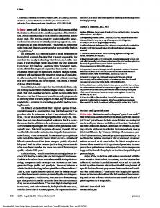
FIG. 3. Relative numbers of spirochetes in infected mouse joints. The animals used for this analysis were the animals described in the legend to Fig. 1. DNA was isolated from one ankle joint of each mouse and then analyzed by PCR for OspA sequences. Sequences from the mouse gene nidogen were amplified to ensure approximately equal loading of DNA into reaction mixtures. The lane labeled BSK shows the lack of OspA sequences in DNA from a mock-infected mouse, which was sacrificed and whose DNA was prepared simultaneously with DNA from infected mice. Lanes 1 to 4 and 5 to 8 show relative OspA levels in the joints of infected C3H/HeJ mice and BALB/c mice, respectively. Lane 9 had a reaction mixture with no DNA present to ensure the purity of PCR reagents. OspA amplification was for 30 cycles.
FIG. 1. Urinary nitrite levels in infected mice. Urine was collected daily from C3H/HeJ mice (closed squares) or BALB/c mice (open circles) and analyzed for nitrite levels as described in Materials and Methods. Mice were infected with 2 3 105 B. burgdorferi cells on day 0. Baseline levels of urinary nitrite were determined for 6 days prior to infection. Nitrite levels were determined from samples of urine from four mice of the same strain, all of which were housed in the same metabolic cage. Values are averages of pooled urinary nitrite based on urine volumes and numbers of mice per cage.
(data not shown and references 7 and 18). C3H/HeJ and BALB/c mice were housed in metabolic cages and were fed a nitrite/nitrate-free diet. Urine samples from these mice were collected daily starting 6 days before infection with B. burgdorferi. Ankles were measured once a week. The level of urinary nitrate (measured as nitrite after conversion with bacterial nitrate reductase) dramatically rose between days 7 and 10 postinfection in both BALB/c and C3H/ HeJ mice (Fig. 1). Nitrate production in BALB/c mice remained elevated, while nitrate production in C3H/HeJ mice dropped after 12 days to just above uninfected levels. This difference in nitrate production in infected mice was correlated
FIG. 2. Ankle swelling of infected mice housed in metabolic cages (mice described in the legend to Fig. 1). Ankle measurements were taken with metric calipers. Closed squares, C3H/HeJ; open circles, BALB/c. Values are the means 6 the standard deviations for all mice at each time point.
with a difference in the severity of arthritis. C3H/HeJ mice exhibited greater ankle swelling at 3 weeks postinfection than did BALB/c mice (Fig. 2). Ankle swelling measurements of arthritis severity were confirmed by histological analysis (data not shown [described in reference 62]). The urinary nitrate levels in infected mice were assessed in three separate experiments for C3H/HeJ mice and two separate experiments for BALB/c mice with similar results. PCR was employed to determine the relative levels of spirochete DNA in hearts and rear ankle joints from the infected mice shown in Fig. 1 and 2 at the week-4 sacrifice. This technique has been used for relative quantification of spirochete numbers in infected mouse tissue and demonstrated the rear ankle joint and heart to be heavily and persistently infected (63). Furthermore, C3H/HeJ mice were found to harbor relatively more B. burgdorferi than BALB/c mice at many time points following infection, with quantitative PCR demonstrating 5- to 10-fold more organisms in the tissues of C3H/HeJ mice than tissues of BALB/c mice at 2 weeks postinfection. Malawista et al. (40) have recently demonstrated a strong correlation between tissues positive for B. burgdorferi infection by PCR and culture positivity; no evidence for the persistence of spirochete DNA in the absence of viable spirochetes was found. The results shown in Fig. 3 at 4 weeks after infection clearly demonstrate a greater intensity of OspA PCR product in DNA from infected C3H/HeJ joints than in that from infected BALB/c joints (63). Similar results were obtained with DNA prepared from the hearts of infected animals (data not shown). Thus, the constraints of the metabolic cage and the special nitrite/nitrate-free diet did not alter the course of B. burgdorferi infection in these mice. The persistence of high levels of urinary nitrate in infected BALB/c mice and its disappearance from C3H/HeJ mice suggested that the high levels of nitric oxide production in vivo were responsible for the lower number of spirochetes found in BALB/c tissues. Conversely, the shutdown of NO in C3H/HeJ mice suggested that a lack of NO production could better allow spirochetes to grow and infect the joints. This hypothesis was directly tested by inhibiting NO production in infected mice of each strain. The inhibitor of NO synthase LMMA was administered to mice twice per day at a dose causing almost complete blockage of NO but not causing generalized toxicity (7, 18). C3H/HeJ mice were allowed to adjust to a nitrite/nitrate-free diet for at least 5 days before infection. The mice were divided into three experimental groups: (i) mock
VOL. 63, 1995
FIG. 4. Suppression of urinary nitrite by LMMA. C3H/HeJ mice given distilled water (closed circles) or C3H/HeJ mice given LMMA (open squares) were housed in metabolic cages, and urine was collected daily for assessment of urinary nitrite levels. Mice were infected with 2 3 103 B. burgdorferi cells on day 0. Baseline levels of urinary nitrite were determined for 3 days prior to infection. Animals were given LMMA or distilled water twice daily by gavage starting approximately 16 h after infection. Administration of LMMA significantly inhibited nitrite production. Values are averages of urinary nitrite based on urine volumes and numbers of mice per cage.
infected, (ii) infected with B. burgdorferi and gavaged twice a day with distilled H2O beginning 24 h postinfection, and (iii) infected with B. burgdorferi and gavaged twice a day with LMMA beginning 24 h postinfection. One mock-infected mouse and four each of the H2O-treated and LMMA-treated mice were sacrificed at weekly time points. Urine was collected daily for assessment of endogenous nitrate production (18). Figure 4 shows that administration of the inhibitor LMMA to infected C3H/HeJ mice suppressed endogenous production of nitrate to uninfected levels, even in infected mice. Overall, LMMA-treated C3H/HeJ mice appeared to be as healthy as H2O-treated infected mice and showed no observable negative side effects of treatment with the inhibitor. The severity of arthritis was assessed by standard histological analysis of one rear ankle joint (the other being used for PCR analysis [see below]). Joints were assessed from the weekly sacrifices, and representative slides were examined in a blinded fashion. Ankles taken at 4 weeks postinfection displayed the most severe inflammation and are shown in Fig. 5. No differences in histological severity between H2O- and LMMAtreated infected C3H/HeJ mice were seen at any time point. The effect of inhibiting NO production on spirochete number in tissues was assessed by PCR (63). DNA was isolated from one rear ankle joint and the heart of each mouse, as the mice were sacrificed at regular intervals following infection. These two tissues were chosen because both have been previously demonstrated by PCR to be heavily infected with spirochetes (63). OspA sequences were amplified to assess relative levels of spirochetes in tissues, and sequences from the mouse nidogen gene were amplified to ensure approximately equal DNA loading. Figure 6 shows the comparison of spirochete infections in the joints of H2O- and LMMA-treated C3H/HeJ mice. Spirochetes were not detected in the joints until 2 weeks after infection. There was no significant difference in the numbers of
NO PRODUCTION DURING MURINE LYME DISEASE
3889
spirochetes found in H2O-treated or LMMA-treated animals at week 1, 2, or 3 postinfection. At 4 weeks postinfection, there were somewhat fewer spirochetes in the joints of LMMAtreated animals. PCR comparison of spirochete sequences in the hearts of LMMA- and water-treated mice at each time point displayed little effect of the inhibitor (shown in Fig. 6), suggesting that the lack of effect of LMMA on ankle infection by B. burgdorferi reflected the systemic effects of inhibiting NO production (data not shown). In a separate experiment, the effect of inhibiting NO production in infected BALB/c mice was determined. This strain maintains high levels of NO throughout 4 weeks of infection and is relatively resistant to arthritis and more effective at regulating spirochete number. The effect of NO inhibition early in infection, prior to the appearance of specific immunity, was of particular interest in this experiment. Therefore, mice were housed in conventional cages, so that more experimental time points could be included. Assessment of serum nitrates required a volume of serum that could be obtained only by sacrificing the mice; thus, samples were taken from individual mice (two to four, depending on recovery of adequate serum volumes) at 4, 7, 10, 14, 21, and 28 days (Fig. 7). These results indicate a trend similar to urinary nitrate (Fig. 1) and demonstrate complete inhibition by orally administered LMMA. Serum nitrate is a measure of the level of nitrate in serum at a single time point, whereas urinary nitrate is a measure of the cumulative production of nitrate during a 24-h period. Thus, differences in the appearance of the graphs in Fig. 1 and 7 for BALB/c mice probably reflect the distinct sampling sources, with urinary nitrate levels giving a more complete report of the animal’s exposure. Arthritis was assessed by weekly ankle measurements and by histological analysis of ankles taken from mice at the indicated sacrifice times. There was no difference in ankle swelling over the 4-week course of the experiment. The histology was analyzed in a blinded fashion (tissues at 4 weeks postinfection shown in Fig. 8A and B) and was mild, which is characteristic of infected BALB/c mice. There was no significant difference between water- and LMMA-treated BALB/c groups; thus, inhibition of NO production did not cause an increase in B. burgdorferi-induced arthritis in BALB/c mice. In BALB/c mice, spirochetes could not be detected by PCR until day 10 postinfection, and then only at an extremely high cycle number (40 cycles) (Fig. 9). At day 10, there were barely detectable OspA bands in DNA from LMMA-treated mice and no detectable DNA from water-treated mice; however, at this time point, the presence of spirochetes was low in both groups. At later time points, there were no significant differences in spirochete numbers in the joints of BALB/c mice given H2O or LMMA (Fig. 9). PCR detection of B. burgdorferi DNA in infected hearts provided similar results (data not shown), again indicating that NO was not involved in regulating spirochetes at either site. The DNA samples from every time point in a single experimental group were run together at the same cycle number to determine a time course for OspA levels. Figure 10 shows the course of infection during the experiment, this time with DNA prepared from the hearts of water-treated (Fig. 10A) or LMMA-treated (Fig. 10B) infected C3H/HeJ mice. The spirochete infection was greatest at approximately 2 weeks, with spirochete levels subsequently decreasing. Treatment with LMMA did not alter the time course of infection in either mouse strain. Similar kinetics of infection in water and inhibitor groups were observed with ankles from C3H/HeJ mice and hearts and ankles of BALB/c mice (data not shown). The kinetics in water-treated animals are consistent with results
FIG. 5. Histology of water- and inhibitor-treated C3H/HeJ mice. The mice depicted were two mice (described in the legend to Fig. 4) sacrificed at week 4. (A) Inflammation and hyperproliferation of the synovium characteristic of rear ankle joints of infected C3H/HeJ mice (this section was from an infected C3H/HeJ mouse given water); (B) same degree of inflammation and proliferation from the rear ankle joint of an infected C3H/HeJ mouse given the NO inhibitor LMMA. Histology from all infected C3H/HeJ animals sacrificed at week 4 is represented.
3890
VOL. 63, 1995
NO PRODUCTION DURING MURINE LYME DISEASE
3891
FIG. 7. Suppression of serum nitrate by LMMA treatment of infected BALB/c mice. BALB/c mice infected with 2 3 103 B. burgdorferi cells were given distilled water (closed squares) or LMMA (closed circles) twice daily by gavage starting 16 h after infection. Uninfected control mice (closed triangles) received water gavage. Mice were housed in conventional cages, and levels of nitrate in serum in mice sacrificed at the indicated times postinfection were determined. Recovery of serum nitrates is described in Materials and Methods. Values are averages 6 the standard deviations for each mouse determined from sera collected from two to four mice in each group, depending on recovery of sufficient serum volume.
FIG. 6. Effect of NO inhibition on spirochete numbers in C3H/HeJ mice. Samples are from mice described in the legend to Fig. 4. One BSK-injected mouse (lane 1) and four water-treated (lanes 2 to 4 for day 7 postinfection; lanes 2 to 5 for all other time points) and four LMMA-treated (lanes 5 to 8 for day 7 postinfection; lanes 6 to 9 for all other time points) mice were sacrificed at days 7, 14, 21, and 28 postinfection. One ankle joint was taken for DNA preparation and PCR analysis. No spirochete DNA was detectable in BSK-injected animals or in the water control of the reaction mixture (lane 9 for day 7 postinfection and lane 10 for all other time points), and OspA sequences in infected joints were not detectable until day 14 postinfection. Comparison of relative levels of OspA sequences showed no significant difference in spirochete levels at day 7, 14, or 21 postinfection. There appeared to be slightly fewer sequences present at day 28 postinfection in LMMA-treated animals. OspA was amplified at 35 cycles for day 7 postinfection and at 30 cycles at all other time points.
from previous experiments from our laboratory and those of others (5, 63). NO has been reported to inhibit the development of some immune responses (20). To determine if there was an effect of inhibiting NO production on the appearance of Borreliaspecific IgG or IgM in infected mice, ELISAs with sonicated B. burgdorferi as antigen were performed. IgG and IgM were detected individually, and concentrations were estimated from a standard curve. No significant differences were seen in IgM levels with either mouse strain. LMMA had no effect on IgG production in C3H/HeJ mice, although it did cause a slight reduction in IgG production by infected BALB/c mice (Fig. 11). DISCUSSION Several previous studies led to the hypothesis that inflammation-induced NO was involved in Lyme disease. These studies suggested that NO could be implicated in either the host defense to B. burgdorferi infection or in the pathological events
associated with arthritis development. Observations supporting involvement of NO in host defenses are as follows: (i) B. burgdorferi stimulates NO production in vitro (37, 58) and is susceptible to killing by NO gas in vitro (37, 44); (ii) many reports have found that B. burgdorferi can enter cells such as endothelial cells and macrophages and persist for several days (11, 34, 38, 45, 55); and (iii) the murine host defense against true intracellular pathogens does require NO production, either early in infection as established for Listeria monocytogenes, or throughout infection as with Leishmania major (16, 18, 36). The observations supporting involvement of NO in the pathology of arthritis are from studies with other animal models of arthritis; NO presence is essential for the development of streptococcal cell wall arthritis in rats (42) and autoimmune arthritis in lpr/lpr mice (59). Further, transgenic mice constitutively producing tumor necrosis factor alpha, which is an inducer of iNOS gene expression, spontaneously develop arthritis (31). The initial observations in this study were made with mice housed in metabolic cages and analyzed for daily urinary nitrate production. This study (shown in Fig. 1 to 3) supported involvement of NO in control of B. burgdorferi infection. BALB/c mice had a sustained elevation of NO production throughout infection. Infected C3H/HeJ mice initially produced NO in response to infection; however, this rapidly returned to baseline levels by 2 weeks postinfection. B. burgdorferi presence in tissues was lower in BALB/c mice than in infected C3H/HeJ mice at the 4-week time point of sacrifice (Fig. 2). This mouse strain difference in infection was previously established with quantitative PCR as characteristic of the entire time course of infection in the two strains of mice (63). The drop in NO to baseline levels observed at 2 weeks postinfection with C3H/HeJ mice in Fig. 1 was concurrent with the peak in infection of this strain of mice, as established in reference 63 and confirmed in the analysis shown in Fig. 10, in which DNA from infected C3H/HeJ mice sacrificed at different time points was subjected to PCR analysis. The correlation between the drop in NO production and the peak in B. burgdorferi infection further supported the concept that NO was part of the host defense to this organism.
FIG. 8. Effect of NO inhibition on severity of arthritis in infected BALB/c mice. Two of the mice described in the legend to Fig. 7 and sacrificed at week 4 are depicted. (A) Mild inflammation characteristic of rear ankle joints of infected BALB/c mice gavaged twice daily with water; (B) similar inflammation from the rear ankle joint of an infected BALB/c mouse given the NO inhibitor LMMA. Histology from all infected BALB/c animals sacrificed at week 4 is represented.
3892
VOL. 63, 1995
NO PRODUCTION DURING MURINE LYME DISEASE
3893
FIG. 10. Appearance of spirochete DNA in infected hearts was not altered by LMMA treatment. Hearts from the infected C3H/HeJ animals described in the legend to Fig. 4 were taken, and DNA was isolated for PCR analysis. Each time point sample was run on the same gel to visualize the time course of infection and relative spirochete numbers. OspA sequences were undetectable until day 14 postinfection (lanes 6 to 9), at which time the number of spirochetes in the heart was maximal. The number of spirochetes gradually began to decrease with time after infection (lanes 10 to 21). No OspA sequences were detectable in the BSK-injected mouse (lane 1) or in the reaction mixture control (lane 22). OspA was amplified at 30 cycles. (A) Water-treated mice; (B) inhibitor-treated mice. dH2O, distilled water.
We also hypothesized that NO could contribute to pathological developments in C3H/HeJ mice, in which elevation of inflammatory cytokines could be greater and of longer duration because of the greater degree of spirochete infection in tissues. Previously, we had found elevation of IL-6 in infected C3H/HeJ mice through 4 weeks of infection (62). In C3H/HeJ mice, we predicted that blocking the spike of NO would result
FIG. 9. Effect of NO inhibition on numbers of spirochetes in BALB/c mice. The mice used in this experiment are described in the legend to Fig. 5B. One BSK-injected mouse (lane 1) and four water-treated (lanes 2 to 5) and four LMMA-treated (lanes 6 to 9) mice were sacrificed at days 4, 7, 10, 14, 21, and 28 postinfection. One ankle joint was taken for DNA preparation and PCR analysis. No spirochete DNA was detectable in BSK-injected animals or in the water control of the reaction mixture (lane 10), and OspA sequences in infected joints were not detectable until day 10 postinfection. Comparison of relative levels of OspA sequences showed no significant difference in spirochete levels at any time point after infection. OspA sequences were amplified for 40 cycles in the day 4 and 7 postinfection samples and for 32 cycles at the other time points.
From the experiment displayed in Fig. 1 to 3, we predicted that NO was a crucial component of the defense of BALB/c mice against B. burgdorferi and that blocking NO production would critically affect the outcome of disease in BALB/c mice. The synthetic analog of L-arginine, LMMA, is thought to function as a competitive inhibitor of the NOS enzymes (7, 18). The dose that was given, 120 mM per day, effectively blocks NO production by iNOS, while having little effect on the other biosynthetic pathways involving arginine, such as protein synthesis (7). Mice received LMMA twice daily by gavage, which ensured that each animal received the same dosage. We predicted that inhibitor-treated BALB/c mice would display more severe infection of ankle joints and hearts and more severe arthritis, similar to that seen with infected C3H/HeJ mice. This did not occur. Spirochete infection in NO-inhibited BALB/c mice was essentially the same as that in control mice at early and late time points (Fig. 9), and arthritis severity was unaffected (Fig. 8).
FIG. 11. IgG and IgM levels in infected mice given distilled water or LMMA. BALB/c mice (A and B) and C3H/HeJ mice (C and D) were infected with 2 3 103 B. burgdorferi cells (closed symbols) or were injected with an equal volume of BSK medium (open squares). Serum was taken at every sacrifice from each mouse and was assessed for IgG and IgM specific for Borrelia antigens. Closed circles, animals treated with distilled water twice daily by gavage; closed triangles, animals treated with LMMA twice daily by gavage. Administration of the NO inhibitor did not significantly affect production of antibody to Borrelia antigens. Values are averages of Ig levels in all mice sacrificed at a single time point.
3894
SEILER ET AL.
in less-severe arthritis. Again, this was not the case, since the histological severity of arthritis was the same in mice receiving the inhibitor of NO production (Fig. 5). Inhibiting the spike in NO production also did not alter the course of B. burgdorferi infection in C3H/HeJ mice, as determined by PCR detection of B. burgdorferi DNA in ankle joints (Fig. 6) and hearts (Fig. 10). This lack of effect of inhibiting NO production in Lyme disease is striking in comparison with an infection in which NO is a crucial component of the host’s defense. For example, in murine leishmaniasis, the mouse strain that is resistant to Leishmania infection is C3H, the mouse strain that is most susceptible to infection with B. burgdorferi. This contrast is carried further with our analysis of urinary nitrate levels. C3H mice maintain high levels of NO throughout the cure phenotype characteristic of their infection with L. major (16). L. major-infected BALB/c mice fail to produce NO, and these animals succumb to infection (16, 36). The contrast is now further extended, since blocking NO production in L. majorinfected C3H mice alters the outcome to resemble that of the susceptible phenotype (BALB/c-like), and the animals die of overwhelming infection. Inhibiting NO production in either BALB/c or C3H/HeJ mice infected with B. burgdorferi had virtually no effect on the clinical course of disease, which is in complete contrast to the L. major model. In another infectious model, mice infected with Toxoplasma gondii, a known stimulant of NO production, display greater resistance to coinfection with intracellular pathogens but no increased resistance to coinfection by extracellular pathogens (23). The importance of NO in controlling infection by viruses, another type of pathogen that replicates within cells, has been recently shown. The gamma interferon-induced inhibition of replication of ectromelia, vaccinia virus, and herpes simplex type 1 viruses in murine macrophages is dependent on NO (12, 30). Furthermore, NO-blocking studies in mice have established that the host defense to ectromelia is dependent on NO in vivo (30). These studies with other pathogens support our interpretation that NO is not an effector molecule for control of B. burgdorferi infection. The facts that infected mice make large amounts of NO and that NO effectively kills B. burgdorferi in culture suggest that these spirochetes are not in the intracellular environment necessary for exposure to the high concentrations of NO required for cytostasis of pathogens. The persistent infection caused by B. burgdorferi has led to speculation that this organism is sequestered in an intracellular location. Importantly, no study has conclusively demonstrated that B. burgdorferi achieves an intracellular site in vivo or can multiply within mammalian cells (11, 34, 38, 45, 55). Our findings support the concept that an intracellular niche is not an essential part of B. burgdorferi infection and persistence. Thus, while B. burgdorferi are susceptible to NO-mediated cytostasis in vitro, they are resistant to the intracellular NO defense mechanism in vivo. Additionally, antibody given at the time of infection can be protective; however, these same antibodies given after infection is established or arising from infection are ineffective at clearing the organism (17, 28, 53). Therefore, B. burgdorferi is also not dealt with effectively in vivo by the classical defenses against extracellular pathogens to which it also succumbs in vitro, i.e., antibody, complement, and opsonophagocytosis by neutrophils and macrophages (10, 35, 49, 50). The paradox of susceptibility to these extracellular defenses in vitro but not in vivo is thought to be crucial to the persistence of infection caused by B. burgdorferi. This must now be extended to the in vivo resistance of B. burgdorferi to NO. B. burgdorferi either must be sequestered in a niche protected from both intracellular and extracellular defenses, or it must produce evasive molecules upon entrance into the mammalian
INFECT. IMMUN.
host which permit survival. These studies point out that this organism possesses a powerful adaptive mechanism for survival that is yet to be defined. ACKNOWLEDGMENTS We thank Chem-Biochem Research, Inc. for LMMA, and Reed Taintor and Renee Christensen for assistance in these studies. This work was supported by grants AI-32223 (J.J.W.) and CA-58248 (J.B.H.) from the National Institutes of Health, by the Department of Veterans Affairs, Washington, D.C. (J.B.H.), and by grant CA-42014 to the University of Utah. REFERENCES 1. Adams, L. B., S. G. Franzblau, Z. Vavarin, J. B. Hibbs, Jr., and J. L. Krahenbuhl. 1991. L-Arginine dependent macrophage effector functions inhibit metabolic activity of Mycobacterium leprae. J. Immunol. 147:1642–1646. 2. Adams, L. B., J. B. Hibbs, Jr., R. R. Taintor, and J. L. Krahenbuhl. 1990. Microbiostatic effect of murine macrophages for Toxoplasma gondii: role of synthesis of inorganic nitrogen oxides from L-arginine. J. Immunol. 144: 2725–2729. 3. Barbour, A. G. 1984. Isolation and cultivation of Lyme disease spirochetes. Yale J. Biol. Med. 57:521. 4. Barthold, S. W., D. S. Beck, G. M. Hansen, G. A. Terwilliger, and K. D. Moody. 1990. Lyme borreliosis in selected strains and ages of laboratory mice. J. Infect. Dis. 162:133–138. 5. Barthold, S. W., D. H. Persing, A. L. Armstrong, and R. A. Peeples. 1991. Kinetics of Borrelia burgdorferi dissemination and evolution of disease after intradermal inoculation of mice. Am. J. Pathol. 139:263–273. 6. Beck, G., J. Benach, and G. S. Habicht. 1989. Isolation of interleukin 1 from joint fluids of patients with Lyme disease. J. Rheumatol. 16:800–806. 7. Boockvar, K. S., D. L. Granger, R. M. Poston, M. Maybodi, M. K. Washington, J. B. Hibbs, Jr., and R. L. Kurlander. 1994. Nitric oxide produced during murine listeriosis is protective. Infect. Immun. 62:1089–1100. 8. Brandt, M. E., B. S. Riley, J. D. Radolf, and M. V. Norgard. 1990. Immunogenic integral membrane proteins of Borrelia burgdorferi are lipoproteins. Infect. Immun. 58:983–991. 9. Burgdorfer, W., A. G. Barbour, S. F. Hayes, J. L. Benach, E. Grunwaldt, and J. P. Davis. 1982. Lyme disease—a tick-borne spirochetosis? Science 216: 1317–1319. 10. Coleman, J. L., R. C. Rogers, and J. L. Benach. 1992. Selection of an escape variant of Borrelia burgdorferi by use of bactericidal monoclonal antibodies to OspA. Infect. Immun. 60:3098–3104. 11. Comstock, L. E., and D. D. Thomas. 1991. Characterization of Borrelia burgdorferi invasion of cultured endothelial cells. Microb. Pathog. 10:137– 148. 12. Croen, K. D. 1993. Evidence for an antiviral effect of nitric oxide. J. Clin. Invest. 91:2446–2452. 13. Defosse, D. L., and R. C. Johnson. 1992. In vitro and in vivo induction of tumor necrosis factor alpha by Borrelia burgdorferi. Infect. Immun. 60:1109– 1113. 14. Ding, A. H., C. F. Nathan, and D. J. Stuehr. 1988. Release of reactive nitrogen intermediates and reactive oxygen intermediates from mouse peritoneal macrophages. J. Immunol. 141:2407–2412. 15. Drapier, J.-C., J. Wietzerbin, and J. B. Hibbs, Jr. 1988. Interferon-g and tumor necrosis factor induce the L-arginine-dependent cytotoxic effector mechanism in murine macrophages. Eur. J. Immunol. 18:1587–1592. 16. Evans, T. G., L. Thai, D. L. Granger, and J. B. Hibbs, Jr. 1993. Effect of in vivo inhibition of nitric oxide production in murine leishmaniasis. J. Immunol. 151:907–915. 17. Fikrig, E., S. W. Barthold, F. S. Kantor, and R. A. Flavell. 1990. Protection of mice against the Lyme disease agent by immunizing with recombinant OspA. Science 250:553–555. 18. Granger, D. L., J. B. Hibbs, Jr., and L. M. Broadnax. 1991. Urinary nitrate excretion in relation to murine macrophage activation. J. Immunol. 146: 1294–1302. 19. Green, S. J., M. S. Meltzer, J. B. Hibbs, Jr., and C. A. Nacy. 1990. Activated macrophages destroy intracellular Leishmania major amastigotes by an Larginine dependent killing mechanism. J. Immunol. 144:278–283. 20. Gregory, S. H., E. J. Wing, R. A. Hoffman, and R. L. Simmons. 1993. Reactive nitrogen intermediates suppress the primary immunologic response to Listeria. J. Immunol. 150:2901–2909. 21. Habicht, G. S., G. Beck, J. L. Benach, J. L. Coleman, and K. D. Leichtling. 1985. Lyme disease spirochetes induce human and murine interleukin-1 production. J. Immunol. 134:3147–3154. 22. Habicht, G. S., L. I. Katona, and J. L. Benach. 1991. Cytokines and the pathogenesis of neuroborreliosis: Borrelia burgdorferi induces glioma cells to secrete interleukin-6. J. Infect. Dis. 164:568–574. 23. Hibbs, J. B., Jr., J. S. Remington, and C. C. Carelton. 1980. Modulation of immunity and host resistance by micro-organisms. Pharmacol. Ther. 8:37–69.
VOL. 63, 1995 24. Hibbs, J. B., Jr., R. R. Taintor, and Z. Vavrin. 1987. Macrophage cytotoxicity: role of L-arginine deiminase activity and imino nitrogen oxidation to nitrite. Science 235:473–476. 25. Hibbs, J. B., Jr., R. R. Taintor, Z. Vavrin, and E. M. Rachlin. 1988. Nitric oxide: a cytotoxic activated macrophage effector molecule. Biochem. Biophys. Res. Commun. 157:87–94. 26. Hibbs, J. B., Jr., Z. Vavrin, and R. R. Taintor. 1987. L-Arginine is required for expression of the activated macrophage effector mechanism causing selective metabolic inhibition in target cells. J. Immunol. 138:550–565. 27. Honarvar, N., U. E. Schaible, C. Galanos, R. Wallich, and M. M. Simon. 1994. A 14,000 MW lipoprotein and a glycolipid-like structure of Borrelia burgdorferi induce proliferation and immunoglobulin production in mouse B cells at high frequencies. Immunology 82:389–396. 28. Johnson, R. C., C. Kodner, M. Russell, and P. H. Duray. 1988. Experimental infection of the hamster with Borrelia burgdorferi. Ann. N. Y. Acad. Sci. 539:258–263. 29. Johnson, R. C., G. P. Schmid, F. W. Hyde, A. G. Steigerwalt, and D. J. Brenner. 1984. Borrelia burgdorferi sp. nov.: etiologic agent of Lyme disease. Int. J. Syst. Bact. 34:496–497. 30. Karupiah, G., Q.-W. Xie, R. M. L. Buller, C. Nathan, C. Duarte, and J. D. MacMicking. 1993. Inhibition of viral replication by interferon-g-induced nitric oxide synthase. Science 261:1445–1448. 31. Keffler, J., L. Probert, H. Cazlaris, S. Georgopoulos, E. Kaslaris, D. Kioussis, and G. Kollias. 1991. Transgenic mice expressing human tumour necrosis factor: a predictive genetic model of arthritis. EMBO J. 10:4025–4031. 32. Kenefick, K. B., J. A. Lederer, R. F. Schell, and C. J. Czuprynski. 1992. Borrelia burgdorferi stimulates release of interleukin-1 activity from bovine peripheral blood monocytes. Infect. Immun. 60:3630–3634. 33. Kenefick, K. B., L. C. L. Lim, J. D. Alder, J. L. Schmitz, C. J. Czurprynski, and R. F. Schell. 1993. Induction of interleukin-1 release by high and low passage isolates of Borrelia burgdorferi. J. Infect. Dis. 167:1086–1092. 34. Klempner, M. S., R. Noring, and R. A. Rogers. 1992. Invasion of human skin fibroblasts by the Lyme disease spirochete Borrelia burgdorferi. J. Infect. Dis. 167:1074–1081. 35. Kochi, S. K., R. C. Johnson, and A. P. Dalmasso. 1991. Complementmediated killing of the Lyme disease spirochete Borrelia burgdorferi: role of antibody in formation of an effective membrane attack complex. J. Immunol. 146:3964–3970. 36. Liew, F. Y., S. Millott, C. Parkinson, R. M. J. Palmer, and S. Moncada. 1990. Macrophage killing of Leishmania parasite in vivo is mediated by nitric oxide from L-arginine. J. Immunol. 144:4794–4797. 37. Ma, Y., K. P. Seiler, K. Tai, L. Yang, M. Woods, and J. J. Weis. 1994. Outer surface lipoproteins of Borrelia burgdorferi stimulate nitric oxide production by the cytokine-inducible pathway. Infect. Immun. 62:3663–3671. 38. Ma, Y., A. Sturrock, and J. J. Weis. 1991. Intracellular localization of Borrelia burgdorferi within human endothelial cells. Infect. Immun. 59:671–678. 39. Ma, Y., and J. J. Weis. 1993. Borrelia burgdorferi outer surface lipoproteins OspA and OspB possess B-cell mitogenic and cytokine-stimulatory properties. Infect. Immun. 61:3843–3853. 40. Malawista, S. E., S. W. Barthold, and D. H. Persing. 1994. Fate of Borrelia burgdorferi DNA in tissues of infected mice after antibiotic treatment. J. Infect. Dis. 170:1312–1316. 41. Mayer, J., M. L. Woods, Z. Vavrin, and J. B. Hibbs, Jr. 1993. Gamma interferon-induced nitric oxide production reduces Chlamydia trachomatis infectivity in McCoy cells. Infect. Immun. 61:491–497. 42. McCartney-Francis, N., J. B. Allen, D. E. Mizel, J. E. Albina, Q.-W. Xie, C. F. Nathan, and S. M. Wahl. 1993. Suppression of arthritis by an inhibitor of nitric oxide synthase. J. Exp. Med. 178:749–754. 43. Miller, L. C., S. Isa, E. Vannier, K. Georgilis, A. C. Steere, and C. A. Dinarello. 1992. Live Borrelia burgdorferi preferentially activate interleukin-1b gene expression and protein synthesis over the interleukin-1 receptor antagonist. J. Clin. Invest. 90:906–912. 44. Modolell, M., U. E. Schaible, M. Rittig, and M. M. Simon. 1994. Killing of Borrelia burgdorferi by macrophages is dependant on oxygen radicals and nitric oxide and can be enhanced by antibodies to outer surface proteins of the spirochete. Immun. Lett. 40:139–146.
NO PRODUCTION DURING MURINE LYME DISEASE
3895
45. Montgomery, R. R., M. H. Nathanson, and S. E. Malawista. 1993. The fate of Borrelia burgdorferi, the agent for Lyme disease, in mouse macrophages. J. Immunol. 150:909–915. 46. Nocton, J. J., F. Dressler, B. J. Rutledge, P. N. Rys, D. H. Persing, and A. C. Steere. 1994. Detection of Borrelia burgdorferi DNA by polymerase chain reaction in synovial fluid from patients with Lyme arthritis. N. Engl. J. Med. 330:229–234. 47. Radolf, J. D., L. L. Arndt, D. R. Akins, L. L. Curetty, M. E. Levi, Y. Shen, L. S. Davis, and M. V. Norgard. 1995. Treponema pallidum and Borrelia burgdorferi lipoproteins and synthetic lipopeptides activate monocytes/macrophages. J. Immunol. 154:2866–2877. 48. Radolf, J. D., M. V. Norgard, M. E. Brandt, R. D. Isaacs, P. A. Thompson, and R. Beutler. 1991. Lipoproteins of Borrelia burgdorferi and Treponema pallidum activate cachectin/tumor necrosis factor synthesis. J. Immunol. 147: 1968–1974. 49. Rittig, M. G., A. Krause, T. Haupl, U. E. Schaible, M. Modelell, M. D. Kramer, E. Lutjen-Drecoll, M. M. Simon, and G. R. Burmester. 1992. Coiling phagocytosis is the preferential phagocytic mechanism for Borrelia burgdorferi. Infect. Immun. 60:4205–4212. 50. Sadziene, A., P. A. Rosa, P. A. Thompson, D. M. Hogan, and A. G. Barbour. 1992. Antibody-resistant mutants of Borrelia burgdorferi: in vitro selection and characterization. J. Exp. Med. 176:799–809. 51. Sigal, L. H. 1989. Lyme disease, 1988: immunologic manifestations and possible immunopathogenetic mechanisms. Semin. Arthritis Rheum. 18:153–167. 52. Sigal, L. H., A. C. Steere, and J. M. Dwyer. 1988. In vivo and in vitro evidence of B cell hyperactivity during Lyme disease. J. Rheumatol. 15:648–654. 53. Simon, M. M., U. E. Schaible, M. D. Kramer, C. Eckerskorn, C. Museteanu, H. K. Muller-Hermelink, and R. Wallich. 1991. Recombinant outer surface protein A from Borrelia burgdorferi induces antibodies protective against spirochetal infection in mice. J. Infect. Dis. 164:123–132. 54. Steere, A. C. 1989. Lyme disease. N. Engl. J. Med. 321:586–596. 55. Szczepanski, A., M. B. Furie, J. L. Benach, B. P. Lane, and H. B. Fleit. 1990. Interaction between Borrelia burgdorferi and endothelium in vitro. J. Clin. Invest. 85:1637–1647. 56. Tai, K.-F., Y. Ma, and J. J. Weis. 1994. Normal human B lymphocytes and mononuclear cells respond to the mitogenic and cytokine-stimulatory activities of Borrelia burgdorferi and its lipoprotein OspA. Infect. Immun. 62:520– 528. 57. Tan, S. S., and J. H. Weis. 1992. Development of a sensitive reverse transcriptase PCR assay, RT-RPCR, utilizing rapid cycle times. PCR Methods Applications 2:137–143. 58. Tatro, J. B., L. I. Romero, D. Beasley, A. C. Steere, and S. Reichlin. 1994. Borrelia burgdorferi and Escherichia coli lipopolysaccharides induce nitric oxide and IL-6 production in cultured rat brain cells. J. Infect. Dis. 169: 1014–1022. 59. Weinberg, J. B., D. L. Granger, D. S. Pisetsky, M. F. Seldin, M. A. Misukonis, S. N. Mason, A. M. Pippen, P. Ruiz, E. R. Wood, and G. S. Gilkeson. 1994. The role of nitric oxide in the pathogenesis of spontaneous murine autoimmune disease: increased nitric oxide production and nitric oxide synthase expression in MRL-lpr/lpr mice, and reduction of spontaneous glomerulonephritis and arthritis by orally administered NG-monomethyl-L-arginine. J. Exp. Med. 179:651–660. 60. Weis, J. J., Y. Ma, and L. F. Erdile. 1994. Biological activities of native and recombinant Borrelia burgdorferi outer surface protein A: dependence on lipid modification. Infect. Immun. 62:4632–4636. 61. Whitmire, W. M., and C. F. Garon. 1993. Specific and nonspecific responses of murine B cells to membrane blebs of Borrelia burgdorferi. Infect. Immun. 61:1460–1467. 62. Yang, L., Y. Ma, R. Schoenfeld, M. Griffiths, E. Eichwald, B. Araneo, and J. J. Weis. 1992. Evidence for B-lymphocyte mitogen activity in Borrelia burgdorferi-infected mice. Infect. Immun. 60:3033–3041. 63. Yang, L., J. H. Weis, E. Eichwald, C. P. Kolbert, D. H. Persing, and J. J. Weis. 1994. Heritable susceptibility to severe Borrelia burgdorferi-induced arthritis is dominant and is associated with persistence of large numbers of spirochetes in tissues. Infect. Immun. 62:492–500.