Sep 5, 1990 - for A/G-Specific Mismatch Repair: Identity of MicA and MutY. JYY-JIH TSAI-WU,' J. .... single EcoRI site (3), fl ALLC3 with a dC-to-dA base substitution, and fl ...... A computer search for homology between this protein and the ...
Vol. 173, No. 6
JOURNAL OF BACTERIOLOGY, Mar. 1991, p. 1902-1910
0021-9193/91/061902-09$02.00/0 Copyright © 1991, American Society for Microbiology
Nucleotide Sequence of the Escherichia coli micA Gene Required for A/G-Specific Mismatch Repair: Identity of MicA and MutY JYY-JIH TSAI-WU,' J. PABLO RADICELLA,2 AND A-LIEN LU1* Department of Biological Chemistry, School of Medicine, University of Maryland at Baltimore, Baltimore, Maryland 21201,1 and Institute of Molecular Biology, University of Oregon, Eugene, Oregon 974032 Received 5 September 1990/Accepted 6 January 1991
The Escherichia coli methylation-independent repair pathway specific for A/G mismatches has been shown to require the gene product of micA. Extracts prepared from micA mutants do not form an A/G mismatchspecific DNA-protein complex and do not contain an A/G mismatch-specific nicking activity. Moreover, a partially purified protein fraction containing both A/G mismatch-specific nicking and binding activities restores repair activity in micA mutant extracts. The DNA sequence of a 2.3-kb fragment containing the micA gene has been determined. There are two open reading frames (ORF) in this DNA fragment: one ORF encodes a 25.7-kDa protein whose function is still unknown, the other ORF codes for a protein with an M, of 39,147, but this ORF can be transcribed and the mRNA can be translated to yield a protein with an apparent Mr of 36 kDa on a sodium dodecyl sulfate-polyacrylamide gel. Deletion analysis showed that this 39.1-kDa ORF is the micA gene as judged by the capacity of the encoded protein to restore the A/G mismatch-specffic nicking activity of micA mutant extracts. Furthermore, our results suggest that micA is the same gene as the closely mapped mutY, which encodes the A/G mismatch-specific glycosylase. In this paper, we show that the A/G mismatch repair pathway is dependent on the micA gene function and involves the A/G mismatch-specific binding and nicking activities. We have also cloned and sequenced the micA gene. Within the cloned DNA fragment, one open reading frame (ORF) for a 39.1-kDa protein defines the micA gene. This ORF is responsible for restoring the A/G mismatch-specific nicking activity in micA as well as mutY mutant extracts in vitro. This result suggests that micA is the same gene as mutY, which codes for the A/G-specific glycosylase. Another ORF upstream of the micA gene is transcribed in the opposite direction and encodes a 25.7-kDa protein whose function is still under investigation.
Mismatch repair directed by adenine (dam) methylation at d(GATC) sequences has been shown to correct most DNA replication errors in Escherichia coli (reviewed in references 7, 25, and 33). This pathway repairs mismatches on the transiently undermethylated daughter strands according to the parental sequences. Gene products of mutH, mutL, and mutS are specifically involved in this repair pathway. In addition, two methylation-independent pathways have been described in E. coli. The T/G-specific pathway repairs T/G mismatches to C/G and is responsible for repairing deaminated 5-methylcytosines (16, 17, 34). An A/G-specific pathway specifically recognizes and repairs A/G mismatches to C/G pairs (18, 32, 38). This repair pathway acts postreplicationally to remove the dA's in A/G mismatches and is not controlled by host mutH, mutL, mutS, uvrE, recF, recJ, recB, recC, recN, mutT, or dnaE gene functions (18, 32, 38, 39a). The repair tract never exceeds 12 nucleotides and the repair synthesis requires DNA polymerase I (39a). Tightly associated A/G mismatch-specific binding and nicking activities have been identified in E. coli cell extracts (19). This pathway involves the action of a DNA glycosylase (2, 19a) followed by excision and resynthesis. The A/G endonuclease makes incisions at the first phosphodiester bond 3' to and the second phosphodiester bond 5' to the apurinic site (19). Two mutators of E. coli, mutY and micA, have been identified independently by two laboratories (28, 32). Both genes mapped to about 64 min on the chromosome, and strains carrying these alleles are defective in A/G-specific repair. In a X heteroduplex assay, the micA mutant is also defective in the correction of dA's in A/C mismatches (32). However, micA- (or mutY)-dependent repair of A/C has not been observed in vitro (18, 38). The MutY protein has been purified and has been shown to be an A/G-specific adenine glycosylase (2). micA and mutY may be the same gene or linked genes involved in the A/G or A/C mismatch repair pathway or in both. *
MATERIALS AND METHODS Bacteria, bacteriophages, and plasmids. E. coli PR8 (or M182; Su- lacZ X74 galU galK Sm) was from E. Signer. Strains PR9 (like PR8 but mutL218::TnJO), PR68 (like PR8 but mutL2J8::TnJO micA68::modified TnJO), and PR70 (like PR8 but micA68::modified TnJO) were constructed in M. S. Fox's laboratory (32). The modified TnJO carries a gene for kanamycin resistance (42). Bacteriophages fl R229 with one single EcoRI site (3), fl ALLC3 with a dC-to-dA base substitution, and fl M28 with a dC-to-dT base substitution at position 5621 have been described before (18, 20). Covalently closed heteroduplexes of fl DNA containing an A/G or T/G mismatch at position 5621 were constructed from the replicative form of fl R229 DNA and single-stranded DNA of fl ALLC3 or fl M28, respectively, as described before (20). A 120-bp HaeII-TaqI fragment (coordinates 5572 to 5671) spanning the A/G or T/G mismatched site was purified from the heteroduplex DNA and 3'-end labeled at the TaqI terminus, using DNA polymerase I Klenow fragment and [a-32P]dCTP (22). A synthetic 40-mer oligonucleotide, 5'AATTGGGCTCCTCGAGGAATTAGCCTTCTGCAGG CATGCC3', was annealed with 5'CCGGGGCATGCCTG CAGAAGGCGAATTCCTCGAGGAGCCC3' to produce a 40-bp oligonucleotide duplex containing an A/G mismatch.
Corresponding author. 1902
VOL. 173, 1991
E. COLI micA GENE FOR A/G-SPECIFIC MISMATCH REPAIR
The resulting oligonucleotide duplex was labeled at the 3' end by using Klenow fragment of DNA polymerase I and [oL-32P]dCTP. The Clarke-Carbon plasmid (6), pLC20-5, was from the E. coli Genetic Stock Center, Department of Biology, Yale University, New Haven, Conn., and the
cloning vector, pT3/T7-2, was purchased from Clontech Laboratories, Inc., Palo Alto, Calif. Mismatch repair assay. The assay scores the conversion of a base-pair mismatch within the EcoRI site of fl heteroduplexes to an intact EcoRI recognition sequence. Repair reactions were performed with E. coli cell extracts prepared as described previously (20). The purified DNA products were digested with EcoRI and BamHI and analyzed by electrophoresis on 1% agarose gels. A/G mismatch-specific nicking assay. E. coli cell extracts used for the nicking assay were prepared as described previously (20) except that the supernatant after ultracentrifugation was used directly or precipitated with 0.277 g of ammonium sulfate per ml. Nicking activity was assayed essentially by the method of Lu and Chang (19). Protein samples were incubated with 0.07 ng (0.9 fmol; 2,000 to 6,000 cpm) of end-labeled 120-bp HaeII-TaqI DNA fragment or a synthesized 40-bp oligonucleotide duplex containing an A/G mismatch in a final volume of 10 pAl. Reactions were carried out in 20 mM Tris hydrochloride (pH 7.6)-80 mM NaCl-1 mM dithiothreitol-1 mM EDTA-2.9% (vol/vol) glycerol for 30 min at 30°C. After incubation, samples were lyophilized and redissolved in 3 ,ul of 90% (vol/vol) formamide-10 mM EDTA-0.1% (wt/vol) xylene cyanol-0.1% (wt/vol) bromophenol blue. DNA was denatured at 90°C for 3 min and applied to a standard 8% polyacrylamide-8.3 M urea sequencing gel for electrophoresis (23). DNA binding analysis. Protein-DNA complexes were analyzed on low-ionic-strength polyacrylamide gels as described before (19). Protein samples were incubated with duplex end-labeled synthetic 40-bp oligonucleotide containing an A/G mismatch as in the nicking assay, except 20 ng of poly(dI-dC) was added. Purification of A/G endonuclease. A/G endonuclease was purified by three chromatographic steps. The first two (phosphocellulose and hydroxylapatite) steps were described by Lu and Chang (19). Fraction IV (23 ml; pooled from the hydroxylapatite column) was dialyzed against 1 liter of buffer A (20 mM KPO4 [pH 7.4], 0.1 mM EDTA, 0.5 mM dithiothreitol, 0.05 M KCl, 0.1 mM phenylmethylsulfonyl fluoride, 10% [vol/vol] glycerol). The dialyzed fraction was applied to a 5-ml heparin-agarose column equilibrated with buffer A and eluted with a 50-ml linear gradient of KCl (0.1 to 0.6 M) in buffer A. Fractions containing the A/G endonuclease activity, which eluted at about 0.38 M KCl, were pooled and dialyzed against buffer A. The dialyzed fraction (fraction V) was stored in small aliquots at -70°C. With these three chromatographic steps, the A/G endonuclease was purified about 1,500-fold. Cloning the micA gene. Since the micA gene in PR70 contained a modified TnJO insertion (32) conferring kanamycin resistance, chromosomal DNA prepared from PR70 (micA68::modified TnJO) was digested with BglII (which fails to cleave the kan gene) and ligated into the BamHI site of pBR322. An ampicillin- and kanamycin-resistant clone containing a 4.8-kb DNA insert with 2.7 kb of chromosome sequence flanking the TnJO was selected (Fig. 1A). The 0.9-kb EcoRI chromosomal fragment adjacent to the transposon DNA was used as a probe to hybridize with the Clarke-Carbon plasmids (6) containing E. coli DNA spanning the 63- to 64-min region of the chromosome. Only a
1903
single 3.5-kb BglII fragment from pLC20-5 hybridized to the probe. This 3.5-kb fragment was purified from an agarose gel by using a GeneClean kit (Bio 101, Inc., La Jolla, Calif.) and cloned into the BamHI site of pBR322 to generate pJTW3-5 and pJTW3-8, which contained the inserts in opposite orientations (Fig. 1B). The micA gene was further subcloned into pT3/T7-2 by cutting pJTW3-5 with SphI. Two clones, pJTW4-20 and pJTW4-21, containing a 2.3-kb DNA insert (including 187 bp of pBR322 sequence; Fig. 1B) were isolated. All of these clones were transformed into PR70, and their extracts were tested for the A/G-specific nicking activity. Unidirectional deletion mutants were produced from pJTW4-20 and pJTW4-21 by using the Stratagene exollI/ mung bean nuclease kit (Stratagene Cloning Systems, La Jolla, Calif.) and following the manufacturer's directions (10). Deletion clones were checked for loss of the ability to restore the A/G mismatch-specific nicking activity of micA mutant (PR70) extracts. DNA sequence determination. Double-stranded plasmid DNAs of pJTW4-20, pJTW4-21, and deletion plasmids (see above) were isolated by passage through Qiagen columns (Qiagen Inc., Studio City, Calif.). The nucleotide sequence of both DNA strands was determined by the dideoxy-chain termination method (36), using Sequenase version 2.0 (United States Biochemical Corp., Cleveland, Ohio). The sequence gaps between deletion plasmids were determined by extension of synthetic oligonucleotide primers. In vivo protein labeling by T7 RNA polymerase system. Because plasmids derived from pJTW4-20 and pJTW4-21 contain the T7 promoter at one end of the inserts, proteins encoded by the inserted DNA can be specifically labeled by the T7 expression system (39). To identify the proteins produced from these DNA fragments, the plasmids were transformed into E. coli K38 harboring plasmid pGP1-2 (39), which contained the T7 RNA polymerase gene under the control of the phage X PL promoter. After heat induction, newly synthesized proteins were labeled with [35S]methionine in the presence of rifampin. The labeled proteins were fractionated on 15% sodium dodecyl sulfate (SDS)-polyacrylamide gels (15) and detected by autoradiography. Nucleotide sequence accession number. The nucleotide sequence reported in this paper has been deposited in the GenBank data base under accession number M59471. RESULTS In vitro mismatch repair in micA mutant extracts. The micA mutant was originally isolated in a mutL Su- background by virtue of its failure to correct X phage DNA heteroduplexes having Pam3 and Pam8O mutations on complementary DNA strands to P+ (32). This mutL micA double mutant (PR68), in a X heteroduplex assay, was defective in a repair pathway that removes the dA's in A/G or A/C mismatches. A micA mutant in a mut+ background (PR70) was constructed and displayed the identical mutator phenotype (32). We have compared the repair activities of extracts from these two micA mutants (PR68 and PR70) with those from the respective isogenic micA+ strains (PR9 and PR8). Phage fl heteroduplex DNA with an A/G mismatch at position 5621, which is repaired well to a C/G base pair by the dam- and mutHLS-independent pathway (18), was used for the in vitro assays. After incubation with cell extracts, the fl DNA was digested with EcoRI and BamHI and analyzed by agarose gel electrophoresis (Fig. 2). When hemimethylated DNA with an A/G mismatch was assayed in
1904
TSAI-WU ET AL.
A S E
Bl
II
I
~ ~I P
E
I
I
J. BACTERIOL.
H m
H
Bl
I
I
m2flmrMrc Ken ---l -11
probe
B
S E
B/BlI
B11/B
P
E
1 I 1
I
I
I I
pJTW3S6
S T3 promoter 1 T7 promoter
T3/T7-2
I
Sphi
Bil/B S E
T3S Bll/B S
T3L.
P
E
I
It
B8
|
Si
S I
E
P
I
lI
39.1kD orf
I
T
pJTW4-21 E S
TL
pJTW4-20
28.7kD orf
1kb
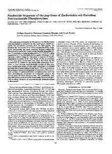
Cloning strategy for the micA gene. (A) Restriction map of the 4.8-kb BgIII fragment from PR70 (micA) chromosome containing a kanamycin resistance gene. This fragment was inserted into the BamHI site of pBR322. The 0.9-kb EcoRI chromosomal DNA fragment (marked as a bar) close to TnlO was isolated as a probe to screen the wild-type gene from the Clarke-Carbon genomic library (6). The modified TnJO DNA is represented by a hatched box with an arrow indicating the kanamycin resistance gene. The chromosomal DNA is shown as an open box. (B) 3.5-kb BglII fragment from pLC20-5 (Clarke-Carbon plasmid containing E. coli chromosomal sequence around 64 min). This fragment was cloned into the BamHI site of pBR322 to generate pJTW3-5 and pJTW3-8. pJTW3-5 was further digested with Sphl, and a 2.3-kb fragment containing 187 bp of pBR322 DNA was subcloned into the multiple cloning sites of vector pT3/T7-2. This step generated pJTW4-20 and pJTW4-21 with the DNA inserts in the opposite orientations. Open boxes represent the chromosomal insert. Filled boxes and thin lines represent pBR322 and pT3/T7-2 DNAs, respectively. The directions of T3 and T7 promoters are indicated by arrows. BI, Bgll; BII, BglII; B/BII, BamHI-BgllI junction; BII/B, BglII-BamHT junction; E, EcoRI; H, HindIII; P, Pstl; S, SphI. FIG.
1.
buffer containing ATP (a condition favorable for the damand mutHLS-dependent repair), the extract from the mutL micA double mutant (PR68) was defective in A/G mismatch repair (Fig. 2, lane 5), whereas the micA (PR70) and mutL (PR9) single mutants were.able to repair AKG mismatches with slightly reduced efficiencies compared with the wild type under the same conditions (Fig. 2, lanes 2 and 4). These observations indicate that there are two independent repair pathways for AKG mismatches: one is the well-characterized mutHLS-dependent pathway, and the other is controlled by micA. It has been shown that the mutHLS-dependent pathway requires ATP and is controlled by dam methylation (20). Mismatch correction is greatly reduced when both DNA strands are methylated. In contrast, the micA-dependent pathway does not require ATP (Fig. 2, lane 11). The A/G repair activity in a micA mutant (PR70) extract is abolished by omitting ATP from the reaction buffer or by using fully methylated DNA as substrate, under which conditions the mutHLS- and dam-dependent pathway is not functional (Fig. 2, lanes 12 and 14). Since the T/G mismatch repair is mainly controlled by the mutHLS-dependent pathway (20,
21), heteroduplex DNA containing a T/G mismatch was used control for the in vitro A/G mismatch repair assay. Hernimethylated DNA containing a T/G mismatch cannot be repaired by mutL (PR9) (Fig. 2, lane 9) and mutL micA (PR68) extracts (Fig. 2, lane 10) but can be repaired by micA (PR70) mutant extract (Fig. 2, lane 8), suggesting that the micA pathway is independent of the mutHLS pathway. Deficiency of A/G mismatch-specific binding and nicking activities in micA mutant extracts. To repair A/G mismatches, the mispaired bases must be recognized so that an endonuclease might act (19). We tested a micA mutant extract for its binding and nicking activities. To assay nicking activity, a 120-bp HaeII-TaqI fragment of fl DNA containing an AKG mismatch was labeled at -the 3' end of the TaqI terminus. This DNA was incubated with protein extracts and fractionated on a denaturing polyacrylamide sequencing gel. In the micA' extracts (PR8 and PR9), the nicking enzyme cut the phosphodiester bond 3' to the mismatched dA (Fig. 3, lanes 1 and 3), whereas this activity was not observed in the PR70 (micA) or PR68 (micA mutL) extracts (Fig. 3, lanes 2 and 4). The A/G-specific binding activity was assayed by the as a
VOL. 173, 1991
* Y__
E. COLI micA GENE FOR A/G-SPECIFIC MISMATCH REPAIR
Extract
Linear -
ATP
Methylatlon
co
n- o%n D-> L 2L L CL >
a.
a.
1 2 3 4 5 6 7 8 9 10 11 12 13
14
1
a
Er
o 0c0 N 0) ( C0 00
o -
co
c
a. oL a:c: acc a.cr-L
_l WS
(JD CO
cc
E
a. a.
0 cc
cc
a
0
2 3 4
5
_
*_
_
.
__ __
-
3.3 kb -_ 3.1 kb -
0
cc
0
1905
.
w __z_
+
+
+
+
+
+
+
/I
+
+
+
-
-
+
+
t+1
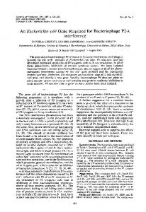
A/G Mismatch A/G T/G FIG. 2. In vitro mismatch repair in micA mutant extracts. The mismatch repair assay (20) used fl heteroduplexes containing an A/G (lanes 1 to 6 and 11 to 14) or T/G (lanes 7 to 10) mismatch at position 5621 within the EcoRI site. Hemimethylated DNA (-/+, lanes 1 to 12) was methylated at d(GATC) sequences on the complementary strand that contained the wild-type EcoRI sequence. Fully methylated heteroduplexes (+/+, lanes 13 and 14) were prepared by in vitro methylation of hemimethylated DNA by using purified dam methylase (11) and S-[methyl-3H]adenosyl-Lmethionine. DNA substrates were incubated with cell extracts (as indicated) with the protein concentration at 7 mg/ml in the presence or absence of ATP (20). After purification, DNA was cleaved by restriction enzymes EcoRI and BamHI, fractionated on 1% agarose gels, and stained with ethidium bromide. Digestion of fl R229 duplex DNA with EcoRI and BamHI yields 3.1- and 3.3-kb fragments. Repair at the mismatched site will restore the EcoRI site; thus, DNA substrate is sensitive to EcoRI cleavage. Lane 1, A/G, -/+, +ATP, PR8; lane 2, AKG, -/+, +ATP, PR70; lane 3, A/G, -/+, +ATP, PR70 plus fraction V; lane 4, AKG, -/+, +ATP, PR9; lane 5, AKG, -/+, +ATP, PR68; lane 6, AKG, -/+, +ATP, PR68 plus fraction V; lane 7, T/G, -/+, +ATP, PR8; lane 8, T/G, -/+, +ATP, PR70; lane 9, T/G, -/+, +ATP, PR9; lane 10, T/G, -/+, +ATP, PR68; lane 11, A/G, -/+, -ATP, PR8; lane 12, AKG, -l+, -ATP, PR70; lane 13, AKG, +/+, +ATP, PR8; lane 14, AKG, +l+, +ATP, PR70.
mobility shift of a labeled 40-bp oligonucleotide duplex containing an A/G mismatch (see Materials and Methods). Using this method, we demonstrated that the micA extract (ammonium sulfate fractions of PR70) failed to form the protein-DNA complex C' (Fig. 4, lane 3), which was evident in the micA+ (PR8) extract prepared in the same manner (Fig. 4, lane 2). As a control, nicking product (Fig. 3, lane 5) and binding complex (Fig. 4, lane 1) were observed with a partially purified fraction V (see Materials and Methods). We conclude, therefore, that both the A/G mismatch-specific nicking and binding activities are absent in the micA mutant extracts.
Complementation of AKG mismatch repair deficiency of the micA mutant extract by the specific nicking and binding activities. Since the micA mutL double mutant is defective in the A/G mismatch repair activity, a complementation assay
FIG. 3. Deficiency of A/G endonuclease activity in micA mutant extracts. A 120-bp HaeII-TaqI fragment with an A/G mismatch at position 5621 and labeled at the 3' end of the TaqI terminus was incubated with different cell extracts (lane 1, PR8; lane 2, PR70; lane
3, PR9; lane 4, PR68) or fraction V (lane 5). Reaction products after denaturation were analyzed on an 8% polyacrylamide DNA sequencing gel. Arrow indicates the cleavage product of the A/G endonuclease. The micA mutant extracts (PR70 and PR68) have no A/G endonuclease activity.
can be developed to purify the micA gene product. To avoid the interference from the mutHLS-dependent repair pathway, the purification of the MicA protein was conducted by using a mutL cell extract. The cell extract from PR9 (mutL) was fractionated on a phosphocellulose column, and fractions were assayed for A/G mismatch nicking activity and their ability to complement the repair deficiency of the micA mutL (PR68) double mutant extract. The A/G mismatch nicking activity and the putative micA gene product (i.e., the activity to complement the mismatch repair deficiency of the micA mutant) coeluted at 0.3 M KCl (data not shown). We also tested a partially purified preparation containing both the A/G mismatch nicking activity and the A/G binding protein for the ability to complement the repair deficiency of the micA cell extract. Fraction V contained both A/G nicking and binding activities and was purified more than 1,500-fold by three chromatographic steps (see Materials and Methods). The mismatch repair activity of PR68 (micA mutL) extract was restored by addition of fraction V (cf. Fig. 2, lanes 5 and 6). Cloning the micA gene. The micA mutation of strain PR70 was defined by the insertion of a modified TnJO that encoded kanamycin resistance (32). Therefore, micA sequence flanking the TnJO was cloned and used as a probe to identify the intact wild-type micA gene from among the Clarke-Carbon plasmids (Materials and Methods). All being putatively
1906
TSAI-WU ET AL.
J. BACTERIOL. 0 co r-
> Q- L C'
CI
-
D '-..
l 2 _C3
we i;.1..
I
c
-
S E
Nickinc Activitv BI Bit/B PR70(micA) MC10(mut)
E
P
25.7kD orf
39.1kD orf
D311 D415 D513 D511 D212
ND
ND
D916
ND
ND
D203 D501 D603 D703 D604 D601 D902 D103 D1003 kb I-
0
F
-
FIG. 4. Deficiency of A/G binding activity in micA mutant extracts. Binding reaction was carried out as in the legend to Fig. 3 except that a labeled 40-bp synthetic oligonucleotide duplex (see Materials and Methods) with an A/G mismatch was incubated with protein extracts from PR8 (lane 2) and PR70 (lane 3) as well as partially purified fraction V (lane 1) in the presence of 20 ng of poly(dI-dC). DNA, after incubation, was fractionated by electrophoresis through a low-ionic-strength 4% polyacrylamide gel. Several distinct bands (A', B', C', D' and E') with lower mobility than the free DNA (F) were observed. Complex C', which is specific for A/G mismatch-containing DNA, was not seen in the extract of PR70.
positive, the recombinant plasmids were tested further for their ability to complement micA mutant extracts by restoring the A/G-specific nicking activity. Two plasmids, pJTW4-20 and pJTW4-21, which contained a 2.3-kb DNA insert in the opposite orientations (Fig. 1), were able to complement the micA mutation (data not shown). Coding region of the micA gene. To define the coding region of the micA gene in the 2.3-kb DNA insert, a series of unidirectional deletions was constructed from pJTW4-20 and pJTW4-21 (see Materials and Methods) and checked for the ability to complement the A/G nicking activity of micA mutant extracts. Figure 5 shows the deletion map and the capabilities of these deletion plasmids to restore the A/Gspecific nicking activity of both micA and mutY mutant extracts. Clone D311, which was deleted about 150 bp from the junction of chromosomal DNA and pBR322 (BglIIBamHI site), still can complement both mutations, whereas D415, which is deleted about 450 bp from the BglII-BamHI site, complemented both mutations poorly. Plasmids (D513 and D511) with deletions of >600 bp from the BglIIBamHI site no longer complemented both micA and mutY mutations. When the DNA fragment was deleted from the other direction (from the SphI site toward the BglI site), the deletion plasmids (D203, D501, D603, D703, D604, and D601) still complemented both mutants. The ability to restore the A/G-specific nicking was reduced in plasmid D902 and was lost when the deletion reached the second EcoRI site (D103 and D1003). From this analysis, the 0.9-kb EcoRIBglI fragment was determined to be the minimal region that could restore the nicking activity of both micA and mutY mutant extracts in vitro. The results also showed the simi-
0.5
1.0
1.5
2.0
2.5
FIG. 5. Complementation of A/G mismatch-specific nicking activities in micA and mutY mutants with the deletion plasmids. A series of deletion plasmids was constructed by exonuclease III and mung bean nuclease treatment. Plasmids pJTW4-21 and pJTW4-20 were used to delete from BglI and SphI ends, respectively. The deletion plasmids were transformed into either micA or mutY mutants. Extracts derived from these cells (without ammonium sulfate precipitation) were assayed for the A/G-specific nicking activity by using a 40-bp oligonucleotide heteroduplex as described in Materials and Methods. Two ORFs of 25.7 and 39.1 kDa are indicated by arrows. Abbreviations for restriction enzyme sites are the same as those in the legend to Fig. 1. ND, Not determined.
larity in complementation of these deletion mutants in micA and mutY mutants. We concluded that micA and mutY had the same coding region. By restriction endonuclease and DNA sequence analyses, we found that the micA70 mutant clone (Fig. 1A) contained a transposon in the 0.9-kb EcoRI-BglI DNA fragment (data not shown). This result is consistent with the above deletion analysis showing that the micA gene is located in the 0.9-kb EcoRI-BglI DNA fragment. Nucleotide sequence of the micA (mutY) gene. The DNA sequence of the cloned fragment was determined on both strands by the standard chain termination method (36). Two possible ORFs with putative control elements were found in this 2.3-kb fragment (Fig. 6). From the deletion analysis (see above), the minimal coding region of micA (or mutY) was located in the 0.9-kb EcoRI-BglI fragment which contained one ORF (positions 1100 to 2149). This ORF with 350 amino acids would define a 39.1-kDa protein which had an extremely high pl of 9.96 (Fig. 6). Potential -35 and -10 sequences (9) are indicated in Fig. 6. Our deletion analysis indicated that the 39.1-kDa protein was the MicA (or MutY) protein. However, the predicted molecular weight of the protein is different from that of the purified MutY protein estimated by SDS-polyacrylamide gel electrophoresis (2). To identify the proteins produced by this DNA fragment, plasmid D703 (Fig. 5) was transformed into E. coli K38 containing a pACYC177-derived plasmid, pGP1-2, which had the T7 RNA polymerase gene under the control of the phage A PL promoter (39; see Materials and Methods). The 39. 1-kDa ORF in D703 was transcribed in the same direction as the T7 promoter. Using the T7 in vivo transcription-translation system (39), we have observed a [35S]methionine-labeled protein specifically expressed from D703 (Fig. 7, lane 1). This labeled protein migrated at the
E. COLI micA GENE FOR A/G-SPECIFIC MISMATCH REPAIR
VOL. 173, 1991
SphI 1
--
1-
-
-------
----__
--_
-_
----------
----------
---------
1907
G R K T D K Y L P W K T P Y Q I D K V W 1141 CGATAAATAC GGGCGAAAAA CTCTGCCCTG GCAAATTGAC AAGACGCCCT ACAAAGTATG
TCGAACGTAC GTCTAGTCTG GAAGGGTCCG GTCTATTGGC GACGGCAGTT TCCGGTCAAA
61.
CAAGCCAAGC TATTGGAGCA ATTATTTTAG TAGTTGCCAA AATAGTTAGA CAAGAAGGCT
F E R L S E Q T Q V I P Y V M L Q V A T 1201 GCTCTCAGAA GTGATGTTGC AACAAACTCA GGTTGCGACC GTTATCCCCT ATTTTGAACG
EcoRI 121. ACATGGAAGG CCCTTAGCGG TACGGTGGCT TTTAGGATTA AGGACCTTAA GCAGCTACAC
181. GTAAAAAAAT GCGTCTGCTG CCGATGCCAA GAAACGGTAA TAAAGTGGGA GAGCTTGTAA
Stop
K
V
R
E
F
M
241. TTCAGGGTAT GAGGCACTGG TTCTGCTACT GGTGCAACAA GTTTAAAGCA GTGGCCTGCA L D W V G H G L R H G R Q E F K T V P R 301
---------- ---------.--
----------
----------
F M A A P L D R F P T L A N V T D E V L 1261 CTTTATGGCG CGCTTCCCGA CGGTGACCGA TCTCGCCAAT GCGCCGCTCG ACGAAGTTCT
H L W T G L G N L H K Y Y A R A R A A Q 1321 CCACTTGTGG ACCGGGCTTG GCTATTACGC CCGCGCGCGC AATCTGCATA AAGCGGCACA
Q V A T L H G A A L F E E V G K F P E T 1381 ACAAGTGGCG ACCTTACACG GCGGTAAATT CCCGGAAACC TTTGAGGAAG TTGCAGCACT
----------
CTACGGCCTG CGCCATGCAT TAGTAACGAG AGACTGTCCA AAAATATTGG CAGTTATCTT S A P R P V Y D N S E S L N K Y G D I S
L S L G P G V A G A I L S K It F G R S T 1441 GCCGGGCGTC GGGCGTTCCA CCGCAGGCGC GATTCTCTCG CTTTCTCTGG GTAAGCAC1T __________
----------
----------
----------
----------
----------
361.
CTGTAGTGAA GTTCGTATAC AAGGCGTATT CCAAGGGTCA GCCAGCGGTA TACCTTATGC S M V E L M H E A Y P E W D T A N H F V
P I L D G N V L A R C Y A V K R V S G W 1501 TCCGATTCTC GACGGTAACG TCAAACGCGT GCTGGCGCGC TGCTATGCTG TAAGCGGCTG __________
----------
----------
----------
----------
----------
PstI 421. GGGGGGTCGA CGTCAAACGA AAAATGGTCA AGCCGTTTGC CGTGGACTTG CTATGCCGCA G G L Q L K S K V L E A F P V Q V I R R
S E Q V T P A W S L P G K K E V E N K L 1561 GCCTGGGAAA AAAGAGGTCG AGAATAAATT ATGGAGTTTG AGCGAGCAGG TGACGCCCGC __________
481. AATAATACCG CGCGAAACAC GGTGCCCAGT K N H R A K H W P D
541
CCCTTTTTCT CGACGTGGTA CGCGTTACTT P F F L Q V M R L S
---------- ---------.-------
----------
----------
----------
----------
----------
----------
C T R E R F N V G V G A N I Q A M D L 1621 GGTTGGCGTG GAACGGTTTA ATCAGGCGAT GATGGATTTG GGTGCGATGA TTTGTACGCG
----------
S
K
P
K
C
L
S
C
P
L
Q
N
G
C
I
A
A
A
N
N
AACAGTCCTT AGTAAAATAC GTCGTGAAGT TGGCGTAGCA CTGTGTAGTG CGCGTCCAAC N D P I M K H L V E V A D H C N V R L N
1681 CTCGAAACCG AAATGTTCGC TCTGTCCGCT ACAAAACGGA TGTATTGCCG CCGCCAACAA
601. GAATTTGGAA GAAGTACGCG TCTTCGGTCC GTGCGTGGTT GCGGGCCACT TACGTGAAGT S L G E E H A S A L C A G V G P S H V E
S W A L Y P G K K P K Q T L P E R T G Y 1741 TAGCTGGGCG CTTTATCCGG GCAAAAAACC GAAACAGACG CTGCCGGAGC GCACCGGCTA
661
TACGGCTCCT TCAGGACGAG TCCCGCTAGA AATCGGTAAC GGTGGTCGCT GCGGGGGTAC I G L F D Q E P R D A V L S K A N A G M 721
-
F L L L Q H E D E V L L A Q R P P S G 1. 1801 CTTTTTGCTA TTACAGCACG AAGATGAAGT ATTGCTGGCG CAGCGTCCGC CGAGCGGATT
GGTTTTGGTT AGAGTTCGCA GTGGCCGCGA AGTGCCGGTT TTTCGCGCCC CTTTAGGTCG E R G G F G I E L T F L A P V P A F D L
W G G L Y C F P Q F A D E E S L R Q W L 1861 GTGGGGCGGT TTATACTGTT TCCCGCAGTT TGCCGACGAA GAAAGTTTGC GGCAGTGGCT
TATAGAAGCG ACTTGAGTTG CGGGTAGTGG CCGGTCATCA AAAGGTCGCG TACAAGGACC
A Q R Q I A A D N L T Q L T A F R 1921 GGCGCAACGG CAGATTGCTG CCGATAACCT GACGCAACTG ACCGCGTTTC
781 M
D
E
S
F
V
E
G
N
V
Y
W
P
N
E
L A
H
E
Q
841
GGAAACCAGT CAGCCGGGGA CCGCCGCGTG TTTTGATGCC TATGCCGCGT CACCCGCCGG L V V A A C S V A C G K T L R G R H A A
IJ
T
F
GtCATACCTr
S H F H L D I V P M W L P V S S F T G C 1981 CAGCCATTTC CACTTAGATA TTGTGCCTAT GTGGCTTCCC GTGTCGTCAT TCACCGGCrG
Bgl I 9 01
----------
----------
CAAAAGTAGT TTAAGGCCAC TTTACTGCAG CAAAAAGTAT TTCCAAATCA T K M 4- orf
GCGAACACTT
--------------
----------
----------
M
2041
D
E
__________
TCACAAGACT TTTGCCCGTA ATAGGTTTCA ATCAACGGCC TACGTTCGTA CTATTCCGGC
_GAGGCA
ACC-------
----------
L
W
Y
N
----------
----------
L
A
Q
----------
P P S V G L A CCGCCGTCAG TTGGCCTAGC ----------
----------
----------
----------
----------
----------
----------
2161 TCGATAAAGA GGATGATTTA TGAGCAGAAC GATTTTTTGT ACTTTCCTGC AACGTGAAGC
-10
-35 1021 ---CTGCGGA AAGTTCCGGT
A
A P V E R L L Q Q L R T G A P V Stop 2101 GGCTCCCGTG GAGCGTTTGT TACAGCAGTT ACGCACTGGC GCGCCGGTTT AGCGCGTGAG __________
-35
-10
N
CATGGATGAA GGCAATGCGC TCTGGTATAA CTTAGCGCAA
25. 7kD 961
G
AAACGCAT-T
--AA-GAGAT
TTACACCCTG CCGTCGCTGT GCTGCAATCT TGCCCCCAAC ----------
----------
----------
---
--
2 221 AGAAGGTCAG GATTTTCAGC TGTACCCCGG CGAGCTGGGA AAACGCATCT ATAACGAGAT 39. lkD Q A S Q F S A Q V L D W Y ECQRI ortf- M 1081 AACAGTCAAT TCGGTGACCA TGCAAGCGTC GCAATTTTCA GCCCAGGTTC TGGACTGGTA _________
--
-
----------
--
-
--
-
----------___
---------
FIG. 6. Nucleotide sequence and predicted protein sequences from the possible ORFs. The numbering is according to the top strand sequence. The first ORF is located in the bottom strand from positions 222 to 908. The transcription direction is shown by arrows. This ORF codes for a 25.7-kDa protein with the initiation codon at positions 906 to 908. The predicted amino acid sequence is shown under the nucleotide sequence. The second ORF is located in the top strand from positions 1100 to 2149. This ORF codes for MicA (or MutY) protein with a molecular weight of 39,100 by calculation. The initiation codon is located at positions 1100 to 1102. The predicted amino acid sequence is shown above the nucleotide sequence. Potential -35 and -10 sequences (9) are indicated.
1908
TSAI-WU ET AL.
1
2
J. BACTERIOL.
3
4
5
6
7
71.044.2-
27.818.3-
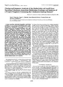
FIG. 7. Proteins produced from the cloned plasmids. The plasmids derived from pJTW4-20 and pJTW4-21 (Fig. 5) were transformed into K38 cells containing pGP1-2, which carries the T7 RNA polymerase gene (39). The proteins produced from the T7 RNA transcripts were labeled by [35S]methionine and fractionated on a 15% SDS-polyacrylamide gel (see Materials and Methods). The molecular weight standards were purchased from Bethesda Research Laboratories. Lane 1, D703; lane 2, D902; lane 3, D103; lane 4, D415; lane 5, D511; lane 6, D212; lane 7, D915.
36-kDa position on the SDS-polyacrylamide gel. This result was not consistent with the size we predicted from the DNA sequence (39.1 kDa). The aberrant mobility is probably due to the extremely high pl of this protein or some other
factor(s) (e.g., posttranslational modification or special protein folding, etc.). Similar in vivo protein labeling experiments showed that plasmid D902, which had the entire 39.1-kDa ORF, could produce the 36-kDa-like protein (Fig. 7, lane 2). In contrast, the deletion plasmid D103 missing the N-terminal 90 bp of the 39.1-kDa ORF could not produce the 36-kDa-like protein (Fig. 7, lane 3). This result indicated that the apparent 36-kDa protein was the MicA protein and was derived from the 39.1-kDa ORF. Sequences upstream of the micA gene contain one ORF. Within the 2.3-kb fragment we sequenced, there was another ORF which started 192 bp upstream of the micA gene (Fig. 6). This ORF, which is transcribed in the opposite direction from micA, is from base numbers 222 to 908 and encodes a 25.7-kDa protein. The putative -35 and -10 sequences (9) are shown in Fig. 6. In a T7 in vivo transcription-translation- system (39), plasmids D415, D511, and D212 containing the intact 25.7kDa ORF (Fig. 5) could produce the 26-kDa protein (Fig. 7, lanes 4, 5, and 6), whereas deletion plasmid D915 with the first 135 bp deleted from the N terminus of the 25.7-kDa ORF could not produce the 26-kDa protein (Fig. 7, lane 7). These results support the notion that the 25.7-kDa ORF is expressed in the cell. Due to its close proximity to the micA gene, we hypothesized that the 25.7-kDa ORF might have some function in the micA-dependent pathway. However, deletion plasmids without an intact 25.7-kDa ORF (D203, D501, D603, D703, D604, D601, and D902) had no effect on complementation of the A/G-specific nicking activity in micA mutant extracts (Fig. 5). Thus far, no evidence implicates the 25.7-kDa protein in an A/G mismatch-specific repair pathway.
DISCUSSION
There are at least two pathways capable of repairing A/G mismatches in E. coli. One is dependent on ATP, dam methylation, and the mutHLS and uvrD gene functions (reviewed in references 7, 25, and 33); the other is controlled by the micA (or mutY) gene and is independent of these three factors (18, 38). The dam-dependent pathway can repair A/G mismatches to either C/G or A/T depending on the state of methylation, whereas the dam-independent pathway repairs A/G mismatches exclusively to C/G base pairs irrespective of methylation state. Radicella et al. (32) have identified a gene function, micA, required for the dam methylationindependent A/G repair pathway by using a bacteriophage X heteroduplex assay. In this work, we show that the dam methylation-independent A/G repair pathway requires the micA gene product in vitro. In E. coli extracts, tightly associated A/G-specific binding and nicking activities have been identified, and efforts to separate these two activities have been unsuccessful (19). Several lines of evidence suggest that these two activities are involved in the micA-controlled repair pathway. The A/G mismatch nicking activity is absent in micA mutant extracts. Extracts of micA fail to form the A/G mismatch-specific binding complex. The A/G-specific binding and nicking activities remain tightly associated with the MicA protein during purification procedures. Furthermore, the 1,500-fold purified preparation (fraction V) containing both the A/G mismatch nicking and binding activities can restore the repair activity absent in a micA mutant extract. The micA gene has been cloned based on its ability to restore the A/G mismatch-specific nicking activity to micA mutant extracts. Nucleotide sequence and deletion analyses indicated that a 39.1-kDa ORF defined the micA gene (Fig. 5 and 6). However, expression of the gene in the T7 RNA polymerase system (39) produced a protein migrating as a 36-kDa protein on an SDS-polyacrylmide gel. The aberrant mobility is probably due to the high pl as deduced from the nucleotide sequence, or perhaps due to posttranslational modifications. The codon usage analysis (14) showed that the micA gene was composed of about 10.3% rare codons and 15.9% infrequently used codons. The uncommon codon usage might explain the poor expression of the micA gene even when directed by the T7 RNA polymerase. The mutY mutant has been shown to increase the C/G-toA/T transversion rate (28), and the mutant extract cannot repair A/G mismatches in vitro (1). The genetic mapping, mutator phenotype, and deficiency in correction of A/G mismatches both in vivo and in vitro suggest that micA and mutY could be the same gene or closely linked genes in the same pathway (28, 32). The DNA we cloned was able to complement the mutY mutant extract by restoring the A/Gspecific nicking activity (Fig. 5). Study of the deletions from the C or N terminus of the 39. 1-kDa ORF indicated that mutY and micA had exactly the same coding regions. This conclusion is supported by the finding that our micA gene sequence is identical to that of the mutY gene (24). Since the micA mutant extract was defective in the A/G mismatchspecific binding activity (Fig. 4), the MicA (MutY) glycosylase may be involved in formation of the A/G mismatchspecific binding complex. When the gene is mutated, there is no specific binding complex and no apurinic intermediate formation. Therefore, no A/G-specific nicking activity can be detected in the mutant extract. No significant homology in DNA or amino acid sequence was found between the MicA (or MutY) glycosylase and the
E. COLI micA GENE FOR A/G-SPECIFIC MISMATCH REPAIR
VOL. 173, 1991
products of ada, alkA, tagA, ung, andfpg from E. coli (4, 13, 26, 27, 35, 37, 41), denV from phage T4 (40), UNGI from Saccharomyces cerevisiae (31), and human uracilDNA glycosylase (29). It has been pointed out by Michaels et al. that MutY has homology to endonuclease III (24). Endonuclease III encoded by the nth gene of E. coli has N-glycosylase and apurinic endonuclease activities which are active on damaged DNA to release ring-saturated and ring-fragmented derivatives of thymine. Sequence analysis suggests that MutY (or MicA) may be an iron-sulfur protein such as endonuclease III (24). The MicA (MutY) glycosylase is restricted to A/G mismatch substrates. Among all eight possible mismatches, the A/G mismatch represents a special type of base pairing. Structural analyses showed that A-G can form hydrogen bonds with three possible configurations: A(anti)CG(anti) gene
A(anti)_G(syn)9 and A(syn)-G(syn), depending on the neighboring
environment (5, 8, 12, 30). MicA (MutY) glycosylase may specifically recognize one of these configurations. It is an interesting finding that the crystal structure of 5'CGC GAATTAGCG3' has shown that A/G mismatches assumed the A(syn)-G(anti) configuration (5). The neighboring sequence of this heteroduplex is similar to that of the DNA substrate used in our assay (see Materials and Methods). One 25.7-kDa ORF was found upstream of micA (or mutY) and could be expressed in a T7 RNA polymerase system (see Results). A computer search for homology between this protein and the proteins in the GenBank indicated that it is a new protein and is not significantly homologous to other known proteins. Deletion of the 25.7-kDa ORF from pJTW4-20 has no effect on restoring the A/G mismatchspecific nicking activity in micA or mutY mutant extracts. Further investigation is needed to identify the function of this protein. ACKNOWLEDGMENTS We thank J. H. Miller and M. L. Michaels for sharing their preprint and information about the mutY gene. We also thank G. Barcak, L. Black, M. S. Fox, and A. Zachary for critical reading of the manuscript and for helpful discussions on this work, and C.-H. Wu for helping us to prepare the manuscript. This work was supported by Public Health Service grant GM 35132 from the National Institute of General Medical Sciences.
REFERENCES 1. Au, K. G., M. Cabrera, J. H. Miller, and P. Modrich. 1988. Escherichia coli mut Y gene product is required for specific A-G C-G mismatch correction. Proc. Natl. Acad. Sci. USA 85:9163-9166. 2. Au, K. G., S. Clark, J. H. Miller, and P. Modrich. 1989. Escherichia coli mutY gene encodes an adenine glycosylase active on G-A mispairs. Proc. Natl. Acad. Sci. USA 86:88778881. 3. Boeke, J. D. 1981. One and two coden insertion mutants of -*
bacteriophage fl. Mol. Gen. Genet. 181:288-291. 4. Boiteux, S., T. R. O'Connor, and J. Laval. 1987. Formamidopy-
rimidine-DNA glycosylase of Escherichia coli: cloning and overproduction of the protein. EMBO J. 6:3177-3183. 5. Brown, T., W. N. Hunter, G. Kneale, and 0. Kennard. 1986. Molecular structure of the G.A base pair in DNA and its implications for the mechanism of transversion mutations. Proc. Natl. Acad. Sci. USA 83:2402-2406. 6. Clarke, L., and J. Carbon. 1976. A colony bank containing synthetic ColEl hybrid plasmids representative of the entire E. coli genome. Cell 9:91-99. 7. Claverys, J.-P., and S. A. Lacks. 1986. Heteroduplex deoxyri-
1909
bonucleic acid base mismatch repair in bacteria. Microbiol. Rev. 50:133-165. 8. Gao, X., and D. J. Patel. 1988. G(syn).A(anti) mismatch formation in DNA dodecamers at acidic pH: pH-dependent conformational transition of G.A mispairs detected by proton NMR. J. Am. Chem. 110:5178-5182. 9. Harley, C. B., and R. P. Reynolds. 1987. Analysis of E. coli promoter sequences. Nucleic Acids Res. 15:2343-2361. 10. Henikoff, S. 1984. Unidirectional digestion with exonuclease III creates targeted breakpoints for DNA sequencing. Gene 28:351359. 11. Herman, G. E., and P. Modrich. 1982. Escherichia coli dam methylase: physical and catalytic properties of the homogeneous, enzymes. J. Biol. Chem. 259:2605-2612. 12. Kan, L.-S., S. Chandrasegaran, S. M. Pulford, and P. S. Miller. 1983. Detection of a guanine.adenine base pair in a decadeoxyribonucleotide by proton magnetic resonance spectroscopy. Proc. Natl. Acad. Sci. USA 80:4263-4265. 13. Kondo, H., Y. Nakabeppu, H. Kataoka, S. Kuhara, S. Kawabata, and M. Sekiguchi. 1986. Structure and expression of the alkB gene of Escherichia coli related to the repair of alkylated DNA. J. Biol. Chem. 261:15772-15777. 14. Konigsberg, W., and N. Godson. 1983. Evidence for use of rare codons in the dnaG gene and other regulatory genes of Escherichia coli. Proc. Natl. Acad. Sci. USA 80:687-691. 15. Laemmli, U. K. 1970. Cleavage of structural proteins during the assembly of the head of bacteriophage T4. Nature (London) 227:680-685. 16. Lieb, M. 1983. Specific mismatch correction in bacteriophage lambda crosses by very short patch repair. Mol. Gen. Genet. 191:118-125. 17. Lieb, M. 1985. Recombination in the lambda repressor gene: evidence that very short patch (VSP) mismatch correction restores a specific sequence. Mol. Gen. Genet. 199:465-470. 18. Lu, A-L., and D.-Y. Chang. 1988. Repair of single base-pair transversion mismatches of Escherichia coli in vitro: correction of certain A/G mismatches is independent of dam methylation and host mutHLS gene functions. Genetics 118:593-600. 19. Lu, A-L., and D.-Y. Chang. 1988. A novel nucleotide excision repair for the conversion of an A/G mismatch to C/G base pair in E. coli. Cell 54:805-812. 19a.Lu, A-L., and D.-Y. Chang. Unpublished data. 20. Lu, A-L., S. Clark, and P. Modrich. 1983. Methyl-directed repair of DNA base pair mismatches in vitro. Proc. Natl. Acad. Sci. USA 80:4639-4643. 21. Lu, A-L., K. Welsh, S. Clark, S.-S. Su, and P. Modrich. 1984. Repair of DNA base pair mismatches in extracts of Escherichia coli. Cold Spring Harbor Symp. Quant. Biol. 49:589-596. 22. Maniatis, T., E. F. Fritsch, and J. Sambrook. 1982. Molecular cloning: a laboratory manual. Cold Spring Harbor Laboratory, Cold Spring Harbor, N.Y. 23. Maxam, A. M., and W. Gilbert. 1980. Sequencing end-labeled DNA with base-specific chemical cleavage. Methods Enzymol. 65:499-560. 24. Michaels, M. L., L. Pham, Y. Nghiem, C. Cruz, and J. H. Miller. 1990. MutY, an adenine glycosylase active on G-A mispairs, has homology to endonuclease III. Nucleic Acids Res. 18:38413845. 25. Modrich, P. 1987. DNA mismatch correction. Annu. Rev. Biochem. 56:435-466. 26. Nakabeppu, Y., T. Miyata, H. Kondo, S. Iwanaga, and M. Sekiguchi. 1984. Structure and expression of the alkA gene of Escherichia coli involved in adaptive response to alkylating agents. J. Biol. Chem. 259:13730-13736. 27. Nakabeppu, Y., and M. Sekiguchi. 1986. Regulatory mechanisms for induction of synthesis of repair enzymes in response to alkylating agents: Ada protein acts as a transcriptional regulator. Proc. Natl. Acad. Sci. USA 83:6297-6301. 28. Nghiem, Y., M. Cabrera, C. G. Cupples, and J. H. Miller. 1988. The mutY gene: a mutator locus in Escherichia coli that generates G:C to T:A transversions. Proc. Natl. Acad. Sci. USA 85:2709-2713. 29. Olsen, L. C., R. Aasland, C. U. Wittwer, H. E. Krokan, and
1910
30.
31.
32.
33. 34.
35.
36.
TSAI-WU ET AL.
D. E. Helland. 1989. Molecular cloning of human uracil-DNA glycosylase, a highly conserved DNA repair enzyme. EMBO J. 8:3121-3125. Patel, D. J., S. A. Kozlowski, S. Ikuta, and K. Itakura. 1984. Deoxyguanosine-deoxyadenosine pairing in the d(C-G-A-G-AA-T-T-C-G-C-G) duplex: conformation and dynamics at and adjacent to the dG.dA mismatch site. Biochemistry 23:32073217. Percival, K. J., M. B. Klein, and P. M. J. Burgers. 1989. Molecular cloning and primary structure of the uracil-DNA glycosylase gene from Saccharomyces cerevisiae. J. Biol. Chem. 264:2593-2598. Radicella, J. P., E. A. Clark, and M. S. Fox. 1988. Some novel mismatch repair activities in E. coli. Proc. Natl. Acad. Sci. USA 85:9674-9678. Radman, M., and R. Wagner. 1986. Mismatch repair in Escherichia coli. Annu. Rev. Genet. 20:523-528. Raposa, S., and M. S. Fox. 1987. Some features of base pair mismatch and heterology repair in Escherichia coli. Genetics 117:381-390. Sakumi, K., Y. Nakabeppu, Y. Yamamoto, S. Kawabata, S. Iwanaga, and M. Sekiguchi. 1986. Purification and structure of 3-methyladenine-DNA glycosylase I of Escherichia coli. J. Biol. Chem. 261:15761-15766. Sanger, F., S. Nicklen, and A. R. Coulson. 1977. DNA sequenc-
J. BACTERIOL.
ing with chain-terminating inhibitors. Proc. Natl. Acad. Sci. USA 74:5463-5467. 37. Steinum, A.-L., and E. Seeberg. 1986. Nucleotide sequence of the tag gene from Escherichia coli. Nucleic Acids Res. 14:37633772. 38. Su, S.-S., R. S. Lahue, K. G. Au, and P. Modrich. 1988. Mispair specificity of methyl-directed DNA mismatch correction in vitro. J. Biol. Chem. 263:6829-6835. 39. Tabor, S., and C. C. Richardson. 1985. A bacteriophage T7 RNA polymerase/promoter system for controlled exclusive expression of specific genes. Proc. Natl. Acad. Sci. USA 82:1074-1078. 39a.Tsai-Wu, J.-J., and A-L. Lu. Unpublished data. 40. Valerie, K., E. E. Henderson, and J. K. DeRiel. 1984. Identification, physical map location and sequence of the denV gene from bacteriophage T4. Nucleic Acids Res. 12:8085-8096. 41. Varshney, U., T. Hutcheon, and J. H. van de Sande. 1988. Sequence analysis, expression and conservation of Escherichia coli uracil DNA glycosylase and its gene (ung). J. Biol. Chem. 263:7776-7784. 42. Way, J. C., M. A. Davis, D. Morisato, D. E. Roberts, and N. Kleckner. 1984. New TnlO derivatives for transposon mutagenesis and for construction of lacZ operon fusions by transposition. Gene 32:369-379.