Department of Microbiology, University of Surrey, Guildford, Surrey G U2 ...
organisms, especially with regard to their taxonomy and pigment chemistry,
remains.
Journal of General Microbiology (1986), 132, 1899-1909.
Printed in Great Britain
1899
Numerical Taxonomy of Red-pigmented Bacteria Isolated from a Lowland River, with the Description of a New Taxon, Rugamonas rubra gen. nov., sp. nov. By D A W N A. AUSTINT A N D M . 0. MOSS* Department of Microbiology, University of Surrey, Guildford, Surrey G U2 5XH, UK (Received 29 November I985 ;revised 3 March 1986)
Twenty red-pigmented bacterial strains, isolated from river water, and 15 named reference cultures were analysed using numerical taxonomic procedures. A simplified dendrogram, based on the simple matching coefficient and unweighted average linkage clustering algorithm, showed that the environmental isolates were recovered in two phenetic groups, defined at or above the 89% similarity level. Nine strains were contained in phenon 1, which exhibited the general characteristics of the Pseudomonadaceae, but did not match the descriptions of any of the existing genera within this family. A new genus and species, Rugamonas rubra, is proposed for these isolates. The 11 red-pigmented environmental strains in phenon 2 were identified as Serratia rubidaea. The red pigment of R. rubra contains at least two compounds related to prodigiosin. The major component produced a mass spectrum compatible with its being 6-methoxy-2-methyl-3-heptylprodigeosene (m/e 35 1). A second component produced a mass spectrum indistinguishable from that of prodigiosin itself (m/e 323). INTRODUCTION
Pigmented bacteria occur frequently in aquatic samples. However, these colourful organisms often arouse only transient interest. Thus knowledge of many of the pigmented microorganisms, especially with regard to their taxonomy and pigment chemistry, remains fragmentary. However, Serratia spp., which produce the red pigment prodigiosin or its derivatives, have been studied in detail. Since the initial characterization of prodigiosin from S.marcescens, a variety of bacterial taxa have been found to contain similar pigments, e.g. ‘ Vibrio psychroerythrus’ (D’Aoust & Gerber, 1974), Vibrio (formerly Beneckea) gazogenes (Harwood, 1978; Allen et af., 1983), Alteromonas rubra (Gauthier, 1976; Gerber & Gauthier, 1979) and several actinomycetes, such as species of Actinomadura (Gerber, 1973), Streptoverticillium rubrireticuli (Gerber & Stahly, 1979, and Streptomyces longisporus ruber (Wasserman et al., 1976). In 1979 during a survey of chromobacteria isolated from freshwater, including the River Wey in Surrey, UK, a tough, membranous red colony was observed on a spread plate of modified Bennett’s agar (see Moss, 1983). This organism was partially characterized and the red pigment was isolated and found to be closely related to prodigiosin (Moss, 1983). At this stage it was considered that the organism comprised a new species, probably within the Pseudomonadaceae, as its general characteristics, i.e. oxidative metabolism, Gram-negative reaction and motile, polarlsub-polarly flagellated rods, matched the family description (Palleroni, 1984). In particular, the cell morphology of the red-pigmented organism strongly resembled that of Zmgloea ramigera because ageing cultures grown in liquid medium showed reduced motility concurrent with floc formation. However, it was considered at the time that insufficient information existed to assign a name to the organism.
t Present address : Department of
EH1 IHX,UK.
0001-3118’0 1986 SGM
~~
Brewing and Biological Sciences, Heriot-Watt University, Edinburgh
Downloaded from www.microbiologyresearch.org by IP: 185.107.94.33 On: Wed, 27 Sep 2017 21:05:39
1900
D . A . A U S T I N A N D M . 0 . MOSS
Table 1. Reference cultures Name as received Janthinobacterium lividurn* Methylobacterium sp. Pseudomonas acidovorans Pseudomonas denitrficans Pseudomonas rhodos Pseudomonas saccharophila* Pseudomonas stutzeri Serratia marcescens Zoogloea rami'era*
Source and collection no.? CCM 160 CCM 3355 NCIB 10409 NCIB 2879 NCIB 9399 CCM 283 NCIB 9496 NCIB 9421 CCM 1980 CCM 2896, CCM 2897 NCTC 8706 NCIB 10706 NCIB 10340, CCM 2456
Comment
Formerly Mycoplana ruber Formerly Protaminobacter ruber Formerly Pseudomonas extorquens
Unz (1971) and NCIB do not consider this strain to be Z. ramigera
* Type strain.
t CCM, Czechoslovak Collection of Micro-organisms, Brno, Czechoslovakia; NCIB, National Collection of
Industrial Bacteria, Aberdeen, UK ; NCTC, National Collection of Type Cultures, Colindale, London, UK.
In this study, red-pigmented bacteria isolated from the River Wey were subjected to numerical taxonomic analyses to ascertain their relationship to the original isolate (MOM 28/2/79), described by Moss (1983). To determine if the red-pigmented isolates resembled existing taxa or constituted a new taxon, they were also compared with a variety of named cultures including Pseudomonas spp., 2. ramigera, S. marcescem and Methylobacterium spp. In addition, the nature of the red pigment was studied further. METHODS
Collection of samples. Water samples were collected from two locations on the River Wey in Surrey, one in Guildford and the other upstream in Shalford. Sampling began in January, 1984, and continued at approximately bi-weekly intervals until the end of May, 1984. The samples were collected by aseptic techniques, from just below the surface, approximately 0.5 m from shore in either pre-sterilized plastic containers (Dippas; Sterilin) or large Duran bottles. All samples were analysed within 1 h of collection. The water temperature was recorded concurrently. Isolation and maintenance of strains. Fivefold serial dilutions up to 1/125 were prepared from the water samples, using 1/4 strength Ringer's buffer (Oxoid) as a diluent, and 0.5 ml samples of appropriate dilutions were spread, in duplicate, on modified Bennett's agar plates (Keeble & Cross, 1977).The plates were incubated at 20 "C for up to 14 d. Tough membranous red-pigmented colonies resembling those described by Moss (1983) were retained for taxonomic analysis. In addition, all other red-pigmented isolates were selected for further examination. The red-pigmented bacteria were streaked five times on Bennett's agar to ensure the purity of cultures. The isolates were stored on Bennett's agar slopes and in Bennett's broth at 20 "C and 10 "C, respectively, with subculturing every 10 d. In addition, each of the isolates was stored at - 70 "C. The isolates were grown in Bennett's broth, with incubation at 20 "C for 3 d. A 1 ml sample of the turbid suspension was placed in a freeze vial (Nunc) to which an equal volume of the following solution was added as a cryoprotectant (g 1-l): K2HP04, 12.6; sodium citrate, 0.9; MgS04.7H20, 0.18; (NH4)$04, 1.8; KH2P04, 3.6; and glycerol, 44 ml. This solution is a double-strength mixture, based on the minimal medium of McFall et al. (1958). The bacterial suspensions and the cryoprotectant were well mixed, before the vials were placed at -70 "C. At the conclusion of the sampling period 20 environmental isolates had been retained for numerical taxonomic analysis. Reference culrures. In addition to the environmental isolates, 15 named cultures (Table 1) were included in the study for reference purposes. Characterization ofthe bacterial strains. Each strain was examined for a total of 147 unit characters. Whenever possible, Bennett's agar was used as the basal medium. Unless stated otherwise, all test media were incubated at 20 "C for three weeks. Tests were done once, and repeated only if inconclusive results were obtained. Downloaded from www.microbiologyresearch.org by IP: 185.107.94.33 On: Wed, 27 Sep 2017 21:05:39
1901
Taxonomy of red-pigmented bacteria
Colony morphology, micromorphology and staining reactions. Colony morphology was monitored daily for up to 14 d. However, characteristics coded for computer analysis were described from 5-d-old cultures. The characters, scored for each strain, were pigmentation (red, pink, yellow, white or purple), elevation (domed, flat, raised or wrinkled), margin (entire or serrated) and production of diffusible pigment. Motility and cell morphology were determined microscopically from wet preparations and heat-fixed smears. Both 24-48 h and 7-d-old cultures were examined, as Moss (1983) observed differences in micromorphology and motility as the cultures aged. Biochemical tests. Tests for arginine, lysine and ornithine decarboxylase production, methyl red, the VogesProskauer reaction, Koser's citrate utilization, nitrate and nitrite reduction, gluconate oxidation, malonate utilization, phenylalanine deaminase, ONPG for P-galactosidase production, indole and phosphatase production were recorded after 7 d, using the methods described in Cowan (1974). The catalase and oxidase tests were determined from 48 h cultures by the procedures outlined in Cowan (1974). Fermentative and oxidative metabolism of glucose were recorded, using the medium of Hugh & Leifson (1953). Degradation characters. Degradation of aesculin, allantoin, casein, starch and urea was determined by the methods outlined in Cowan (1974). Degradation of tyrosine, xanthine, hypoxanthine and elastin and the hydrolysis of Tween 20, 40, 60 and 80 were tested using the procedures described by Gordon (1967) and Sierra (1957), respectively. Gelatin hydrolysis was detected by the method of Smith & Goodner (1958). Degradation of cellulose, chitin, DNA and RNA was determined using methods which have been outlined previously (Goodfellow et al., 1976). All degradation tests were checked at 7 d intervals for up to three weeks, with the exception of chitin and cellulose, which were recorded after five weeks. Physiology. The ability to grow on sodium hippurate (Thirst, 1957), potassium tellurite, sodium azide (0.04%, w/v) agar, MacConkey agar and crystal violet (0.001 %, w/v) agar was recorded on appropriate media. Growth on 0,0-5, 1, 3 and 5 % (w/v) NaCl was tested in appropriately modified Bennett's agar. Growth at 4, 10, 20, 30 and 37 "C was recorded after incubation for 28, 7 and 3 d. Growth at pH 4, 5, 6, 8 and 9 was tested by adjusting Bennett's broth to the required pH. Anaerobic growth was tested using Bennett's agar and incubation at 25 "C,in an anaerobic chamber (Raven Scientific). Enzymeproduction. API Zym test strips (API Laboratory Products) were used to screen the isolates for activity of the following enzymes: alkaline phosphatase, esterase, esterase-lipase, leucine arylamidase, valine arylamidase, cystine arylamidase, trypsinase, chymotrypsinase, acid phosphatase, phosphoamidase, a- and 8glucosidase, a- and P-galactosidase, Pglucuronidase, N-acetyl-P-glucosaminidase, a-mannosidase and afucosidase. Utilization of substrates as sole sources of carbon for energy and growth. Carbon utilization was tested using the medium described by Stevenson (1967). The compounds tested were : DL( -)-alanine, L( +)-arabinose, DL( -)arginine monohydrochloride, adonitol, cellobiose, ethanol, D( - )-fructose, D( +)-galactose, glucose, L( - )histidine monohydrochloride, i-erythritol, meso-inositol, sodium DL-lactate, lactose, sodium malonate, maltose, D( +)-mannose, D( - )-melibiose, D( - )-melezitose, D(-)-raffinose, L( - )-rhamnose, D( - )-ribose, DL-serine, sodium acetate, trisodium citrate, D( - )-sorbitol, sucrose, trehalose, DL-valine and D( )-xylose. The carbon compounds as 20% (w/v) solutions were sterilized by filtration (0.22pm porosity filter) and added to the supporting medium to give a final concentration of 0.2% (w/v). Utilization of the compounds was determined by visually examining the media after 14 d and comparing the amount of growth, if any, with a negative control. Sensitivity to antibiotics. 'Multodisks' (Oxoid) were used to test sensitivity to ampicillin (2 and 25 pg), chloramphenicol (10 pg), chlortetracycline (10 pg), cloxacillin (5 pg), colistin sulphate (10 pg), erythromycin (10 pg), furazolidone (50 pg), gentamicin (10 pg), kanamycin (10 pg), neomycin (10 pg), nitrofurantoin (200 pg), novobiocin (5 pg), oxytetracycline (10 pg), penicillin G (1-5 i.u.), streptomycin (10 pg), sulphafurazole (100 and 500 pg), tetracycline (10 and 50 pg) and cotrimoxazole (25 pg). Briefly,the procedure entailed inoculation, using presterilized swabs, of overnight broth cultures onto Bennett's agar plates. These were air dried at room temperature to absorb excess moisture, and multodisks were then placed on the agar surface. The plates were incubated at 25 "C and recorded as soon as growth was observed. Sensitivity or resistance was determined using a growth zone template (Oxoid). Coding of data. The characters were coded '1' for positive or present and '0'for negative or absent. The final n x t matrix contained 35 strains and 119 unit characters. Computer analyses. The data were analysed using the simple matching (SSM;Sokal & Michener, 1958) and Jaccard ( S J ;Sneath, 1957) coefficients. Clustering was by unweighted average linkage (UMPGA ; Sneath 8c Sokal, 1973). These methods were contained on the CIustan 1C program package (Wishart, 1969), available on the University of Surrey Prime computer Electron microscopy. To examine cell morphology, particularly flagellar arrangement, preparations were made from strain MOM 28/2/79, using 24 h and 1 I-d-old cultures. The strain was grown in Bennett's broth incubated at 25 "C, centrifuged gently at 3500 r.p.m., washed three times in sterile distilled water and fixed with 10% (v/v) formalin, followed by negative staining with 3% (w/v) phosphotungstic acid. Alternatively it was grown in 1/4 strength Bennett's broth for 48 h at 15 "C,and drops of culture were placed directly onto Formvar-carbon coated grids and allowed to attach for 30 s. before removal of excess culture fluid and rinsing in filter-sterilized (0.22 pm
+
Downloaded from www.microbiologyresearch.org by IP: 185.107.94.33 On: Wed, 27 Sep 2017 21:05:39
1902
D. A . A U S T I N AND M . 0 . MOSS
porosity filters) distilled water. After drying over a desiccant at room temperature, grids were shadowed with gold/palladium. The specimens were examined on a Philips EM 4007 transmission electron microscope at 80 kV. D N A base composition. DNA from representative strains was extracted and purified by the method of Mandel et al. (1971). The base composition was determined from T, values (Marmur & Doty, 1962). Pigment analysis. Freeze-dried culture material scraped from six plates of Bennett's agar was extracted with dichloromethane (50 ml) in a Soxhlet extractor overnight. The extract was washed twice with an equal volume of 1 M-NaOH followed by an equal volume of 1 M-HCl.After concentration to a dark oil on a rotary evaporator under vacuum it was treated with light petroleum (b.p. 68-80 "C) and cooled to - 5 "C to yield a dark maroon precipitate (22.6 mg). Pigments were purified by preparative TLC on sodium acetate buffered silica gel C plates with ethyl acetate/chloroform/acetic acid (30 :80 : 2, by vol.). Chromatography was done in subdued light and the pigments were recovered immediately by scraping the appropriate bands off the plate into small chromatography columns and eluting with the minimum volume of chloroform.
RESULTS
All 35 strains included in the study gave a positive response for the following tests: rod-shaped cells, motility, production of round colonies, growth at 10, 20 and 30 "C and at pH 6, 8 and 9, growth on 0%NaCl and utilization of ethanol and glucose as the sole source of carbon for energy and growth. Negative responses for all strains were obtained for presence of coccoid cells, growth on sodium azide agar (0.04 %, w/v), production of b-galactosidase, a-galactosidase, pglucuronidase, N-acetyl-p-glucosaminidase,a-mannosidase and a-fucosidase, degradation of cellulose, starch hydrolysis and indole production. All isolates were sensitive to kanamycin (10 pg) but resistant to penicillin G (1.5 i.u.) and cloxacillin (5 pg). Since the characters listed above were non-discriminatory they were excluded from the n x t matrix. Clustering of the strains Six phena and seven single member clusters were defined at, or above, the 83% similarity (S) level when the S,, coefficient was used (Fig. 1). The 20 red-pigmented environmental isolates clustered in phena 1 and 2 (Fig. 1). Nine of the isolates, including MOM 28/2/79, clustered in phenon 1 at the 89% S level. The remaining 11 environmental isolates, contained in phenon 2, clustered at 95 % similarity. The high S levels obtained indicate that the organisms within each of the clusters formed very homogeneous groups. Phenon 3 contained Pseudomonas acidovorans and Ps. saccharophila,phenon 4 comprised two strains of Zoogloea ramigera, each related at 83 % similarity, phenon 5 comprised two strains of Ps.stutzeri, which joined at 84% similarity, and phenon 6 included two strains of Methylobacterium sp. (formerly Mycoplana ruber and Ps. extorquens) at the 87% S level (Fig. 1). The remaining seven reference cultures occurred as single-member clusters. Neither of the two phena that contained environmental isolates was substantially related to any of the named reference cultures included in the study. However, the highest similarity (79.5%) among the clusters was between phenon 2, later identified as Serratia rubidaea (synonym S . marirwrubra), and the reference cultures of S. marcescens (Fig. 1). The SJ coefficient gave similar results albeit at lower S levels. Thus, the isolates in phenon 1 clustered at 83.5% similarity. However, the strains in phenon 2 retained an extremely high S level at 93%. Three of the clusters containing the reference cultures listed above grouped at S levels 12% lower than those observed for the S,, coefficient, i.e. phena 3 and 4 formed at 71 % and phenon 5 at 72% similarity. Phenon 6, however, showed only 59% similarity when the SJ coefficient was used. Characterization and identiJicationof the environmental isolates The nine red-pigmented strains in phenon 1 possessed the general traits of the family Pseudomonadaceaedescribed by Palleroni (1984), the G C of the DNA of the type strain of this phenon, MOM 28/2/79, being 66.5mol%. The isolates were relatively active in standard bacteriological test media, giving positive reactions for Koser's citrate, gluconate oxidation, reduction of nitrate to nitrogen, j?-galactosidase and phosphatase production, and the degradation of aesculin, chitin, DNA, RNA, gelatin, Tween 20,40,60 and 80, tyrosine and urea.
+
Downloaded from www.microbiologyresearch.org by IP: 185.107.94.33 On: Wed, 27 Sep 2017 21:05:39
1903
Taxonomy of red-pigmented bacteria Percentage similarity
50
60
I
70
80
90
100
I
Phenon
Identification
No. of strains
1
Rugamonas rubra
9
2
Serratia rubidaea
11
S . marcescens
1
Janthinobacterium lividurn*
1
3
Pseudomonas acidovorans Ps. saccharophila.
2
4
Zoogloea ramigera
2
Ps. denitrijcans
1
J . lividum
1
Ps. stutzeri
2
Z . ramigera*
1
Methy lobacterium sp.
1
Methylobacterium sp.
2
5
6
I Ps. rhodos
1
Fig. 1. Simplified dendrogram based on the simple matching coefficient and unweighted average linkage clustering.
They utilized a wide range of compounds as a sole source of carbon for energy and growth, including the amino acids alanine, arginine, histidine and serine, and also a variety of carbohydrates such as arabinose, cellobiose, fructose, galanose, glucose, inositol, maltose, mannose, raffinose, ribose, sorbitol and trehalose. Some interesting properties of these organisms included an extreme sensitivity to NaCl, even on a medium such as nutrient agar (Oxoid) containing only 0.5 % (w/v) NaCl, and an inability to grow at temperatures above 30 "C. Perhaps the most outstanding features of these bacteria, however, were their distinctive colonial and cellular morphology when grown in Bennett's medium. On Bennett's agar, the isolates initially formed white, shiny, round colonies within 48 h incubation at 20 "C. These colonies developed a pink colour at 4 d which progressed to deep red/maroon, shiny, raised, wrinkled colonies, 3-4 mm in diameter, with serrated margins and a tough, membranous, rubbery texture after 5 d. Two of the strains also produced a light brown diffusible pigment on Bennett's agar. Often, when the cultures reached the age of 7 d or more, a metallic sheen on the initial streak of growth was noted. In Bennett's broth the cultures were uniformly turbid up to 4-5 d incubation at 20 "Cbut, with ageing, pink flocs developed, which sank into the medium. Cells from 24-48 h cultures consisted of motile, Gram-negative rods, approximately 2.5-4-0 pm in length. At approximately 7 d, motility was considerably reduced, many pleomorphic cells could be seen and the culture contained well-developed pink flocs with a morphology reminiscent of Z . ramigera. Although the strains in phenon 1 possessed the overall characteristics of the Pseudomonaduceae, they could not be assigned to any of the existing genera within the family as described in Bergey's Manual of Systematic Bacteriology (Krieg, 1984). Neither did the organisms demonstrate sufficient similarity to any of the reference strains included in the study, which Downloaded from www.microbiologyresearch.org by IP: 185.107.94.33 On: Wed, 27 Sep 2017 21:05:39
Downloaded from www.microbiologyresearch.org by IP: 185.107.94.33 On: Wed, 27 Sep 2017 21:05:39
Colony morphology Domed elevation Wrinkled elevation Entire margin Serrated margin Diffusible pigment Biochemical tests Lysine decarboxylase Ornithine decarboxylase Koser's citrate Fermentative metabolism Malonate Nitrate reduction Oxidase Phenylalanine deaminase Phosphatase Vogues-Proskauer reaction Degradation of: Allantoin Gelatin Hy poxant hine Tween 80 Xanthine Growth at: 37 "C PH 4 PH 5 100 100
0 0 78
100
55
18 82 100 100
0 0 100
0
0 100 0 78 0
100 0
100 11
0 0 100 100 82
56 67 89 0 0 0 100
100 0 100 0 0
2
Phenon
0 100 0 100 22
1
Phenon
+
+
Anaerobic growth Growth on: NaCl, 0.5% (w/v) NaCl, 1-5% (w/v) Sodium hippurate agar MacConkey agar Enzyme production Alkaline phosphatase Esterase Esterase-lipase Leucine arylamidase Phosphoamidase P-Glucosidase Utilization, as sole source of carbon, of: Lactose D( )-Melibiose D( - FMelizitose D( -)-Sorbit01 D( )-Xylose Sensitivity to: Ampicillin (2 pg) Ampicillin (25 pg) Chloramphenicol (10 pg) Erythromycin (10 pg) Neomycin (10 pg) Novobiocin (5 pg) Sulphafurazole (1 00 pg) Sulphafurazole (500 pg) Cotrimoxazole (25 pg) 78 11 100 100 100 0
100
0 67
22 0 0 100 0
89 100 0
100
67
100
78 0 0 0
0
1
Phenon
0 0 91
64
0 0
100 0
82
0 100
100
100
100
64 91 36
91 0 82
100 100 I00 100
100
Phenon 2
Table 2. Differential characteristics of the environmental isolates in terms of percentage positive responses
tl
z
P
P
0
c \o
Taxonomy of red-pigmented bacteria
1905
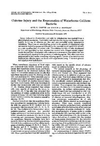
Fig. 2. Transmission electron micrographs of strain MOM 28/2/79. (a, 6) Gold/palladium shadowed preparation from a 48 h culture. (c, d ) Negatively stained preparation from an 1 I-d-old culture. Bars, 2 pm.
would have assisted in their tentative identification. Due primarily to the distinctive morphology of the isolates in phenon 1, it was concluded that they comprise a new taxon at the generic and specific levels, for which the name Rugamonas rubra is proposed. The 11 red-pigmented river water isolates contained in phenon 2 were identified as Serratia rubidaea, by using the API 20E identification system and conventional bacteriological testing regimes. Thus, the isolates comprised Gram-negative, red-pigmented, fermentative motile rods, which produced catalase, but not oxidase, indole and arginine, and lysine and ornithine decarboxylases; aesculin, DNA, gelatin and Tween 20, 40 and 80, but not starch, were hydrolysed ; and growth occurred at 37 "C. Arabinose, melibiose, xylose, arginine, cellobiose, Downloaded from www.microbiologyresearch.org by IP: 185.107.94.33 On: Wed, 27 Sep 2017 21:05:39
1906
D . A. AUSTIN AND M . 0 . MOSS
(a) R
(b) R
= (CH,),.CH3 = (CH2),.CH3
Fig. 3. Structure of (a) prodigiosin, (b) 6-methoxy-2-methyl-3-heptylprodigeosene and (c) the common ion at m/e 266.
lactate, lactose and raffinose, but not rhamnose or sorbitol, were utilized as sole sources of carbon. However, chitin was degraded, which is in disagreement with the species description of S. rubidaea (see Grimont & Grimont, 1984). The isolates formed round, domed, entire, shiny, bright red colonies, approximately 3 mm in diameter, on Bennett's agar after incubation for 3 d at 20 "C. Differential characteristics of the environmental isolates have been included in Table 2.
Electron microscopy Gold/palladium shadowed preparations of 48 h cultures of strain MOM 28/2/79, examined by transmission electron microscopy, showed a single polar flagellum and, frequently, a number of sub-polar and peritrichous flagella, which were thinner and had a shorter wavelength (Fig. 2a, b). Such variable flagellation has been recorded in a number of species of bacteria, including chromobacteria (Leifson, 1956; Moss & Ryall, 1981) and Vibrio aZginoZyticus (DeBoer et al., 1975). Negatively stained preparations of ageing cultures showed the presence of intracellular granules within highly pleomorphic cells (Fig. 2c, d ) . Deeply pigmented granules were often visible when unstained bacteria from an ageing culture were examined with the light microscope. Pigment analysis The major band of material obtained by preparative TLC had A,, (EtOH HCl) 535.5, 291 nm; A,, (EtOH NaOH), 465 nm. Although it gave a single spot centred at RF 0.45, it produced a mass spectrum characteristic of a mixture of prodigiosin and a related compound differing by an additional C2H4.A sample of prodigiosin prepared from S . marcescens had an RFof 0.46 and a mass spectrum with a parent ion at m/e 323 with a major peak at m/e 266 and a metastable at m/e 219, confirming the loss of C4H9from the parent ion (Fig. 3). The mass spectrum of the pigment from R . rubra had a parent ion at m/e 351,the parent ion of prodigiosin at m/e 323,the base peak at m/e 266 and metastables at 201.5 and 219,confirming that the base peak was formed by the loss of C6HI3from m/e 351 and C4H9from 323.These results, and other aspects of the mass spectrum, suggest that the pigment of R. rubra contains prodigiosin itself and (Fig. 3). the higher homologue 6-methoxy-2-methyl-3-heptylprodigeosene Preparative TLC showed a number of minor compounds well separated from the material described above and it is possible that one of these corresponds to nonylprodiginine as discussed by Moss (1983).
+
+
DISCUSSION
The red-pigmented bacteria recovered from the River Wey belong to two distinct families, the Pseudomonadaceae and the Enterobacteriaceae. It is interesting that the strains in both of these groups were consistently isolated in low numbers from the river water samples throughout the sampling period (January to May, 1984). Although this suggests that these bacteria were continually present in the river system, it is not possible to conclude whether they are Downloaded from www.microbiologyresearch.org by IP: 185.107.94.33 On: Wed, 27 Sep 2017 21:05:39
Taxonomy of red-pigmented bacteria
1907
autochthonous members of the river micro-flora. For instance the strains in phenon 1, named as Rugamonas rubra, were usually isolated when rain had fallen 24-48 h before collection of samples, an indication that these organisms may be washed into the river with land runoff. Also, their nutritional capabilities, i.e. the utilization of a wide variety of the sugars and amino acids tested as sole carbon sources, hint at a potential for these organisms to exploit a range of environments. These diverse metabolic abilities are similar to those of the pseudomonads, which are ubiquitous in the natural environment, occurring in water and soil and in association with plants. Likewise, the majority of the isolates in phenon 2 (64%), identified as Serratia rubidaea, were recovered from samples collected within 24-72 h of previous rainfall. These strains also utilized a wide range of the compounds tested as sole carbon sources for energy and growth. In a survey of the River Wey in April, 1984, water was collected from five additional sites upstream of the usual two sampling locations described previously. No R . rubra strains were recovered. However, an isolate which subsequentlyclustered in phenon 2 (S. rubidaea) was found, providing tentative evidence that this species is distributed throughout the river. In view of this, it is relevant to note that Grimont & Grimont (1984) reported that S. rubidaea was rarely isolated, from either the natural environment or human clinical specimens. Clearly, further work is necessary to elucidate the distribution and ecological role of both these groups of redpigmented organisms in the natural environment. The isolates in phenon 1 have been characterized as Gram-negative, motile, rod-shaped bacteria with a respiratory metabolism, which produce prodigiosin and related pigments and possess distinctive morphological traits. Their properties distinguish them from any previously described prodigiosin-producingbacteria, which include several actinomycetes, Serratia spp., Alteromonas rubra and ‘Pseudomonas magnesiorubra’, and ‘Vibrio psychroerythrus’. Recently, a red-pigmented Gram-negative rod-shaped organism has been isolated from the water of a swimming pool (Sly & Hargreaves, 1984). Although oxidase- and catalase-positive, it was described as a budding organism and differs in other respects from the organisms described here. Although the general characteristics of phenon 1 are in agreement with the description of the family Pseudomonadaceae, the strains cannot be placed into any of the existing recognized genera in this family, on the basis of this study. In particular, the isolates are differentiated from the genus Pseudomonas by their floc-formingability in liquid medium, their flagella pattern and prodigiosin-pigment production. Also, none of the reference cultures of Pseudomonas spp., i.e. Ps. stutzeri, Ps. saccharophila, Ps. acidovorans, Ps. denitrijicans or Ps. rhodos, included in the taxonomic analysis showed sufficient relatedness to the strains in phenon 1 to provide evidence for a generic affinity. Although the genus Flavobacterium includes some species which are red, the pigments from members of this genus so far characterized are carotenoids. Flavobacterium includes isolates which are non-motile or peritrichously flagellate. Although the cell morphology of the strains is similar to that of Zoogloea ramigera (Moss, 1983), the present study shows that, with a similarity between phenon 1 and the type strain of 2. ramigera of only 56%, the strains should not be included in the genus Zoogloea. Thus, due to the differences from the genera contained in the Pseudomonadaceae, it is proposed that the isolates in phenon 1 be placed in a new genus, Rugamonas gen. nov., and MOM 28/2/79 named as the type strain of the type species, Rugamonas rubra sp. nov. Description of Rugamonas gen. nov. Ru.ga.mo’nas L. fem.n. ruga wrinkle, Gr. fem.n. monas unit, monad M.L. fem.n. Rugamonas wrinkle(d) unit. Gram-negative rod-shaped cells, with rounded ends. Motile by one or more sub-polar/polar flagella. Colonies on agar medium develop a wrinkled elevation and rubbery consistency after incubation for 5 d at 20 “C. Flocs are formed in liquid culture after 7 d; cells become pleomorphic after 5 d. Chemoorganotrophic, metabolism is respiratory, never fermentative. Catalase and oxidase are produced. Type species: Rugamonas rubra gen. et sp. nov. Downloaded from www.microbiologyresearch.org by IP: 185.107.94.33 On: Wed, 27 Sep 2017 21:05:39
1908
D . A . A U S T I N A N D M . 0 . MOSS
Description of Rugamonas rubra sp. nov. rub'ra. L. fem.adj. rubra red. Rods, approximately 2.4-4.0 x 0.8-0.9 pm in size with rounded ends. Intracellular granules of prodigiosin develop in old cells, i.e. 2 7 d. Young cells are motile by one or more sub-polar/polar flagella, older cultures have reduced motility. Cultures in Bennett's broth form pinklred flocs after 7 d. Colonies on Bennett's agar at 20 "C are white, shiny and circular after 2-3 d, become pink after 4 d, and deep red, wrinkled and rubbery in consistency after 5 d, with a diameter of about 3 mm. Nitrates are reduced to nitrogen. Temperature range fdr growth: 4 "C to 30 "C, but not 37 "C. Optimum temperature is about 25 "C. Growth occurs in 0% to 0.5% (w/v) NaCl, and at pH 5-9. Arginine decarboxylase is not produced. Lysine and ornithine decarboxylases are produced by some strains. Aesculin, chitin, DNA, RNA, gelatin, lecithin, Tween 20,40,60and 80, tyrosine and urea are degraded but not allantoin, cellulose, elastin, hypoxanthine, starch and xanthine. P-Galactosidase and phosphatase are produced but H2S and phenylalanine deaminase are not. The gluconate and Koser's citrate tests but not the Voges-Proskauer, methyl red and malonate tests are positive. The following enzymes are produced : alkaline phosphatase, acid phosphatase, esterase (some strains), esterase-lipase, leucine arylamidase and phosphoamidase. Sensitive to chloramphenicol (10 pg), chlortetracycline (10pg), erythromycin (10 pg), furazolidone (50pg), gentamicin (10 pg), kanamycin (30pg), novobiocin (5 pg), oxytetracycline (10 pg), streptomycin (10 pg), sulphafurazole (500 pg), tetracycline (10pg) and cotrimoxazole (25 pg) but resistant to ampicillin (2 pg), cloxacillin (5 pg), colistin sulphate (10 pg), neomycin (10 pg), nitrofurantoin (200pg) and penicillin G (1.5 i.u.). Utilizes D( -)-alanine, L( )-arabinose, DL-arginine, meso-inositol, sodium DL-lactate, cellobiose, D( -)-fructose, D( +)-galactose, L(-)-histidine, maltose, D( +)-mannose, D( -)raffinose, D( -)-ribose, m-serine, sodium acetate, trisodium citrate, D(-)-sorbitol, sucrose and trehalose as sole carbon sources for energy and growth, but not adonitol, i-erythritol, sodium malonate, D( )-melibiose, D(-)-melezitose, D(-)-rhamnose, DL-valine and D( )-xylose. Lactose is utilized by some strains. The G C ratio of the DNA of the type strain is 66.7 f 0.1 mol%. Isolated from river water. The type strain is MOM 28/2/79;a culture of this strain has been deposited with the American Type Culture Collection, Rockville, Md, USA, as ATCC 43154, and the Czechoslovak Collection of Micro-organisms as CCM 3730.
+
+
+
+
We thank Dr T. N. Bryant for providing the computer analysis, Mr J. Delderfield for obtaining the mass spectra, and Mr T. Baker for excellent technical assistance. We are grateful to Dr M. F. Lang for helpful advice on Latin usage in connection with the naming of the new genus. REFERENCES
ALLEN,G. A., REICHELT, J . L. & GRAY,P. P. (1983). Influence of environmental factors and medium composition on Vibrio gazogenes growth and prodigiosin production. Applied and Environmental Microbiology 45, 1727-1 732. COWAN, S. T.(1974). Cowan and SteelS Manual for the IdentiJcution of Medical Bacteria, 2nd edn. Cambridge : Cambridge University Press.
D'AOUST,J. Y. & GERBER, N . N. (1974). Isolation and purification of prodigiosin from Vibrio psychroerythrus. Journal of Bacteriology 118, 756-757. DEBOER,W. E., GOLTEN,C. & SCHEFFERS, W. A. (1975). Effects of some chemical factors on flagellation and swarming of Vibrio alginolyticus. Antonie van Leeuwenhoek 41, 385-403. M. J. (1976). Alteromonas rubrasp. qov., a GAUTHIER,
Downloaded from www.microbiologyresearch.org by IP: 185.107.94.33 On: Wed, 27 Sep 2017 21:05:39
Taxonomy of red-pigmented bacteria new marine antibiotic-producing bacterium. International Journal of Systematic Bacteriology 26, 459466. GERBER,N. N. (1973). Minor prodiginine pigments from Actinomadura madurae and Actinomadura pelletieri. Journal of Heterocyclic Chemistry 10, 925-929. GERBER,N. N. & GAUTHIER,M. J. (1979). New prodigiosin-like pigment from Alteromonas rubra. Applied and Environmental Microbiology 37, 11761179. GERBER,N. N. & STAHLY,D. P. (1975). Prodiginine (prodigiosin-like) pigments from Streptoverticillium rubrireticuli,an organism that causes pink staining of polyvinyl chloride. Applied Microbiology 30, 807810. GOODFELLOW, M., AUSTIN,B. & DICKINSON, C. H. (1976). Numerical taxonomy of some yellow pigmented bacteria isolated from plants. Journal of General Microbiology 97, 219-233. GORDON,R. E. (1967). The taxonomy of soil bacteria. In The Ecology of Soil Bacteria, pp. 293-321, Edited by T. R. G. Gray & D. Parkinson, Liverpool: Liverpool University Press. GRIMONT, P. A. D. & GRIMONT, F. (1984). Genus VIII. Serratia Bizio 1823, 288. In Bergey’s Manual of Systematic Bacteriology, vol. 1, pp. 477-484. Edited by N. R. Krieg & J. G. Holt. Baltimore & London: Williams & Wilkins. C. S. (1978). Beneckeaguzogenes sp. nov., a HARWOOD, red, facultatively anaerobic marine bacterium. Current Microbiology l, 233-238. HUGH, R. & LEIFSON,E. (1953). The taxonomic significance of fermentative versus oxidative metabolism of carbohydrates by various Gram negative bacteria. Journal of Bacteriology 66, 24-26. KEEBLE,S. R. & CROSS,T. (1977). An improved medium for the enumeration of Chromobacterium in soil and water. Journal of Applied Bacteriology 43, 325-327. KRIEG, N. R. (editor) (1984). Bergey’s Manual of Systematic Bacteriology, vol. 1, pp. 140-219. Baltimore & London: Williams & Wilkins. LEIFSON,E. (1956). Morphological and physiological characteristics of the genus Chromobacterium. Journal of Bacteriology 71, 393-400. MCFALL,E., PARDEE,A. B. & STENT,G. S. (1958). Effects of radio phosphorus decay on some synthetic capacities of bacteria. Biochimica et biophysica acta 21, 282-297. MANDEL,M., LEADBETTER, E. R., PFENNIG,N. & TRUPER,H. G. (1971). Deoxyribonucleic acid base composition of phototrophic bacteria. International Journal of Systematic Bacteriology 21, 222-230.
1909
MARMUR, J. & DOTY,P. (1962). Determination of the base composition of deoxyribonucleic acid from its thermal denaturation temperature. Journal of Molecular Biology 5, 109- 1 18. Moss,M. 0. (1983). A note on a prodigiosin-producing pseudomonad isolated from a lowland river. Journal of Applied Bacteriology 55, 373-375. MOSS, M. 0. & RYALL, C. (1981). The genus Chromobacterium. In The Prokaryotes, vol. 2, pp. 1355-1364. Edited by M. P. Starr, H. Stolp, H. G. Truper, A. Balows & H. G. Schlegel. Berlin: Springer-Verlag. PALLERONI, N. J. (1984). Family I. Pseudomonadaceae W inslow, Broadhurst, Buchanan, Krumwiede, Rogers and Smith, 1917, 555. In Bergey’s Manual of Systematic Bacteriology, vol. 1, p. 141. Edited by N. R. Krieg & J. G. Holt. Baltimore & London: Williams & Wilkins. SIERRA, G. (1957). A simple method for the detection of lipolytic activity of microorganisms and some observations on the influence of the contact between cells and fatty substrates. Antonie van Leeuwenhoek 23, 15-22. SLY,L. I. & HARGREAVES, M. H. (1984). Two unusual budding bacteria isolated from a swimming pool. Journal of Applied Bacteriology 56, 479-486. SMITH,H. L. & GOODNER,K. (1958). Detection of bacterial gelatinases by gelatin agar plate methods. Journal of Bacteriology 16, 662-665. SNEATH, P. H. A. (1957). The application of computers to taxonomy. Journal o j General Microbiology 17, 202-226. SNEATH,P. H. A. & SOKAL,R. R. (1973). Numerical Taxonomy. London : W. H. Freeman. SOKAL,P. R. & MICHENER, C. D. (1958). A statistical method for evaluating systematic relationships. University of Kansas Science Bulletin 38, 1409- 1438. STEVENSON, I. L. (1967). Utilization of aromatic hydrocarbons by Arthrobacter spp. Canadian Journal of Microbiology 13, 205-21 1. (1957). Hippurate hydrolysis in KlebTHIRST,M. siella-Cloaca classification. Journal of General Microbiology 17, 390-395. UNZ,R. F. (1971). Neotype strain of Zoogloea ramigera Itzigsoh - request for an opinion. International Journal of Systematic Bacteriology 21, 9 1-99. WASSERMAN, H. H., RODGERS, G. C. & KEITH,D. D. (1 976). Undecylprodigiosin. Tetrahedron 32, 18511854. WISHART, D. (1969). Fortran I1 programs for 8 methods of cluster analysis. In Computer Contributions 38. University of Kansas : State Geological Survey.
Downloaded from www.microbiologyresearch.org by IP: 185.107.94.33 On: Wed, 27 Sep 2017 21:05:39