L E T T E R S
OBSERVATIONS Selective Screening for Gestational Diabetes in Chinese Women
T
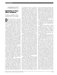
he American Diabetes Association (ADA) has recommended selective screening for gestational diabetes mellitus (GDM) (1). Pregnant women aged ⬍25 years, who have normal body weight, no family history of diabetes, and are not members of an ethnic/racial group with a high prevalence of diabetes, are regarded as a low-risk group for GDM and need not be screened. The effect of selective screening guidelines has been investigated in Caucasian populations using a 100-g 3-h oral glucose tolerance test (OGTT) (2,3). We report here the effect of the selective screening protocol. A total of 9,471 pregnant women in Tianjin, China, took part in a universal screening program from December 1998 to December 1999. The screening test consisted of a 50-g 1-h glucose test and was carried out at 26- to 30-weeks’ gestation. A total of 888 (9.4%) women had a glucose reading ⬎7.8 mmol/l, of whom 701 undertook a further 75-g 2-h OGTT using the WHO diagnostic criteria for GDM (baseline: ⱖ7.0 mmol/l; 2-h: ⱖ7.8 mmol/l) (4). A total of 171 women were confirmed to have GDM (prevalence 1.8%). Age, prepregnancy BMI, and family history of diabetes were risk factors for GDM in this cohort. The prevalence of overweight (BMI ⱖ25 kg/m2) was low (10%), and family history of diabetes was uncommon (8%). Furthermore, the onechild policy has resulted in a cohort of 98% (9,240/9,471) of nulliparas. Twenty-eight percent of women were ⬍25 years of age. The application of the ADA selective screening guideline in this study would exclude 24% (2,248/9,469) of women from the screening test. An estimated 12% of women with confirmed GDM under the WHO criteria would otherwise have been denied the opportunity for early detection. These findings differ substantially from reports using the ADA rec796
ommendations: exclusion of 10% of women in the screening and oversight of 4% of GDM women (3). We adopted a similar, although slightly later, approach to the initial screening (26to 30- vs. 24- to 28-weeks’ gestation) (1). The WHO diagnostic criteria have been shown to give a higher estimation of GDM prevalence (5,6) in comparison with the ADA criteria. However, the prevalence of GDM in our study population was low. As our subjects are deemed a high-risk group (of Asian backgrounds) under the ADA selective screening guidelines, the lack of other risk factors is an important determinant of GDM prevalence. The greater proportion of Chinese women with GDM who would fail to be identified using selective screening, compared with the proportion shown in other studies, is unlikely to be explained by either the delay in screening or the use of the WHO criteria. The low frequency of risk factors for GDM in this cohort was associated with a low prevalence of GDM. However, young and lean women were not immune from the development of GDM. We conclude that if selective screening is to be considered in this population, different age and BMI cutoff points are required, and other risk factors for GDM (such as stature) may need to be considered for inclusion in any revised selective screening recommendation. XILIN YANG, PHD1 BRIDGET HSU-HAGE, PHD1 LICHUN YU, MD2 DAVID SIMMONS, MD1 From the 1Department of Rural Health, University of Melbourne, Melbourne, Australia; and the 2Tianjin Institute for Women’s Health, Tianjin, China. Address correspondence to Bridget Hsu-Hage, Department of Rural Health, Faculty of Medicine, University of Melbourne, PO Box 6500, Shepparton, Victoria 3632, Australia. E-mail: bhhage@unimelb. edu.au. ● ● ● ● ● ● ● ● ● ● ● ● ● ● ● ● ● ● ● ● ● ● ●
References 1. The Expert Committee on the Diagnosis and Classification of Diabetes Mellitus: Report of the Expert Committee on the Diagnosis and Classification of Diabetes Mellitus. Diabetes Care 20:1183–1197, 1997 2. Danilenko-Dixon DR, Van Winter JT, Nelson RL, Ogburn PL Jr: Universal versus selective gestational diabetes screening: application of 1997 American Diabetes Association recommendations. Am J Obstet Gynecol 181:798 – 802, 1999
3. Williams CB, Iqbal S, Zawacki CM, Yu D, Brown MB, Herman WH: Effect of selective screening for gestational diabetes. Diabetes Care 22:418 – 421, 1999 4. Alberti KG, Zimmet PZ: Definition, diagnosis and classification of diabetes mellitus and its complications. Part 1: diagnosis and classification of diabetes mellitus provisional report of a WHO consultation [see comments]. Diabet Med 15:539 – 553, 1998 5. Deerochanawong C, Putiyanun C, Wongsuryrat M, Serirat S, Jinayon P: Comparison of National Diabetes Data Group and World Health Organization criteria for detecting gestational diabetes mellitus. Diabetologia 39:1070 –1073, 1996 6. Pettitt D, Narayan K, Bennett P, Knowler W, Hanson R: Comparison of World Health Organization and National Diabetes Data Group procedures to detect abnormalities of glucose tolerance during pregnancy. Diabetes Care 17:1264 –1268, 1994
Cigarette Smoking Affects Glycemic Control in Diabetes
T
ight glycemic control in diabetes is one of the cornerstones of management. Glycemic control is usually assessed with HbA1c. The Diabetes Control and Complications Trial showed that in type 1 diabetes, decreasing HbA1c from 9.0 to 7.2% resulted in a 50 –75% reduction in retinopathy, nephropathy, and neuropathy (1). The U.K. Prospective Diabetes Study (UKPDS) showed that in type 2 diabetes, decreasing HbA1c from 7.9 to 7.0% decreased retinopathy and nephropathy (2). Increased insulin resistance occurs in smokers with and without diabetes (3,4). Smoking is also a risk factor for development of type 2 diabetes. The aim of this study was to determine whether smoking cessation was associated with improved glycemic control. This study was a prospective cohort design and included 34 patients who ceased smoking, were followed for 1 year, and continued not to smoke at 1 year. Two control groups were randomly selected from patients participating in a prospective diabetes complications study (DCS). A DCS includes history, examination, ophthalmological review, and invesDIABETES CARE, VOLUME 24, NUMBER 4, APRIL 2002
Letters
Beneficial Effect of Diabetes on Acute Intermittent Porphyria
A
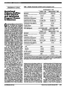
Figure 1—HbA1c before and after cessation of cigarette smoking in 34 patients.
tigations, including HbA1c. The control groups were 34 never smokers and 34 current smokers. Smoking status was assigned based on patient self report. HbA1c was measured by high-performance liquid chromatography, with 3.5–5.7% as the normal range. Results are presented as means ⫾ 1 SD. The study was approved by the institutional ethics committee. Average consumption before cessation was 24 cigarettes per day. Mean age for the group who stopped smoking was 60 ⫾ 15 years. A total of 7 subjects had type 1 diabetes, and 27 had type 2 diabetes. Mean HbA1c was 7.7 ⫾ 2.2% while smoking and 7.0 ⫾ 1.6% after smoking cessation (P ⫽ 0.048) (Fig. 1). Thus, the change in HbA 1c was ⫺0.70% (P ⬍ 0.0001 vs. expected change of 0%). Mean change was 0.07% for never smokers (P ⫽ 0.036) and 0.12% for continuing smokers (P ⬍ 0.01). Some studies have shown increased insulin resistance in association with cigarette smoking (3,4). It is possible that cigarette smoking may affect glycosylation of hemoglobin, although no studies were found with a literature search. It is possible that some patients may report smoking cessation when they continue to smoke. However, it seems unlikely that patients with diabetes would report continuing to smoke after stopping. Patients incorrectly reporting nonsmoking would tend to make the observed difference smaller than the true difference. To our knowledge, this is the first study documenting improvement in HbA1c with smoking cessation. It may reflect a change in lifestyle factors, with the decision to stop smoking, or a direct effect. The 0.7% decrease is clinically significant, approaching the 0.9% in the UKPDS (2). The small but significant inDIABETES CARE, VOLUME 24, NUMBER 4, APRIL 2002
creases in other groups parallel the 0.12% rise per year in the UKPDS metformin group (2). These results constitute yet another reason why patients with diabetes should not smoke. JENNY E. GUNTON, MBBS, FRACP LINDA DAVIES, BAPPSC ERROL WILMSHURST, MBBS, MRACP, MD, FRACP
GREG FULCHER, MBBS, MD, FRACP AIDAN MCELDUFF, MBBS, PHD, FRACP From the Department of Endocrinology, Royal North Shore Hospital, Sydney, Australia. Address correspondence to Dr. Jenny Gunton, Department of Endocrinology, Royal North Shore Hospital, St. Leonards 2065, Australia. E-mail:
[email protected].
● ● ● ● ● ● ● ● ● ● ● ● ● ● ● ● ● ● ● ● ● ● ●
References 1. Diabetes Control and Complications Trial: The effect of intensive treatment of diabetes on the development and progression of long-term complications in insulin-dependent diabetes mellitus. N Engl J Med 329:977–986, 1993 2. UK Prospective Diabetes Study: Intensive blood-glucose control with sulphonylureas or insulin compared with conventional treatment and risk of complications in patients with type 2 diabetes (UKPDS 33). Lancet 352:837– 853, 1998 3. Targher G, Alberiche M, Zenere MB, Bonadonna RC, Muggeo M, Bonora E: Cigarette smoking and insulin resistance in patients with noninsulin-dependent diabetes mellitus. J Clin Endocrinol Metab 82: 3619 –3624, 1997 4. Ronnemaa T, Ronnemaa EM, Puukka P, Pyorala K, Laakso M: Smoking is independently associated with high plamsa insulin levels in nondiabetic men. Diabetes Care 19:1229 –1232, 1996
cute intermittent porphyria (AIP) is characterized by attacks of abdominal pain and neuropsychiatric symptoms. In northern Sweden, about half of those patients carrying the gene encoding for this condition have experienced attacks with abdominal pain, more frequently and more severely affecting women. In biochemical terms, AIP is an autosomal hereditary metabolic aberration resulting from a partial defect in the activity of the third-step enzyme (porphobilinogen deaminase [PBGD]) during the course of heme synthesis (1). Carbohydrate ingestion blocks d-aminolevulinic acid (ALA)synthase, as has been demonstrated in numerous clinical and experimental studies. However, the mechanisms by which carbohydrates modulate the components of porphyrins and heme synthesis are highly complex and only partially elucidated to date (2). The main long-term complications of AIP are polyneuropathy (3), hepatocellular carcinoma (HCC) (4), and renal insufficiency (5). Treatment of AIP patients entails treating both the symptoms and the complications, but also requires an endeavor to reverse the fundamental disease by prescribing a carbohydrate-rich diet and by treating the attacks with intravenous infusions of glucose (2) or heme (6). We conducted a population-based study on AIP patients aged ⱖ18 years living in northern Sweden, in which 319 participated (95%). A total of 16 patients (5 women) with AIP and type 2 diabetes were found, with a mean age of 67 years. Eight of these patients had AIP symptoms, with three patients suffering severe, recurring attacks. After the onset of their diabetes, no patient suffered attacks or any other AIP symptoms (7). During several weeks in the hospital ward, it was possible to closely examine and study the course of developing diabetes in a 52-year-old woman who had previously been hospitalized at the Department of Medicine due to AIP attacks on 46 occasions. At 4 years after 797
Letters
menopause, her AIP was still unremitting, with recurrent attacks. However, after she had developed a metabolically rather mild type 2 diabetes, the AIP symptoms subsided. Mean values on 13 occasions of testing for urinary porphobilinogen (PBG) during 6 months before and on 11 occasions during the first 4 months after she developed diabetes were 65.9 and 10.2 mol/l, respectively (reference levels 1.3–11.0 mol/l) (P ⫽ 0.0001). Now, 5 years later, she is still free from AIP symptoms, the levels of ALA and PBG in her urine fluctuate around normal or just above the upper reference levels, and she leads “a good life.” More than 2 decades ago on the hospital ward, I noticed two patients, both with AIP and HCC, which are rare diseases in the Western world. To test whether this was a random finding, a retrospective study of HCC in deceased AIP patients was performed using records of 206 carriers ⬎15 years of age. The diagnosis was based on a clearly depressed activity of PBGD. Statistically, only 1 patient with HCC was expected, yet 11 (7 women) were found. This coincidence was highly significant. The findings were verified in the autopsy records. The mean age was 67 years (8). Two municipalities in northern Sweden, Arjeplog (population 3,800) and Arvidsjaur (population 8,080), have the highest prevalence of AIP patients in the world (1–2%). In a retrospective population-based study of mortality during the period 1978 –1990, AIP patients (n ⫽ 33) were compared with all other individuals who had died during this period (n ⫽ 2,089). In the AIP group, nine patients (27%) had HCC, whereas in the other group, only four patients (0.2%) had HCC (⬍0.0001) (9). In an attempt to elucidate the molecular mechanism of the carcinogenesis in this HCC, archived specimens were collected (9). Selective mutations at codons 249 and 166 of the p53 gene have been described in HCC, associated with aflatoxin and hepatitis B virus (10). HCC in AIP, on the other hand, is characterized by an absence of the genes mentioned and by only mild or no liver cirrhosis. A diminished free heme pool and an increased concentration of ALA could lead to an increase in reactive oxygen radicals and may, in a longer perspective, cause cancer (11). Recurrant AIP attacks ceased when 798
the patients became diabetic. None of the 16 diabetic patients with AIP had HCC. Of all the 30 AIP patients with HCC registered, none had diabetes, whereas in a population-based group of individuals in southern Sweden (mean age 67 years), diabetes was found in 12.8% of the men and 15.0% of the women (12). This suggests that diabetes also counteracts HCC in AIP patients, probably by normalization of ALA (11). FOLKE LITHNER MD, PHD From the Department of Internal Medicine, University Hospital, Umea, Sweden. Address correspondence to Dr. Folke Lithner, Department of Internal Medicine, University Hospital, Umea, Sweden S-90185. E-mail: folke.lithner @medicin.umu.se.
● ● ● ● ● ● ● ● ● ● ● ● ● ● ● ● ● ● ● ● ● ● ●
References 1. Strand LJ, Meyer UA, Felsher BF, Redeker AC, Marver HS: Decreased red cell uroporphyrinogen 1 synthetase activity in intermittent acute porphyria. J Clin Invest 51:2530 –2536, 1972 2. Doss M, Verspohl F: The “glucose effect” in acute hepatiac porphyrias and in experimental porphyria. Klin Wschr 9:727– 735, 1981 3. Wikberg A, Andersson C, Lithner F: Signs of neuropathy in the lower legs and feet of patients with acute intermittent porphyria. J Intern Med 248:27–32, 2000 4. Andersson C, Bjersing L, Lithner F: The epidemiology of hepatocellular carcinoma in patients with acute intermittent porphyria. J Intern Med 240:195–201, 1996 5. Andersson C, Wikberg A, Stegmayr B, Lithner F: Renal symptomatology in patients with acute intermittent porphyria: a population-based study. J Intern Med 248: 319 –325, 2000 6. Mustajoki P, Normann Y: Early administration of heme arginate for acute porphyric attacks. Arch Intern Med 153:2004 –2008, 1993 7. Andersson C, Bylesjo¨ I, Lithner F: Effects of diabetes mellitus on patients with acute intermittent porphyria. J Intern Med 245: 193–197, 1999 8. Lithner F, Wetterberg L: Hepatocellular carcinoma in patients with acute intermittent porphyria. Acta Med Scand 215:271– 274, 1984 9. Bjersing L, Andersson C, Lithner F: Hepatocellular carcinoma in patients from
northern Sweden with acute intermittent porphyria. Cancer Epidemiol Biomarkers Prev 2:393–397, 1996 10. Hsu JC, Metcalf RA, Sun T, Welsh JA, Wang NJ, Harris CC: Mutational hotspot in the 53p gene in hepatocellular carcinomas. Nature (Lond) 350:427– 428, 1991 11. Battle AM: Porphyrins, porphyrias, cancer and photodynamics: a model for carcinogenesis. J Photochem Photobiol B 20:5– 22, 1993 12. Andersson DKG, Sva¨ rdsudd K, Tibblin G: Prevalence and incidence of diabetes in a Swedish community 1972– 87. Diabet Med 8:428 – 434, 1991
Risk Factors of Developing Proliferative Retinopathy in Type 1 Diabetic Patients Role of BMI
I
n the July issue of Diabetes Care, we read with interest the study by Zangh et al. (1). They revisited material from the Diabetes Control and Complications Trial database in order to search for potential prognostic factors of developing retinopathy in patients with extreme (either good or poor) metabolic control, but who were free of the complication at baseline. The development and progression of retinopathy was defined by a three-step change or more, according to the Early Treatment Retinopathy Study protocol, on stereoscopic color fundus photographs. Their conclusion was that other than duration of diabetes and metabolic control, BMI had a significant predictive value. We found a similar influence of BMI in our population of young diabetic subjects when we examined the risk factors of progression from background retinopathy to proliferative retinopathy. In 1991, we published a longitudinal study (2), begun in the 1970s (3), in order to detect the initial retinal abnormalities at fluorescein angiography in 161 type 1 diabetic children and adolescents. Of these patients, 69 developed an early retinopathy. We showed, in several studies covering ⬎20 years (summary in 4), that the development of microaneurysms, which are irreversible lesions, can be preceded by fluorescein leakage due to dis-
DIABETES CARE, VOLUME 24, NUMBER 4, APRIL 2002
Letters
ruption of the blood-retinal barrier (5), and that risk factors for early retinopathy are diabetes duration, age at diagnosis (with younger children having longer times to retinopathy), puberty and sex (with onset 1 year earlier in girls than in boys), and glycated hemoglobin (GHb) measured over several years. Retinopathy was not found in children ⬍12 years of age and was detected only after at least 3 years of diabetes. The mean age at which early abnormalities occurred was 16 years, after a mean duration of diabetes of 8 years (2). In the year 2000, 32 subjects (15 women and 17 men), among the 69 patients who developed early retinopathy (2), were always treated by our team. Their mean age was 33 years, and mean diabetes duration was 26 years. Some potential risk factors (GHb, total cholesterol, blood pressure, BMI, insulin dose, frequency of home blood glucose monitoring and of clinic attendance, tobacco, and presence of other complications), measured during the whole follow-up, were analyzed in relation with the evolution of retinopathy to the proliferative stage using fluorescein angiography. In the statistical analysis, stepwise logistic regression was used to identify the most important parameters for the development of proliferative retinopathy. Proliferative retinopathy was diagnosed in six patients (19%), three men and three women. Its occurrence was significantly related to poor glycemic control during the preceding years (cumulated GHb was 143 vs. 120%; 100% is the upper normal limit, P ⫽ 0.049), cumulated cholesterol levels (⬎200 mg/dl, P ⫽ 0.014), higher BMI (27 vs. 22 kg/m2, P ⫽ 0.035), and the presence of other complications (P ⫽ 0.029). However, higher levels of LDL cholesterol are related to poor metabolic control (6). In conclusion, our data suggest that the risk factors for developing proliferative retinopathy are poor long-term metabolic control, which is well known, and an elevated BMI, which is novel, as noticed by Zhang et al. (1). They confirm the importance of always maintaining a GHb level ⬍120% of the upper normal limit, as we have shown in homozygous diabetic twins (7), which is possible in diabetic children and adolescents (8). HARRY DORCHY, MD, PHD CATHERINE CLAES, MD CLAIRE VEROUGSTRAETE, MD DIABETES CARE, VOLUME 24, NUMBER 4, APRIL 2002
From the Diabetology Clinic, University Children’s Hospital Queen Fabiola, Brussels, Belgium; and the Ophthalmology Clinic, University Hospital Brugmann, Brussels, Belgium. Address correspondence to Harry Dorchy, Clinique de Diabe´ tologie, Hoˆ pital Universitaire des Enfants Reine Fabiola, Ave. JJ Crocq 15, B-1020 Bruxelles, Belgium. E-mail:
[email protected]. ● ● ● ● ● ● ● ● ● ● ● ● ● ● ● ● ● ● ● ● ● ● ●
References 1. Zangh L, Krentowski G, Albert A, Lefebvre PJ: Risk of developing retinopathy in Diabetes Control and Complications Trial type 1 diabetic patients with good or poor metabolic control. Diabetes Care 24: 1275–1279, 2001 2. Verougstraete C, Toussaint D, De Schepper J, Haentjens M, Dorchy H: First angiographic abnormalities in childhood diabetes: types of lesions. Graefes Arch Clin Exp Ophthalmol 229:24 –32, 1991 3. Dorchy H, Toussaint D, De Vroede M, Ernould C, Loeb H: Diagnostic de la re´ tinopathie diabe´ tique infantile par angiographie fluoresce´ inique: description des le´ sions initiales. Nouv Presse Med 6:345–347, 1977 4. Dorchy H: De´ pistage des complications subcliniques chez les jeunes diabe´ tiques: expe´ rience bruxelloise. Ann Pediatr (Paris) 45:585– 606, 1998 5. Dorchy H: Characterization of early stages of diabetic retinopathy: importance of the breakdown of the blood-retinal barrier (Letter). Diabetes Care 16:1212–1213, 1993 6. Willems D, Dorchy H: Taux des lipoprote´ ines et des apolipoprote´ ines chez les jeunes diabe´ tiques insulinode´ pendants: relations avec l’he´ moglobine glycosyle´ e et la fructosamine. Presse Med 19:17–20, 1990 7. Verougstraete C, Libert J, Dorchy H: Discordant diabetic retinopathy in homozygous twins: the importance of good metabolic control. J Pediatr 134:658, 1999 8. Dorchy H, Roggemans M-P, Willems D: Glycated hemoglobin and related factors in diabetic children and adolescents under 18 years of age: a Belgian experience. Diabetes Care 20:2– 6, 1997
Is Type 1 Diabetes Transmissible by Bone Marrow Allograft?
A
review of the medical literature suggests that type 1 diabetes in a bone marrow allograft donor may be transmitted to the recipient after a suc-
cessful transplant (1,2). We report a 21year follow-up after a successful allograft for aplastic anemia. The donor had type 1 diabetes at the time of transplant. The recipient has developed a number of antibodies against pancreatic islet cells, and although these have persisted for a number of years, she has not developed diabetes. A 6-year-old Caucasian girl presented in 1976 with aplastic anemia. She showed a partial response to androgens, but this was not sustained, and in 1979 she received a bone marrow transplant (BMT) from her HLA-matched male sibling, who was 3 years older and had been diagnosed with type 1 diabetes 7 years earlier. Both patients were group O Rh (D) positive and HLA-A1, -2, -B8, and MLC nonreactive. The transplant procedure and the progress post-BMT have already been published (3). Full donor engraftment of the hemopoietic system has been documented on a number of occasions over the past 21 years. Mild acute graft versus host disease (GVHD) was followed by chronic GVHD, which was severe in the mouth. There was also a mild but persistent patchy fibrosis of the skin. Oral GVHD eventually responded to dental clearance and immunosuppressive therapy. She required prednisone, usually 10 mg/day, from June 1979 to June 1987. Azathioprine, 100 mg/day, was started in August 1979 and stopped in January 1986. She has had no immunosuppressive therapy since June 1987. Evidence of hyposplenism judged by blood film appearances was noted in 1983, but she has not experienced any significant sepsis to date. At the time of this report, she remains well and has had two healthy children. Both pregnancies and deliveries were uneventful, and the children are in good health. The recipient was negative for islet cell antibodies (ICAs) by indirect immunofluorescence until 1994, when a positive result of 10 Juvenile Diabetes Foundation units was recorded. The development of high titer anti-nuclear antibodies since 1996 has made the detection of ICAs by indirect immunofluorescence virtually impossible. Recipient sera revealed high titer anti-GAD antibodies, determined using Diaplets Anti-GADPLUS ELISA (Roche Diagnostics) in 1994, 1997, 1998, 1999, and 2000 (maximum 3,086 ng/ml, normal range 0 –32). However, neither anti–IA-2 antibodies (deter799
Letters
mined using Diaplets Anti-IA-2 ELISA, Roche Diagnostics) nor insulin antibodies (measured by radioimmunoassay) were detected at any time during follow-up. Fasting glucose measurements in 1999 and 2000 were 4.0 and 5.4 mmol/l, respectively. The diabetic donor was ICAnegative at the time of transplant and was negative for ICA, anti-GAD, and anti– IA-2 on follow-up testing in 1999. Evidence of autoimmunity would be expected at or soon after diagnosis with type 1 diabetes but was predictably absent a long time after presentation. HLA-DQ␣ and - allele typing was performed using DNA extracted from buccal epithelial and white blood cells in the recipient and in DNA from white blood cells in the donor. The recipient showed HLA identity with the donor at these loci. Both individuals were characterized by 0103/0501 at the HLA-DQ␣ locus and 0201/0602, three at the HLA-DQ locus. Endomysial antibodies, anti-myeloperoxidase, tissue antibodies, and antibodies against proteinase-3 and double stranded DNA were not detected. Both IgG and IgA antibodies to Gliadin were positive in 1999 but were not detected at follow-up testing in 2000. Transfer of type 1 diabetes by BMT from a donor with type 1 diabetes was first reported in 1993 (1). The recipient developed type 1 diabetes–associated antibodies and developed diabetes 4 years after BMT from an HLA-identical brother. Subsequently, a review of the International Bone Marrow Transplant Registry revealed that 15 transplants had been undertaken from bone marrow donors with diabetes, including nine donors with confirmed diagnoses of type 1 diabetes (2). Of the three patients who had completed more than 2 years follow-up, two recipients had progressed to type 1 diabetes, all having received transplants in 1989. This report describes a BMT that took place in 1979. The donor had been diagnosed with type 1 diabetes 7 years before the transplant and was negative for ICAs at the time of transplant. The recipient has over time developed ICAs and high levels of anti-GAD antibodies. HLA identity together with ICA and/or anti-GAD confer a high positive predictive value for development of type 1 diabetes in siblings of children with type 1 diabetes (4). However, the patient also had chronic GVHD, a condition in which multiple autoantibodies are commonly found (5). We can800
not find any report that looked for ICAs, anti-GAD, or insulin autoantibodies in chronic GVHD. It is possible that these antibodies have developed in our patient as part of the spectrum that occurs secondary to GVHD and might not be related to the fact that the donor had type 1 diabetes. Alternatively, the autoimmune process observed in the recipient may have been modified by the Azathioprine therapy, which was in place for 6 years immediately post-transplant. Blood glucose measurements over the past 21 years have shown no evidence of type 1 diabetes. Follow-up of patients after BMT for ⬎20 years is rare, and in particular, there is very little information on the long-term outcome in patients when the donor has type 1 diabetes. Follow-up is needed to ensure this individual remains glucose tolerant. MICHAEL E. BEARD, MD1 JINNY A. WILLIS, PHD2 RUSSELL S. SCOTT, MD2 JEFF W. NESBIT, BSC2 From the 1Department of Haematology, Christchurch Hospital, Christchurch, New Zealand; and the 2Lipid and Diabetes Research Group, Hagley Building, Christchurch Hospital, Christchurch, New Zealand. Address correspondence to Dr. Jinny Willis, Lipid and Diabetes Research Group, Hagley Building, Christchurch Hospital, Christchurch, New Zealand. E-mail:
[email protected]. ● ● ● ● ● ● ● ● ● ● ● ● ● ● ● ● ● ● ● ● ● ● ●
References 1. Lampeter EF, Homberg M, Quabeck K, Schaefer UW, Werner P, Bertrams J, GrosseWilde H, Gries FA, Kolb H: Transfer of insulin-dependent diabetes between HLAidentical siblings by bone marrow transplantation. Lancet 341:1243–1244, 1993 2. Lampeter EF, McCann SR, Kolb H: Transfer of insulin-dependent diabetes by bone marrow transplantation. Lancet 351:568 – 569, 1998 3. Darlow BA, Abbott GD, Beard ME, Fox HWJ, Hamer JW, Heaton DC: Bone marrow transplantation for severe aplastic anaemia. N Z Med J 91:86 – 89, 1980 4. Kulmala P, Savola K, Reijonen H, Veijola R, Vahasalo P, Karjalainen J, Tuomilehto-Wolf E, Ilonen J, Tuomilehto J, Akerblom HK, Knip M, the Childhood Diabetes in Finland Study Group: Genetic markers, humoral autoimmunity, and prediction of type 1 diabetes in siblings of affected children. Diabetes 49: 48 –58, 2000 5. Quaranta S, Shulman H, Ahmed A, Shoenfeld Y, Peter J, McDonald GB, Van
de Water J, Coppel R, Ostlund C, Worman HJ, Rizzetto M, Tsuneyama K, Nakanuma Y, Ansari A, Locatelli F, Paganin S, Rosina F, Manns M, Gershwin ME: Autoantibodies in human chronic graftversus-host disease after hematopoietic cell transplantation. Clinical Immunology 91:106 –116, 1999
Comorbidity of Type 1 Diabetes and Anorexia Nervosa in a 6-Year-Old Girl
A
high prevalence of eating disorders has been described in female adolescents with type 1 diabetes as being almost twice as high as that found in their nondiabetic peers (1–3). Weight gain caused by insulin therapy, dietary restraint, and food preoccupation may predispose diabetic girls to develop a clinical or subclinical eating disorder (4 – 6); intentional insulin omission is the most common symptom underlying an eating disorder (7). The coexistence of these conditions could lead to poor metabolic control and increase the risk of microvascular complications (8). Very few reports have been published on eating disorders in childhood (9,10), and in only one report this condition was also described in diabetic children, starting from the age of 8 years (4). We describe the successful treatment of a 6-year-old girl with comorbid type 1 diabetes and anorexia nervosa. This case underlines that children with diabetes, as well as adolescents, may be prone to develop a disorder of eating behavior, and that an early diagnosis and multidisciplinary approach are fundamental for a positive outcome. R.F. was diagnosed to be affected by type 1 diabetes at the age of 4.5 years at the Department of Pediatrics of University Federico II in February 1995. She presented a partial remission of diabetes, lasting 16 months; she was treated with two daily doses of insulin (0.2– 0.3 units 䡠 kg⫺1 䡠 day⫺1), and her HbA1c ranged from 5.0 to 6.2% (normal values ⬍6%). In July 1996, when her HbA1c rose to 9.6%, she was treated with 0.8 units 䡠 kg⫺1 䡠 day⫺1 of insulin in three daily doses. Her height and weight were in the normal range (50th percentile according to Tanner growth charts). In October DIABETES CARE, VOLUME 24, NUMBER 4, APRIL 2002
Letters
1996, her HbA1c was 6.6%. One month later, she began to first refuse several foods and then any kind of food. Consequently, she presented severe weight loss and frequent hypoglycemic crises; therefore, her insulin dose was reduced. Chronic bowel diseases, tumors, and other endocrine or autoimmune diseases were excluded. Diagnosis of anorexia nervosa was performed according to the DSM IV criteria. The personality profile was characterized by a defensive system set up against depression, which hindered the girl from recognizing the state of the disease through a denial and splitting mechanism. In addition, her mother was affected by depression and required pharmacological treatment, and the parental couple was in violent conflict. Medical treatment of anorexia was based on enteral nutrition from January to June of 1997. Subsequently, the girl accepted and maintained a normal and complete oral nutrition, which has persisted until present (10.5 years of age). She presents a normal growth and a good control of diabetes (HbA1c 6.9 –7.5%). Psychiatric treatment consisted of intensive individual psychoanalysis that the patient is still receiving. In the first year, she presented a progressive decrease of anguish relative to weight gain and food ingestion; anorexia completely disappeared. Successively, she expressed the fear of “intrusion inside her body” by very severe obsessive-phobic symptoms. At the present time, she has several fantasies connected to diabetes acceptance. Treatment is actually addressed to give support to the “self.” Diabetes is a complex disease that affects all aspects of the daily life, in particular, eating behaviors. Dietary restraint and food preoccupation break into all of the daily events of life, leading to important changes in the aspects of conviviality and social relationships. Food may become the main enemy of the child or the main reason of contrast with parents. In our patient, symptoms of anorexia started immediately after the “honeymoon” period, when dietary regimen is often poorly stressed and parental surveillance on diabetic management is less stringent. Successively, recurrence of hyperglycemia and the increased insulin requirement might have been a trigger to precipitate an eating disorder in a prone subject. Moreover, the environmental deDIABETES CARE, VOLUME 24, NUMBER 4, APRIL 2002
mands related to the concomitant beginning of schooling may have exerted an additional role. Anorexia in a very young child does not lead to insulin omission, as in adolescents, because children are still under their parents’ surveillance. On the contrary, undernutrition is the main clinical problem at this age, causing recurrent hypoglycemic crises and troubles in insulin management. Furthermore, whereas in otherwise healthy children appropriate evaluation and treatment may precociously stop an eating disorder, in diabetic children treatment is complicated by the lifelong persistence of diabetes, with its rules regarding food regulation, multiple glycemic controls, and insulin injections. However, early diagnosis of eating disorder and long-term multidisciplinary treatment are very important in a diabetic child in order to avoid the risk of persistence until adolescence or adulthood, when it may be potentially fatal. 1
ADRIANA FRANZESE, MD GIULIANA VALERIO, MD, PHD2 PIETRO BUONO, MD2 ENZA MOZZILLO, MD1 ANTONELLA GRITTI, MD3 MARIA ANTONIETTA LUCARIELLO, MD3 From the 1Department of Pediatrics, University “Federico II,” Naples, Italy; the 2Department of Experimental and Clinical Pathology and Medicine, University of Udine, Udine, Italy; and 3Cattedra di Neuropsichiatria Infantile, Second University of Naples (SUN), Naples, Italy Address correspondence to Adriana Franzese, MD, Department of Pediatrics, via S. Pansini 5, 80131 Napoli, Italy. E-mail:
[email protected]. ● ● ● ● ● ● ● ● ● ● ● ● ● ● ● ● ● ● ● ● ● ● ●
References 1. Jones JM, Lawson ML, Daneman D, Olmsted MP, Rodin G: Eating disorders in adolescent females with and without type 1 diabetes: cross sectional study. BMJ 320: 1563–1566, 2000 2. Meltzer LJ, Johnson SB, Prine JM, Banks RA, Desrosiers PM, Silverstein JH: Disordered eating, body mass, and glycemic control in adolescents with type 1 diabetes. Diabetes Care 24:678 – 682, 2001 3. Hoffmann RP: Eating disorders in adolescents with type 1 diabetes: a close look at a complicated condition. Postgrad Med 109:67– 69, 2001 4. Striegel-Moore RH, Nicholson TJ, Tamborlane WV: Prevalence of eating disorder symptoms in preadolescents and adolescent girls with IDDM. Diabetes Care 15: 1361–1368, 1992
5. Peveler RC, Faiburn CG, Boller I, Dunger D: Eating disorders in adolescents with IDDM: a controlled study. Diabetes Care 15:1356 –1360, 1992 6. Verrotti A, Catino M, De Luca FA, Morgese G, Chiarelli F: Eating disorders in adolescents with type 1 diabetes mellitus. Acta Diabetol 36:21–25, 1999 7. Crow SJ, Keel PK, Kendall D: Eating disorders and insulin dependent diabetes mellitus. Psycosomatics 39:233–243, 1998 8. Herpetz S, Albus C, Wagener R, Kocnar M, Wagner R, Henning A, Foerster H, Sculze Schleppinghoff B, Thomas W, Kohle K, Mann K, Senf W: Comorbidity of diabetes and eating disorders. Does diabetes control reflect disturbed eating behavior? Diabetes Care 21:1110 –1116, 1998 9. Sokol MS, Steinberg D, Zerba KJ: Childhood eating disorders. Curr Opin Pediatr 10:369 –377, 1998 10. Robb AS: Eating disorders in children. Psychiatr Clin North Am 24:259 –270, 2001
Childhood Abuse and Diabetes in the Community
D
iabetes is a significant pubic health problem among children and adolescents, and its prevalence has rapidly increased in recent years (1). There is some evidence to suggest that chronic stress is associated with onset of physical illness, such as diabetes (2). Childhood abuse is thought to be one source of severe, persistent stress. Preclinical data suggest that stress is associated with onset of diabetes (3). No previous study has investigated this hypothesis in a epidemiologic sample of humans. To obtain preliminary evidence in support of this hypothesized etiologic factor, we sought to determine the association between childhood emotional and physical abuse and the odds of self-reported diabetes among adults in the general population. Multivariate logistic regression analyses were used to analyze data from the Midlife Development in the U.S. Survey (MIDUS) (n ⫽ 3,032) (4), a representative sample of adults aged 25–74 years in the U.S., in order to compute odds ratios (ORs) (with 95% CIs) of the association between history of childhood abuse and self-reported diabetes. Our results revealed that self-reported diabetes occurred in 4.8% of the adult population (aged 25–74 years). Childhood abuse was 801
Letters
associated with a significantly increased odds of self-reported diabetes (OR 2.0 [95% CI 1.04 –2.8]), which persisted after adjusting for differences in sociodemographic characteristics and current common mental disorders. Specifically, individuals who reported maternal emotional abuse (OR 3.4 [95% CI 1.9 – 6.4]), maternal physical abuse (2.8 [1.2– 6.4]), and severe maternal physical abuse (3.4 [1.2–9.4]) had significantly higher rates of self-reported diabetes. Our findings provide preliminary support for the hypothesis that childhood abuse may be associated with increased likelihood of self-reported diabetes among adults in the community. Limitations of our analyses include lack of information on type of diabetes, family history, and self-reported diabetes without data on physiological measures. Replication of these findings is needed. Future studies that use longitudinal data with more detailed information on diabetes type, age of onset, childhood abuse experience, and family history may help to improve our understanding of the mechanism of this observed association. RENEE D. GOODWIN, PHD STUART P. WEISBERG, BS From the 1Department of Psychiatry and Epidemiology, Columbia University College of Physicians and Surgeons, New York, New York; and the 2Department of Medicine, Columbia University College of Physicians and Surgeons, New York, New York. Address correspondence to Dr. Renee D. Goodwin, Department of Psychiatry, Columbia University College of Physicians and Surgeons, 1051 Riverside Dr., Unit 43, New York, NY 10032. Email:
[email protected].
● ● ● ● ● ● ● ● ● ● ● ● ● ● ● ● ● ● ● ● ● ● ●
References 1. Larkin M: Diabetes on the rise worldwide and webwide (Letter). Lancet 10:815, 2001 2. Fava GA, Sonino N: Psychosomatic medicine: emerging trends and perspectives. Psychother Psychosom 69:184 –197, 2000 3. Surwit RS, Feinglos MN, Livingston EG, Kuhn CM, McCubbin JA: Behavioral manipulation of the diabetic phenotype in ob/ob mice. Diabetes 33:616 – 618, 1984 4. Wang PS, Berglund P, Kessler RC: Recent care of common mental disorders in the United States: prevalence and conformance with evidence-based recommendations. J Gen Intern Med 15:284 –292, 2000
802
Postischemic Microcirculatory Blood Flow Correlates Negatively and Independently With Plasma C-Reactive Protein in Longstanding Type 1 Diabetes
M
oderately increased plasma Creactive protein (CRP), an acute phase protein produced by the liver and an exquisitely sensitive marker of inflammation, is associated with an increased risk of cardiovascular disease (1). It is not known how systemic inflammation leads to atherosclerosis, but experimental (2) and clinical (3) studies have suggested that it may do so by causing endothelial dysfunction, a process central to the development of atherosclerosis. Reactive hyperemia (RH) is an endothelium-dependent, physiological, and probably protective response to tissue ischemia that is reduced in patients with atherosclerosis or risk factors for atherosclerosis. The forearm vascular bed, which is readily accessible and rarely affected by atheromatous change, is a useful site to study RH using noninvasive techniques. A close relationship has been shown between coronary endothelial function and RH in the human forearm (4). To further investigate the relationship between inflammation and endothelial dysfunction, we have studied RH and circulating CRP in 50 subjects (28 men), all of whom had type 1 diabetes for at least 25 years. Urinary albumin/creatinine ratio (ACR), total and HDL cholesterol, and HbA1c were all measured as previously described (5). CRP was measured by enzyme-linked immunosorbent assay (ELISA) (within-assay coefficient of variation 8%). Forearm RH was measured using the method of Tagawa et al. (6). The patients had a mean age of 47 ⫾ 2 years and a mean BMI of 27 ⫾ 1 kg/m2. Mean total and HDL cholesterol were 5.5 ⫾ 0.2 and 1.4 ⫾ 0.1 mmol/l, respectively. Mean systolic and diastolic blood pressure were 136 ⫾ 3 and 75 ⫾ 2
mmHg, respectively. Eight patients had microalbuminuria (ACR ⬎3 mg/mmol), and three had established nephropathy (urinary albumin excretion rate ⬎300 mg/24 h). Mean baseline forearm bloodflow (FBF) was 2.8 ⫾ 0.2 ml 䡠 100 ml–1 䡠 min–1 and did not correlate with any measured variable. Mean peak FBF was 7.6 ⫾ 0.5 ml 䡠 100 ml–1 䡠 min–1. Peak FBF was higher in men and correlated negatively with BMI (r ⫽ ⫺0.31, P ⬍ 0.05) and log CRP (r ⫽ ⫺0.38, P ⬍ 0.02). To determine whether the relationship between CRP and RH was independent of other variables, stepwise linear regression analysis was performed. Possible predictor variables were entered into the model if a significant bivariate relationship was observed between the measure of RH and the predictor variable. This analysis revealed CRP to be the only independent predictor of peak bloodflow ( ⫽ ⫺0.135, P ⬍ 0.02). The findings were not altered when patients with microalbuminuria or nephropathy were excluded from analysis. Although the cross-sectional nature of this study does not permit a causeeffect relationship to be determined, the negative relationship between RH and CRP is consistent with the hypothesis that inflammation contributes to endothelial dysfunction in diabetic patients. Schalwijk et al. (7) recently studied CRP in a similar population of long-standing type 1 diabetic subjects and demonstrated increased CRP as compared with a wellmatched control population. By assessing the glycation pattern of ␣-1-acid glycoprotein, they demonstrated that this effect represented a chronic rather than acute hepatic inflammatory response. Furthermore, in concordance with our findings, they also demonstrated an inverse relationship between CRP and plasma markers of endothelial function. These observations have potentially important therapeutic implications. Data from the Cholesterol and Recurrent Events (CARE) study have demonstrated that hydroxymethylglutaryl (HMG) CoA reductase inhibitors reduce CRP independently of their effects on plasma lipids (8). It is conceivable that these or other agents, which reduce CRP, might improve endothelial function and reduce development of vascular disease in diabetes. In summary, CRP correlates negatively and independently with forearm RH in patients with longstanding type 1 diabetes. DIABETES CARE, VOLUME 24, NUMBER 4, APRIL 2002
Letters
Prospective studies are warranted to determine whether inflammation precedes endothelial dysfunction and whether therapeutic strategies that reduce inflammation would also reduce development of vascular disease in diabetes. JAMES GIBNEY, 1 UTE WEIS, 2 BEN TURNER, 1 DARYL R. MEEKING, 2 JANE CANSFIELD, 2 GERALD F. WATTS, 3 KEN M. SHAW, 2 MICHAEL H. CUMMINGS, 2 From 1St. Thomas’ Hospital, London, U.K.; 2Queen Alexandra Hospital, Portsmouth, U.K.; and the 3 University Department of Medicine, University of Western Australia, Royal Perth Hospital, Perth, Australia. Address correspondence to Dr. James Gibney, Pituitary Research Unit, Garvan Institute of Medical Research, 384 Victoria St., Darlinghurst, Australia. E-mail:
[email protected]. ● ● ● ● ● ● ● ● ● ● ● ● ● ● ● ● ● ● ● ● ● ● ●
References 1. Ridker PM, et al: Inflammation, aspirin, and the risk of cardiovascular disease in apparently healthy men. N Engl J Med 336: 973–979, 1997 2. Vane JR, Anggard EE, Botting RM: Regulatory functions of the vascular endothelium. N Engl J Med 323:27–36, 1990 3. Fichtlscherer S, Rosenberger G, Walter DH, Breuer S, Dimmeler S, Zeiher AM: Elevated C-reactive protein levels and impaired endothelial vasoreactivity in patients with coronary artery disease. Circulation 102:1000 –1006, 2000 4. Anderson TJ, Uehata A, Gerhard MD, Meredith IT, Knab S, Delagrange D, Lieberman EH, Ganz P, Creager MA, Yeung AC, et al: Close relation of endothelial function in the human coronary and peripheral circulations. J Am Coll Cardiol 26:1235–1241, 1995 5. Powrie JK, Watts GF, Ingham JN, Taub NA, Talmud PJ, Shaw KM: Role of glycaemic control in development of microalbuminuria in patients with insulin dependent diabetes. BMJ 309:1608 – 1612, 1994 6. Tagawa T, Imaizumi T, Endo T, Shiramoto M, Harasawa Y, Takeshita A: Role of nitric oxide in reactive hyperemia in human forearm vessels. Circulation 90: 2285–22890, 1994 7. Schalkwijk CG, Poland DC, van Dijk W, Kok A, Emeis JJ, Drager AM, Doni A, van Hinsbergh VW, Stehouwer CD: Plasma concentration of C-reactive protein is increased in type I diabetic patients without clinical macroangiopathy and correlates with markers of endothelial dysfunction:
DIABETES CARE, VOLUME 24, NUMBER 4, APRIL 2002
evidence for chronic inflammation. Diabetologia 42:351–357, 1999 8. Ridker PM, Rifai N, Pfeffer MA, Sacks F, Braunwald E: Long-term effects of pravastatin on plasma concentration of C-reactive protein. The Cholesterol and Recurrent Events (CARE) Investigators. Circulation 100:230 –235, 1999
COMMENTS AND RESPONSES Two-hour 75-g Oral Glucose Tolerance Test Early in Pregnancy Detects Most Cases of Gestational Diabetes
W
e congratulate Schmidt et al. (1) for their excellent work on gestational diabetes mellitus (GDM), published in Diabetes Care. We would like to make several comments. We believe that testing for GDM early in pregnancy would help to detect cases and has the potential to improve pregnancy outcomes. Also, World Health Organization criteria for the diagnosis of GDM received further endorsement at the Fourth International Workshop-Conference on Gestational Diabetes Mellitus in 1977 (2); therefore, 2-h 75-g glucose may be a better diagnostic test than fasting glucose alone (3) or the currently recommended American Diabetes Association criteria for GDM. We tested 564 patients attending the antenatal clinic of Ibra Regional Referral Hospital of Oman for glucose intolerance by glucose tolerance test using 75 g anhydrous glucose. Oral glucose tolerance tests (OGTTs) were performed at booking. If the results were normal, the test was repeated two or three times at 2-month intervals, the last being at the 7th month of pregnancy. We found that 21.3% of pregnant women had abnormal glucose tolerance in Oman. A small proportion (1.1%) had high fasting values (⬎6 mmol/l in venous plasma), whereas many (20.2%) had high post-glucose values (⬎7.8 venous plasma). Over 88% of the patients with GDM were diagnosed
before the 7th month of pregnancy. Hence, a large proportion of cases were detected by a test early in pregnancy rather than the usual recommended time of screening for GDM at 7 months of pregnancy. In our study, 10% of women required a second test, and 2.5% were diagnosed only at the third test. Of the children born to the GDM subjects, 10% had a complication or abnormality. Birth weight of the children in the GDM group was 3.13 kg (SD 0.54) compared with 2.90 kg (0.44) in the nondiabetic group (P ⬍ 0.01). Three women with abnormal glucose tolerance gave birth to large babies, weighing ⬎4 kg, compared with one large baby in the control group of 388 subjects (relative risk 10.2, P ⫽ 0.0017). Three babies in the abnormal glucose tolerance group had congenital abnormalities compared with one in the control group (P ⫽ 0.013). Early and multiple screening for GDM has the potential to increase detection of GDM and to favorably influence pregnancy outcome. We recommend a more aggressive screening program for GDM in Oman and in other places with high prevalences of diabetes early in pregnancy. UMESH DASHORA, MD, MRCP VANDANA DASHORA, MS LEE KENNEDY, MD, FRCP From the Royal Sunderland Hospital, Sunderland, U.K. Address correspondence to Dr. Umesh Dashora, Royal Sunderland Hospital, Kayll Road, Sunderland, U.K. SR4 7 TP. E-mail:
[email protected]. ● ● ● ● ● ● ● ● ● ● ● ● ● ● ● ● ● ● ● ● ● ● ●
References 1. Schmidt MI, Duncan BB, Reichelt AJ, Branchtein L, Matos MC, Costa e Forti A, Spichler ER, Pousada JMDC, Teixeira MM, Yamashita T: Gestational diabetes mellitus diagnosed with a 2-h 75-g oral glucose tolerance test and adverse pregnancy outcomes. Diabetes Care 24:1151– 1155, 2001 2. Metzger BE, Coustan DR, the Organizing Committee: Summary and recommendations of the Fourth International Workshop-Conference on Gestational Diabetes Mellitus. Diabetes Care 21 (Suppl. 2): B161–B167, 1998 3. Perucchini D, Fischer U, Spinas GA, Huch R, Huch A, Lehmann R: Using fasting plasma glucose concentrations to screen for gestational diabetes mellitus: prospective population based study BMJ 319: 812– 815, 1999
803
Letters
Response to Dashora et al.
tolerance test to those judged to be positive, with a diagnosis of GDM to be made if FPG is ⱖ110 mg/dl or 2-h plasma glucose is ⱖ140 mg/dl; 3) after week 20, to screen previously negative women again; 4) to obtain immediate diagnostic confirmation of a screening FPG ⱖ110 mg/dl, at any moment. Until more data are available on the cost-effectiveness of various screening strategies, detection of GDM must be based on local guidelines that take into account available resources and health care priorities.
W
e thank Dashora et al. (1) for their comments regarding our study (2). Their observations corroborate our findings about the validity of using a 2-h 75-g OGTT in pregnancy to detect gestational diabetes mellitus (GDM). In their study, the 2-h 75-g OGTT was done at booking and, if normal, was repeated two or three times at 2-month intervals. Using World Health Organization (WHO) criteria, they found a 21.3% prevalence of GDM, with over 88% of cases being diagnosed before the 7th month of pregnancy. This high prevalence of diagnosis may reflect a truly higher prevalence or may in part be the result of their more intensive screening policy. They concluded that early and multiple screening is important and recommended more aggressive screening in Oman and other settings with a high prevalence of diabetes. In our study, the prevalence of GDM was 7.2% by WHO criteria. With this prevalence, population-attributable fractions for adverse pregnancy outcomes associated with a diagnosis of GDM were small (⬍8%). Assuming that the relative risks we found are applicable to those with GDM in Oman, diagnosed as described, and considering the prevalence of GDM (21.3%) found by Dashora et al., population-attributable fractions would be 12% for macrosomia, 23% for preeclampsia, and 13% for perinatal death. When establishing the intensity of
804
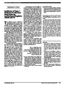
Figure 1—Screening protocol for GDM proposed by the Brazilian Diabetes and Pregnancy Working Party, 2001. PG, plasma glucose.
GDM screening, other factors need to be taken into account, especially because evidence to support GDM screening is largely based on observational studies and consensus. An important factor is the effectiveness of alternative strategies and competing priorities for prevention of perinatal morbimortality. Such evaluation requires modern cost-effectiveness analyses. Given the uncertainties regarding these issues in Brazil, the second meeting of Diabetes in Pregnancy held last April in Porto Alegre, Brazil, recommended the following screening protocol (Fig. 1): 1) to screen all women at booking with fasting plasma glucose (FPG) using, as a cut point, a value of 85 or 90 mg/dl, depending on the priority established for GDM in the local setting; 2) at weeks 20 –24, to apply a diagnostic 2-h 75-g oral glucose
MARIA I. SCHMIDT, MD BRUCE B. DUNCAN, MD ANGELA J. REICHELT, MD FOR THE BRAZILIAN STUDY OF GESTATIONAL DIABETES STUDY GROUP From the Department of Social Medicine, School of Medicine, Federal University of Rio Grande do Sul, Porto Alegre, Brazil. Address correspondence to Maria I. Schmidt, Av. Luiz Manoel Gonzaga, 630/8, Port Alegre, RS 90470-280 Brazil. E-mail:
[email protected] .br. ● ● ● ● ● ● ● ● ● ● ● ● ● ● ● ● ● ● ● ● ● ● ●
References 1. Dashora U, Dashora V, Kennedy L: Twohour 75-g oral glucose tolerance test early in pregnancy detects most cases of gestational diabetes (Letter). Diabetes Care 25: 803, 2002 2. Schmidt MI, Duncan BB, Reichelt AJ, Branchtein L, Matos MC, Costa e Forti A, Spichler ER, Pousada JMDC, Teixeira MM, Yamashita T: Gestational diabetes mellitus diagnosed with a 2-h 75-g oral glucose tolerance test and adverse pregnancy outcomes. Diabetes Care 24:1151– 1155, 2001
DIABETES CARE, VOLUME 24, NUMBER 4, APRIL 2002