L E T T E R S
OBSERVATIONS
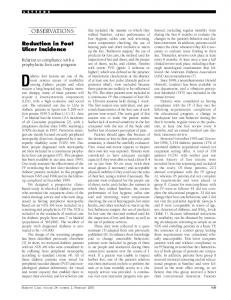
Table 1—Success and failures to iloprost by ascending supine tcpCO2 tertiles (n ⴝ 31 limbs) Tertiles
Transcutaneous Gases Determination in Diabetic Critical Limb Ischemia
T
ranscutaneous oxygen tension (tcpO2) quantifies oxygen delivery through skin capillaries as a function of two main physiological variables, the effective rate of skin blood flow and skin resistance to oxygen permeation, represented mainly by stratum-corneum permeability (1). TcpO2 is an accepted measure of nutritive skin perfusion (1) and predicts the therapeutic outcome in critical limb ischemia (CLI) (2), a serious complication of type 2 diabetes, a disease characterized by the coexistence of macro- and microvascular alterations (3). Transcutaneous carbon dioxide tension (tcpCO2), another tensiometric parameter, is sensitive to severe limb ischemia (4) and correlates closely with HCO⫺3 depletion, H⫹ accumulation, and acidotic milieu (5). Thus, tcpCO2, by providing an indication of the local acid-base balance, might improve the clinical management of patients on CLI, but its prognostic potential in that context is unknown. We addressed this issue in 31 critically ischemic limbs (n ⫽ 26 type 2 diabetic patients, n ⫽ 5 Fontaine’s stage III, n ⫽ 21 Fontaine’s stage IV) on iloprost treatment (6-h intavenous administration, 1–2 ng 䡠 kg⫺1 䡠 min⫺1 daily for 4 weeks) using a drug that is useful for treating CLI not responsive to surgery (6). Iloprost, an analog of epoprostenol (prostacyclin, a potent but short-lived endothelial-derived prostanoid), mimics the pharmacodynamic properties of this compound, namely inhibition of platelet aggregation, vasodilatation, and, as yet illdefined, cytoprotection (7). Infusion rates were titrated in each individual to reach the maximum tolerated dose without hypotension, tachycardia, and other common side effects (facial flushing, headache, nausea, vomiting, abdominal cramping, and diarrhea). Outcome variable was pain relief evaluated by a visual analog scale (1, no pain; 10, intolerable pain). Success (pain relief of ⬎75% from DIABETES CARE, VOLUME 28, NUMBER 8, AUGUST 2005
1 2 3 Total
tcpCO2 (mmHg)
tcpCO2 (kPa)
Failure
Success
⬍40 40–53 ⬎53
⬍5.33 5.33–7.07 ⬎7.07
2 (13) 3 (20) 10 (67) 15 (100)
8 (50) 7 (44) 1 (6) 16 (100)
Data are n (%), unless otherwise indicated.
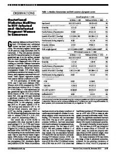
baseline) was obtained in 16 limbs (52%) and failure in 15 limbs (48%). No patient needed surgery or amputation during that short-term study interval. Supine tcpO2 and tcpCO2 were bilaterally recorded on preheated (44°C) dorsal skin between the first and second metatarsal. Response to iloprost (either success or failure) was stratified by tcpO2 and tcpCO2 tertiles, and likelihood ratios (LRs) quantified the predicting power for success (percentage of successes divided by percentage of failures) or failure (percentage of failures divided by percentage of successes) of those two tensiometric parameters (8). LRs are diagnostic statistics that quantify the increased likelihood of an event according to different results of the diagnostic test under evaluation (8). Tertile cutoffs rather than threshold values derived from established literature sources were used because there is no consensus on the definition of normal tcpCO2 that is valid for diagnostic and prognostic use. Iloprost failed in 10 limbs and succeeded in 1 limb in the upper tcpCO2 tertile (cutoff 53 mmHg, 7.07 kPa; LR 10.7) (Table 1). Corresponding data for the bottom tcpO2 tertile (cutoff 1 mmHg, 0.13 kPa; LR 3.3) were 10 vs. 3, respectively (Table 2). All limbs in the upper tcpO2 tertile (cutoff 23 mmHg, 3.07 kPa; LR 17.8) (Table 2) responded to the drug in contrast to only 8 of 10 in the bottom tcpCO2 tertile (cutoff 40 mmHg, 5.33 kPa; LR 3.8) (Table 1). Thus, the upper end of tcpCO2 values pre-
dicted failure to iloprost treatment, and LR analysis confirmed that conclusion. In fact, the further LRs are from 1, the stronger their clinical impact, and when ⬎10 (or ⬍0.1), they should influence therapeutic and diagnostic attitudes (8). Furthermore, pretreatment supine tcpCO2 ⬎53 mmHg (7.07 kPa) predicted treatment failure threefold more efficiently (LR 10.7 vs. 3.3) than supine tcpO2 ⬍1 mmHg (0.133 kPa), an established prognostic marker for limb salvage procedures (2), particularly at those tensions too low to be detectable by the sensor system. Conversely, therapeutic success was 17.8-fold more likely when pretreatment tcpO2 was ⬎23 mmHg (3.07 kPa), which, according to available nomograms (8), corresponds to a 0.95 posttest probability (i.e., close to the certainty of success). Thus, combined use of tcpCO2 and tcpO2 allows more precise prognostic stratification and therefore more rational treatment strategies in diabetic patients with CLI. However, this conclusion awaits further support from studies carried out in samples larger than the present one. Follow-ups longer than our 4-week interval are also needed to evaluate whether elevated tcpCO2 predicts with similar efficiencies clinical end points more significant than pain relief, such as limb amputation. ELIO MELILLO, MD1 MAURO FERRARI, MD1,2 ALBERTO BALBARINI, MD1 ROBERTO PEDRINELLI, MD1
Table 2—Success and failures to iloprost by ascending supine tcpO2 tertiles (n ⴝ 31 limbs) Tertiles 1 2 3 Total
tcpO2 (mmHg)
tcpO2 (kPa)
Failure
Success
⬍1 1–23 ⬎23
⬍0.13 0.13–3.06 ⬎3.07
10 (67) 5 (33) 0 (0)* 15 (100)
3 (19) 4 (25) 9 (56) 16 (100)
Data are n (%), unless otherwise indicated. *LR calculated by adding 0.5 to each of the four component cells, i.e. 关(9.5/16.5)/(0.5/15.5)兴 ⫽ 17.8.
2081
Letters From the 1Dipartimento Cardio Toracico, Universita` di Pisa, Pisa, Italy; and the 2Dipartimento Chirurgia Vascolare, Universita` di Pisa, Pisa, Italy. Address correspondence to Roberto Pedrinelli, MD, Dipartimento Cardio Toracico, Universita` di Pisa, Via Paradisa 2, Pisa, Italy, 56100. E-mail:
[email protected]. © 2005 by the American Diabetes Association. ● ● ● ● ● ● ● ● ● ● ● ● ● ● ● ● ● ● ● ● ● ● ●
References 1. Lubbers DW: Theoretical basis of the transcutaneous blood gas measurements. Crit Care Med 9:721–733, 1981 2. Ubbink DT, Spincemaille GH, Reneman RS, Jacobs MJ: Prediction of imminent amputation in patients with non-reconstructible leg ischemia by means of microcirculatory investigations. J Vasc Surg 30: 114 –121, 1999 3. Beckman JA, Creager MA, Libby P: Diabetes and atherosclerosis: epidemiology, pathophysiology, and management. JAMA 287:2570 –2581, 2002 4. Melillo E, Catapano G, Dell’Omo G, Iabichella L, Berchiolli R, Ferrari M, Pedrinelli R: Transcutaneous oxygen and carbon dioxide measurement in peripheral vascular disease. Vasc Surg 29:273– 280, 1995 5. McBride ME, Berkenbosch JW, Tobias JD: Transcutaneous carbon dioxide monitoring during diabetic ketoacidosis in children and adolescents. Paediatr Anaesth 14:167–171, 2004 6. Loosemore TM, Chalmers TC, Dormandy JA: A meta-analysis of randomized placebo control trials in Fontaine stages III and IV peripheral occlusive arterial disease. Int Angiol 13:133–142, 1994 7. Grant SM, Goa KL: Iloprost: a review of its pharmacodynamic and pharmacokinetic properties, and therapeutic potential in peripheral vascular disease, myocardial ischaemia and extracorporeal circulation procedures. Drugs 43: 889 –924, 1992 8. Deeks JJ, Altman DJ: Diagnostic tests 4: likelihood ratios. BMJ 329:168 –169, 2004
Statin Neuropathy Masquerading as Diabetic Autoimmune Polyneuropathy
S
tatin-induced neuropathy is increasingly described. Proposed mechanisms include an alteration in cholesterol synthesis, producing a disturbance in the cholesterol-rich neuronal 2082
membrane, or in the activity of ubiquinone (coenzyme Q10), a mitochondrial respiratorychainenzymeinhibitedbystatins leading to neuronal damage (1). The entire class is implicated, and both polyneuropathy and mononeuropathy have been described with improvement or even complete resolution occurring with cessation of therapy (1). In all cases, clinical improvement occurred soon after statins were discontinued, and in the absence of specific clinical, biochemical, or electrophysiological characteristics, this has become the key diagnostic feature of statininduced neuropathy. To date, autonomic features accompanying symmetrical neuropathy have not been described. We present an 18-year-old white female with type 1 diabetes for 5 years who, over several months, developed restless legs followed by parasthesias, nocturnal diarrhea, fecal incontinence, early satiety, and weight loss. Examination revealed loss of pinprick sensation to the upper arms and thighs accompanied by areflexia and loss of vibration sense, a fixed tachycardia, and orthostatic hypotension. Autoimmune demyelinating polyneuropathy was initially suspected due to the relatively rapid onset of symptoms and the combination of peripheral and autonomic findings. Neuroelectrophysiological studies showed evidence of axonal, sensory, and motor polyneuropathy but did not meet criteria for demyelination. Supportive therapy included gabapentin, clonazepam, metoclopramide, metronidazole, cholestyramine, and fludrocortisone. Before further investigations began, it was noted that the subject had been taking atorvastatin despite very low lipid levels, and it was discontinued. Within 1 week, her symptoms improved dramatically. Within 6 months, the postural hypotension, diarrhea, and symptoms of gastroparesis had resolved, and all medicines other than insulin were discontinued. There remained a minimal decrease in vibration sense, areflexia, and loss of sensation to the wrist and ankle. Isolated cases of statin-associated neuropathy have been reported since 1994 (1). Epidemiological and casecontrol studies from the U.K. and Denmark suggest elevated odds ratios (ORs) of 2.5 (95% CI 0.3–14.2) to 3.7 (1.8 – 7.6), respectively, for the development of neuropathy while on statin therapy (2,3). The OR jumped to 26.4 (7.8⫺45.4) in
patients with confirmed neuropathy taking statins for ⬎2 years (3). The key to diagnosing statin-induced neuropathy is to discontinue the statin and observe for potential improvement. In conclusion, statins can infrequently cause an idiosyncratic somatic and autonomic neuropathy that, in the diabetic patient, will almost invariably be attributed to diabetes. Awareness of this association and a trial removal of the statin could result in restoration of neurological function and a much-improved quality of life in the diabetic patient. TOM BROOKS VAUGHAN, MD DAVID S.H. BELL, MD Address correspondence and reprint requests to Tom Brooks Vaughan, MD, Division of Endocrinology, University of Alabama School of Medicine, Faculty Office Tower, Suite 758, 510 20th St. S., Birmingham, AL 35294. E-mail:
[email protected]. D.S.H.B. has received honoraria from AstraZeneca. © 2005 by the American Diabetes Association. ● ● ● ● ● ● ● ● ● ● ● ● ● ● ● ● ● ● ● ● ● ● ●
References 1. Backes JM, Howard PA: Association of HMG-CoA reductase inhibitors with neuropathy. Ann Pharmacother 37:274 –278, 2003 2. Gaist D, Rodriguez LA, Huerta C, Hallas J, Sindrup S: Are users of lipid-lowering drugs at increased risk of peripheral neuropathy? Eur J Clin Pharmacol 56:931– 933, 2001 3. Gaist D, Jeppesen U, Andersen M, Garcia Rodriguez LA, Hallas J, Sindrup SH: Statins and risk of polyneuropathy: a casecontrol study. Neurology 58:1333–1337, 2002
Characteristics of California Children With Single Versus Multiple Diabetic Ketoacidosis Hospitalizations (1998 –2000)
D
iabetic ketoacidosis (DKA) is a frequent reason for hospital admission of children with newly diagnosed diabetes (1,2) and the most frequent cause for rehospitalization of children with poorly controlled diabetes (3). DKA is an ambulatory care-sensitive condition
DIABETES CARE, VOLUME 28, NUMBER 8, AUGUST 2005
Letters
for which timely and appropriate outpatient intervention may reduce the need for hospitalization (4,5). We examined DKA hospitalizations for children 0 –18 years using 1998 –2000 California Office of Statewide Health Planning and Development (COSHPD) hospital discharge data to determine resource use and prevalence of DKA hospitalization for hospitalized pediatric patients. Using ICD-9 codes, we identified 4,957 DKA hospitalizations. Pediatric DKA accounted for 14,279 hospital days (median length of stay 2 days; mean 2.9 ⫾ 2.3 days). Hospital charges were reported for 83.6% of hospitalizations. Median charges per hospitalization were $8,440 (mean 11,615 ⫾ $15,537), with an estimated yearly cost of $20 million for pediatric DKA care. Only 28% of these DKA hospitalizations occurred in institutions designated as hospitals providing highly specialized care to children by the National Association of Children’s Hospitals and Related Institutions. Record linkage numbers, a COSHPD encrypted data element enabling linkage of hospitalizations and thus analyses at the child level, were available for 3,409 (69%) of hospitalizations. We sorted hospitalizations with record linkage numbers into two groups: one DKA hospitalization (1,694) and two or more DKA hospitalizations (1,715). Compared with children with one DKA hospitalization (n ⫽ 1,694), children with DKA recurrence (n ⫽ 499) were older (14.4 ⫾ 3.5 vs. 12.0 ⫾ 4.9 years; P ⬍ 0.001), female (65.1 vs. 49.2%; P ⬍ 0.0001), nonHispanic blacks (16.0 vs. 11.1%; P ⬍ 0.001), and publicly insured (49.7 vs. 42.7%; P ⬍ 0.01); fewer (27.6 vs. 35.3%; P ⬍ 0.001) received care at a designated children’s hospital. Using public insurance as a marker of poverty (6), we further examined the association of race/ethnicity and health insurance type for children with one versus two or more DKA hospitalizations using goodness-of-fit statistics. Among children with one DKA hospitalization, the proportion of minority children who were publicly insured (non-Hispanic black 59.9%, Hispanic 53.2%, other race 61.1%) was higher than that of nonHispanic white children (29.4%; P ⬍ 0.0001). The same trend was true for children who had recurrent DKA hospitalizations (non-Hispanic black 52.5%, Hispanic 59.8%, other race 64.8% vs. DIABETES CARE, VOLUME 28, NUMBER 8, AUGUST 2005
non-Hispanic white 40.5%, P ⬍ 0.001). Thus, ⬎50% of minority children in both DKA groups were publicly insured, suggesting an association between poverty and DKA. In addition, the proportion of publicly insured non-Hispanic white children was higher in the recurrent DKA group (40.5 vs. 29.4%), further supporting this association. Children with poorly controlled diabetes are at risk for long-term complications of diabetes (7–9) and psychiatric comorbidity (10). Comparison of our findings with those of Cohn et al. (11) using 1991 COSHPD data illustrate that both the frequency and demographic characteristics of children hospitalized for DKA events remain essentially unchanged despite implementation of Diabetes Control And Complications Trial Research Group recommendations and improvements in insulin delivery options for pediatric patients over the past decade. Children with recurrent DKA may benefit from comprehensive care provided by a diabetes team including pediatric endocrinologists, diabetes educators, mental health professionals, and social workers to reverse the chain of events resulting in poor self-care. Yet, our data show that only 28% of children with recurrent DKA were hospitalized in designated children’s hospitals. Similarly, Curtis et al. (12) observed that the highest DKA rates were found in regions in Ontario, Canada, lacking academic pediatric centers. DKA hospitalizations are costly. Because access to diabetes teams may decrease the incidence of recurrent DKA, specialist visits may actually be cost saving to society. Compared with singleepisode DKA, recurrent DKA is highest among adolescent girls, minority children, and those publicly insured. Opportunities exist to reduce DKA hospitalizations for children with diabetes with clinical and policy interventions targeted to this population. ARLENE SMALDONE, DNSC, CPNP, CDE1,2 JUDY HONIG, EDD, CPNP3 PATRICIA W. STONE, PHD, RN3 RAYMOND ARONS, DRPH4 KATIE WEINGER, EDD, RN1,2 From the 1Behavioral and Mental Health Research, Joslin Diabetes Center, Boston, Massachusetts; the 2 Departments of Medicine and Psychiatry, Harvard Medical School, Harvard University, Boston, Massachusetts; the 3Columbia University School of Nursing, New York, New York; and the 4Mailman School
of Public Health, Columbia University, New York, New York. Address correspondence to Arlene Smaldone, DNSc, CPNP, CDE, Department of Behavior and Mental Health Research, Joslin Diabetes Center, One Joslin Place, Boston, MA 02215. E-mail: arlene.
[email protected]. © 2005 by the American Diabetes Association.
Acknowledgments — This study was supported by National Institutes of Health Training Grant 2T32DK007260-26, National Institute of Diabetes and Digestive and Kidney Diseases Grant DK07260, the Joslin Diabetes Center (A.S.), and the Juliet Wright Grady Research Fund (K.W.). ● ● ● ● ● ● ● ● ● ● ● ● ● ● ● ● ● ● ● ● ● ● ●
References 1. Neu A, Willasch A, Ehehalt S, Hub R, Ranke MB: Ketoacidosis at onset of type 1 diabetes mellitus in children: frequency and clinical presentation. Pediatr Diabetes 4:77– 81, 2003 2. Smith CP, Firth D, Bennett S, Howard C, Chisholm P: Ketoacidosis occurring in newly diagnosed and established diabetic children. Acta Paediatr 87:537–541, 1998 3. Glasgow AM, Weissberg-Benchell J, Tynan WD, Epstein SF, Driscoll C, Turek J, Beliveau E: Readmissions of children with diabetes mellitus to a children’s hospital. Pediatrics 88:98 –104, 1991 4. Gadomski A, Jenkins P, Nichols M: Impact of a Medicaid primary care provider and preventive care on pediatric hospitalization. Pediatrics 101:E1, 1998 5. Parker JD, Schoendorf KC: Variation in hospital discharges for ambulatory caresensitive conditions among children. Pediatrics 106 (Suppl. 4):942–948, 2000 6. Keenan HT, Foster CM, Bratton SL: Social factors associated with prolonged hospitalization among diabetic children. Pediatrics 109:40 – 44, 2002 7. Svensson M, Eriksson JW, Dahlquist G: Early glycemic control, age at onset, and development of microvascular complications in childhood-onset type 1 diabetes: a population-based study in northern Sweden. Diabetes Care 27:955–962, 2004 8. Harvey JN, Allagoa B: The long-term renal and retinal outcome of childhood-onset type 1 diabetes. Diabet Med 21:26 –31, 2004 9. Olsen BS, Sjolie A, Hougaard P, Johannesen J, Borch-Johnsen K, Marinelli K, Thorsteinsson B, Pramming S, Mortensen HB: A 6-year nationwide cohort study of glycaemic control in young people with type 1 diabetes: risk markers for the development of retinopathy, nephropathy and neuropathy. Danish Study Group of Diabetes in Childhood. J Diabetes Complications 14:295–300, 2000 10. Kovacs M, Goldston D, Obrosky DS, Bo-
2083
Letters
nar LK: Psychiatric disorders in youths with IDDM: rates and risk factors. Diabetes Care 20:36 – 44, 1997 11. Cohn BA, Cirillo PM, Wingard DL, Austin DF, Roffers SD: Gender differences in hospitalizations for IDDM among adolescents in California, 1991: implications for prevention. Diabetes Care 20:1677–1682, 1997 12. Curtis JR, To T, Muirhead S, Cummings E, Daneman D: Recent trends in hospitalization for diabetic ketoacidosis in Ontario children. Diabetes Care 25:1591– 1596, 2002
Acute Presentation of Fetal Hypertrophic Cardiomyopathy in a Type 1 Diabetic Pregnancy
T
he incidence of intrauterine fetal death in type 1 diabetic patients is significantly higher than in the general population (1). Although vascular disease, poor glycemic control, polyhydramnios, fetal macrosomia, and preeclampsia are associated with a higher incidence of fetal death, the etiology of the increased stillbirth rate remains unknown. Besides other complications, infants of diabetic mothers have long been recognized to be at risk of having hypertrophic cardiomyopathy, a condition that is characterized by thickening of the interventricular septum and ventricular walls, and by systolic and diastolic dysfunction of the neonatal heart. This condition is normally asymptomatic in utero and may only result in congestive heart failure in the immediate postnatal period, although this is uncommon and transient (2). A 30-year-old woman, gravida 3, para 0, abortus 2, was referred to our unit for the assessment of suspected fetal macrosomia at 35 weeks of gestation. She had been diagnosed with type 1 diabetes at age 13 years. In the current pregnancy, her glycemic control had been suboptimal, as confirmed by an HbA1c (A1C) value of 7.6% obtained at 31 weeks. The ultrasound scan performed in our unit showed fetal abdominal growth above the 95th percentile. There was associated polyhydramnios, and the fetal heart showed features of hypertrophic cardiomyopathy. The umbilical artery showed
2084
an abnormal pattern of flow. There were no signs of fetal hydrops, but the fetus showed reduced movements. In view of these findings, reevaluation of the fetus was planned in 4 – 6 h to decide further management. Within a few hours, the patient started complaining of uterine contractions. An external fetal monitor was applied, showing a fetal heart baseline rate of 160 bpm, with reduced variability and repetitive late decelerations, indicating an ominous outcome if untreated. An emergency Caesarean section was performed, delivering a female infant of 3,575 g (weight above 95th percentile for gestation) with an Apgar score of 4, 7, and 10 at 1⬘, 5⬘, and 10⬘, respectively. No signs of abruption were noted clinically or at pathological examination of the placenta. The newborn needed nasal continuous positive air pressure for the first 12 h for stabilization. Postnatal echocardiography confirmed the diagnosis of hypertrophic cardiomyopathy. Treatment with propranolol was started, and the neonate was discharged on day 7. A follow-up visit at 3 months after delivery showed resolution of the cardiac hypertrophy. One previous report has described a case of stillbirth at 37 weeks of gestation associated with previously undiagnosed hydrops fetalis and hypertrophic cardiomyopathy in the fetus of a diabetic mother (3). The same authors suggested that unexplained fetal deaths described in earlier reports (4 – 6) might be attributable to hypertrophic cardiomyopathy. In the present case, the fetus showed no signs of hydrops or cardiac failure, but the abnormal umbilical flow suggested a frail state near to decompensation. The increased cardiac work requirement brought on by the onset of uterine contractions was sufficient to induce acute fetal distress, as documented by a grossly abnormal fetal heart rate pattern. These findings indicate that diabetic hypertrophic cardiomyopathy can present with acute fetal distress even in absence of hydrops and suggest that this condition might be one of the causes of the increased stillbirth rate in pregnancies complicated by type 1 diabetes. FEDERICO PREFUMO, MD1 CLAUDIO CELENTANO, MD2 FRANCESCA PRESTI, MD3 PIERANGELA DE BIASIO, MD1 PIER LUIGI VENTURINI, MD1
From the 1Department of Obstetrics and Gynaecology, Institute G. Gaslini, University of Genova, Genova, Italy; the 2Department of Obstetrics and Gynaecology, San Massimo Hospital, Penne, Italy; and the3Department of Obstetrics and Gynaecology, Sacred Heart Hospital, Negrar, Italy. Address correspondence to Dr. Federico Prefumo, Department of Obstetrics and Gynaecology, Institute G. Gaslini, Largo Gaslini, 5, 16147 Genova, Italy. E-mail:
[email protected]. © 2005 by the American Diabetes Association. ● ● ● ● ● ● ● ● ● ● ● ● ● ● ● ● ● ● ● ● ● ● ●
References 1. Jensen DM, Damm P, Moelsted-Pedersen L, Ovesen P, Westergaard JG, Moeller M, Beck-Nielsen H: Outcomes in type 1 diabetic pregnancies. Diabetes Care 27:2819 – 2823, 2004 2. Rizzo G, Capponi A, Romanini C: Fetal echocardiography in the diagnosis of obstetric pathology. In Textbook of Fetal Cardiology. Allan LD, Hornberger LK, Sharland G, Eds. London, Greenwich Medical Media, p. 453– 470, 2000 3. Sardesai MG, Gray AA, McGrath MM, Ford SE: Fatal hypertrophic cardiomyopathy in the fetus of a woman with diabetes. Obstet Gynecol 98:925–927, 2001 4. Gutgesell HP, Speer ME, Rosenberg HS: Characterization of the cardiomyopathy in infants of diabetic mothers. Circulation 61:441– 450, 1980 5. Leslie J, Shen SC, Strauss L: Hypertrophic cardiomyopathy in a midtrimester fetus born to a diabetic mother. J Pediatr 100: 631– 632, 1982 6. Girz BA, Divon MY, Merkatz IR: Sudden fetal death in women with well-controlled, intensively monitored gestational diabetes. J Perinatol 12:229 –233, 1992
Use of Glargine Insulin Before and During Pregnancy in a Woman With Type 1 Diabetes and Addison’s Disease
W
e report a case of a 31-year-old Caucasian woman (weight 50 kg, height 148 cm) with type 1 diabetes diagnosed 27 years ago and Addison’s disease discovered 5 years ago, who had a pregnancy with normal outcome treated with lispro and glargine insulin. For years, the patient has followed a regimen of multiple daily injections of lispro before meals, NPH at bedtime, and 75 mg/day cortone acetate. Her metabolic
DIABETES CARE, VOLUME 28, NUMBER 8, AUGUST 2005
Letters
control has been constantly altered (April 2002 A1C, 8.4%), and she has had frequent episodes of mild and at times severe hypoglycemia, especially at night. At morning, glycemia was often high. When NPH was changed to insulin glargine, hypoglycemic episodes were drastically reduced, and metabolic control improved (December 2002 A1C, 6.9%). In August 2003, the patient discovered her pregnancy at the 18th week of gestation (A1C 6.2%, ACTH 6 pg/ml). We informed her that the use of glargine and lispro had not been previously evaluated during pregnancy, but considering that the critical organogenic period had already ended and that metabolic control was good, we suggested that it would be possible to continue use of them throughout pregnancy. The patient gave her written informed consent. The pregnancy proceeded regularly, and the patient’s weight increased from 50 to 63 kg. At the beginning, the patient took 70 IU/day of insulin (1.4 IU/kg) divided as follows: lispro 42 IU (0.84 IU 䡠 kg⫺1 䡠 day⫺1) and glargine 28 IU (0.56 IU 䡠 kg⫺1 䡠 day⫺1). At the end, lispro had to be increased to 88 IU (1.76 IU 䡠 kg⫺1 䡠 day⫺1), whereas glargine remained unchanged with a total of 116 IU/day (2.32 IU/kg). The patient did not have any important hypoglycemic episodes. A1C and ACTH ranged between 6.2 and 5.9% and 6 and 12 pg/ml, respectively. Use of cortisone was unchanged. No diabetes complications developed
DIABETES CARE, VOLUME 28, NUMBER 8, AUGUST 2005
during pregnancy, and blood pressure was always normal. Ultrasound examination showed a normal fetus, smaller for gestational age. Placental flow was normal. A Cesarean section was performed at the 38th week because of the small size of the patient’s pelvis. A healthy baby girl was born with no malformations: weight 2,150 g (under 10th percentile birth weight), height 44 cm, and Apgar score 9/10. Patient and neonate had no complications during postpartum. Therefore, in our patient, use of glargine and lispro did not induce any fetal malformations notwithstanding their use from start of pregnancy. It is not known why the fetus was so small for gestational age, since the mother neither smoked nor had any history of drug abuse. Indeed, the mother is small sized, as is her husband, but that is not enough to justify low birth weight. There are many reports about safe use of lispro in pregnancy, and a recent study demonstrated in vitro that it does not cross the human placenta (1,2). However, little has been written on the use of glargine in pregnancy; one study showed that use after the 3rd month of pregnancy resulted in no fetal malformations (3), whereas two trials report use since the beginning of pregnancy (4,5). Regarding our patient, it is likely that optimal control of glycemia with this insulin favored a long sought after pregnancy. However, additional studies are
needed before stating that glargine is completely safe in pregnancy. MARIA DOLCI MARY MORI FABIO BACCETTI From the Diabetes Service, Massa e Carrara, Italy. Address correspondence and reprint requests to Fabio Baccetti, Consultant in Endocrinology and Diabetology, Diabetes Service Massa e Carrara, Via Sottomonte 1 54100, Massa e Carrara, Italy. E-mail:
[email protected]. © 2005 by the American Diabetes Association.
● ● ● ● ● ● ● ● ● ● ● ● ● ● ● ● ● ● ● ● ● ● ●
References 1. Bhattacharyya A, Brown S, Hughes S, Vice PA: Insulin lispro and regular insulin in pregnancy. QJM 94:255–260, 2001 2. Boskovic R, Feig DS, Derewlany L, Knie B, Portinoi G, Koren G: Transfer of insulin lispro across human placenta. Diabetes Care 26:1390 –1394, 2003 3. Devlin JT, Hothersall L, Wilkis JL: Use of insulin glargine during pregnancy in a type 1 diabetic woman (Letter). Diabetes Care 25:1095–1096, 2002 4. Holstein A, Plaschke A, Egberts EH: Use of insulin glargine during embryogenesis in a pregnant woman with type 1 diabetes (Letter). Diabet Med 20:779 –780, 2003 5. Di Cianni G, Volpe L, Lencioni C, Chatzianagnostou K, Cuccuru I, Ghio A, Benzi L, Del Prato S: Use of insulin glargine during the first weeks of pregnancy in five type 1 diabetic women (Letter). Diabetes Care 28:982–983, 2005
2085