Human Reproduction vol.15 no.12 pp.2496–2503, 2000
Oestrous cycle and pregnancy alter the reactivity of the rat uterine vasculature
J.J.Dalle Lucca1, A.S.O.Adeagbo2 and N.L.Alsip1, 3 1Center
for Applied Microcirculatory Research and 2Department of Physiology, Health Sciences Center, A1115, University of Louisville School of Medicine, Louisville, KY 40292, USA 3To
whom correspondence should be addressed. E-mail:
[email protected]
Isolated uterine vascular beds from virgin and pregnant rats were used to assess vascular reactivity and the ability of nitric oxide (NO), prostanoids and endothelium-derived hyperpolarizing factor (EDHF) to modulate these responses. One uterine horn from female rats in each oestrous cycle day and gestation day 17 was removed and perfused with physiological saline solution. Tone was induced with cirazoline (1 µmol/l), and concentration– response curves to acetylcholine (ACh) generated. Responsiveness to ACh was tested in the presence of N-nitro-Larginine (L-NA), ibuprofen (IBU) and tetrabutylammonium (TBA), to inhibit NO synthase, cyclo-oxygenase and K⍣ channels respectively. Cirazoline-induced tone was smaller in the pregnant compared with the proestrous group. Sensitivity to ACh was cycle day and pregnancy dependent with pregnant > dioestrous day-1 > dioestrous day-2 > proestrous and oestrous. L-NA shifted the curve to the right in all groups except dioestrous day-1. IBU inhibited the ACh response in the pregnant group only. TBA virtually abolished the response in all groups. These results suggest that in the uterine vascular bed from pregnant rats, EDHF, along with NO and a dilator prostanoid mediate ACh-induced dilatation. In contrast, in the dioestrous day-1 group, only EDHF seems to be released by ACh in this vascular bed. In the oestrous, dioestrous day-2 and proestrous groups, ACh releases both EDHF and NO. Key words: acetylcholine/oestrous cycle/pregnancy/uterus/vascular reactivity
Introduction In females, the fluctuations of sex hormones during the reproductive cycle and pregnancy are known to exert a dramatic impact on the homeostasis of the cardiovascular system. During normal pregnancy, there is a profound remodelling of the cardiovascular system, characterized by an increase in plasma volume and cardiac output that is accompanied by a decrease in vascular resistance. The uterine vascular control mechanisms are poorly understood, especially how these mechanisms are 2496
affected by the changing hormonal milieu. The changes in vascular reactivity during the reproductive cycle have not been studied to a great extent, but it does appear that there are alterations. For example, in rats in proestrous, maximal contraction of the aorta to noradrenaline is reduced compared with rats on the other days of the oestrous cycle (Zamorano et al., 1994). Our first goal was to determine if vascular reactivity in the isolated, perfused uterine vascular bed is altered by the oestrous cycle phase or pregnancy. The control mechanisms underlying vascular smooth muscle tone involve the release of contractile and dilative factors from the vascular endothelium. Among them, nitric oxide (NO) and dilator prostanoids acting via cGMP and cAMP respectively, have been shown to participate in the control of uterine microvessels (Kimura et al., 1995; Saha et al., 1998). Endothelium-derived hyperpolarizing factor (EDHF) is another factor that is released by the endothelium. EDHF relaxes vascular smooth muscle cells by opening K⫹ channels (Feletou and Vanhoutte, 1999). Its involvement in controlling the uterine microvasculature is unknown, although it is active in the uterine artery (Jain et al., 1999). NO activity appears to be enhanced in the vasculature of the pregnant animal (Nathan et al., 1995), possibly as a result of the increase in oestrogen, since chronic administration of oestrogen increases NO and prostaglandin activity in both humans and experimental mammals (Farhat et al., 1996). EDHF activity may also be increased with pregnancy. Ring segments from the abdominal, but not thoracic, aorta have an enhanced release of EDHF in the pregnant compared with the virgin rat (Bobadilla et al., 1997). In addition, in the intact animal EDHF appears to be more active in some vascular beds during pregnancy (Keyes et al., 1998). We postulated that uterine vascular reactivity is sensitive to the hormonal changes observed during the oestrous cycle and pregnancy, and that NO activity would show a correlation with oestrogen concentrations. The present study was designed to evaluate the response to acetylcholine (ACh) in the isolated, perfused uterine horn from virgin female rats at various stages of the oestrous cycle (oestrous, dioestrous day-1, dioestrous day-2 and proestrous), and in pregnant rats (gestation day 17). The contribution of the endothelium-derived products—NO, prostanoids and EDHF—as mediators of ACh-induced vascular relaxation was also explored. In pregnant females, there is reduced contraction to vasoconstrictors which is thought to be secondary to increased synthesis of NO and prostaglandin I2 (PGI2) (Magness et al., 1992; Kim et al., 1994) and possibly EDHF (Keyes et al., 1998). Therefore, the ability of a selective α1-adrenergic agonist, cirazoline (CRZ), to induce tone was © European Society of Human Reproduction and Embryology
Uterine vascular reactivity in oestrous cycle and pregnancy
also assessed, as well as the influence of NO, prostanoids and EDHF on this CRZ-induced tone. Materials and methods Virgin female Sprague-Dawley rats weighing 200–225 g (Harlan, Indianapolis, IN, USA) were housed in a temperature- and humiditycontrolled room of our AALAC approved animal care facility with a 12 h light:12 h dark cycle. The animals had access to distilled water and rat chow ad libitum. A vaginal smear was performed daily before 9:00 a.m. to determine the stage of oestrous cycle. Vaginal smears were examined and vaginal cytology used to determine oestrous stage by the commonly used method (Baker, 1979). Animals were monitored for at least two oestrous cycles, and only those animals exhibiting regular 4-day oestrous cycles were used in this study. An animal was mated when her vaginal smear indicated that she was in proestrous. The day that spermatozoa were present in the vaginal smear was considered to be day 1 of pregnancy. Female virgin rats on each day of the oestrous cycle (oestrous, dioestrous day-1, dioestrous day-2, proestrous) and pregnant rats on gestation day 17 were used in this study. Following anaesthesia with an i.p. injection of ketamine (37.5 mg/kg) combined with xylazine (5.0 mg/kg), the abdominal cavity was opened and one uterine horn isolated from any connective tissues. The anterior iliac artery, hypogastric artery and uterine neck and vesical artery branches of the uterine artery were ligated, and the uterus perfused through a catheter placed in the common iliac artery. The ovarian artery and vein were cut at the neck of the uterus. The uterine horn was flushed with heparinized physiological saline solution (PSS) and transferred to a warmed chamber. The isolated uterine horn was then perfused with PSS (2 ml/min) using a peristaltic pump (Masterflex, ColePalmer Instruments Co., Vernon Hills, IL, USA). Changes in perfusion pressure were recorded via a Statham pressure transducer (model P23XL; Grass, Quincy, PA, USA) coupled to a Grass polygraph recorder (model 7H; Grass). This surgery was modified from that described previously (Langer et al., 1993). The composition of our PSS (in mmol/l) was: NaCl 118, KCl 4.7, CaCl2 2.5, KH2PO4 1.2, MgSO4 1.2, NaHCO3 12.5, glucose 11.1. The pH of the PSS was maintained at 7.4 after saturation with carbogen (95% O2/5% CO2). Tissues were allowed to equilibrate for at least 30 min before the start of the protocol. All protocols involved measuring vascular reactivity in the absence, and in the presence of inhibitors of nitric oxide synthase (NOS), cyclo-oxygenase and K⫹ channels, or combinations of these agents. Vascular tone in the isolated, perfused uterine vascular bed was generated by continuous infusion of CRZ, an α1-adrenergic agonist. CRZ produces stable tone for many hours in this preparation (Adeagbo and Triggle, 1993). Protocol 1 The purpose of this series of experiments was two-fold. The first was to determine the pressor effect of CRZ infusion and the relative modulation of this by NO, prostanoids and K⫹ channels (EDHF). NOS was inhibited with N-nitro-L-arginine (L-NA, 100 µmol/l), cyclo-oxygenase inhibition was achieved with ibuprofen (IBU, 10 µmol/l), and K⫹ channel blockade with tetrabutylammonium (TBA, 100 µmol/l). The second purpose of these experiments was to determine the influence of the oestrous cycle and pregnancy on AChinduced dilatation and the relative contribution of NO, prostanoids and EDHF in mediating this response. To achieve this goal, one uterine horn was removed and perfused as described above. Following the equilibration period, CRZ (1 µmol/l) was added to the perfusion medium and retained for the entire protocol. Once the
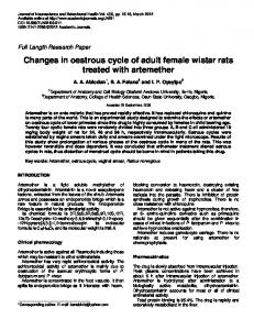
Figure 1. Representative tracing from a time control experiment. The responsiveness to acetylcholine (ACh) was tested repeatedly in an isolated, perfused uterine vascular bed taken from a rat in dioestrous day-1. Tone was induced with cirazoline (1 µmol/l) during the entire protocol. Segment A is analogous to the control response in Figures 3–5; segment B was obtained 20 min after the highest concentration of ACh in segment A; segment C was obtained 20 min after the highest concentration of ACh in segment B; segment D was obtained 40 min after the highest concentration of ACh in segment C. Sodium nitroprusside (SNP) was given as a bolus injection after the last injection of ACh. Similar results were obtained in tissues taken from pregnant animals. Scale of tracing is in mmHg. developed tone had plateaued, a concentration–response relationship to ACh (10–9 to 10–5 µmol) was established by giving bolus injections (0.1 ml) of ACh into the perfusion tube. After the tissue had recovered from the highest concentration of ACh, L-NA was added to the perfusion solution (PSS) and co-infused with CRZ. After 20 min, the bolus injections of ACh were repeated. IBU was then added to the perfusion solution already containing CRZ and L-NA. After the tone reached a plateau (usually 20 min), ACh injections were given for a third time. Finally, TBA was added to the perfusion solution along with the CRZ, L-NA and IBU and again after tone levelled off (usually 40 min), the ACh injections repeated. Sodium nitroprusside (SNP, 1 µmol/l) was administered at the end of each protocol to test the viability of the vessels. The preparation was always allowed to return to its pre-injection pressure before another concentration of ACh or an inhibitor was given. One group of virgin rats on dioestrous day-1 and one group of pregnant rats were subjected to the protocol without addition of L-NA, IBU or TBA to the perfusion medium. These groups served as time controls, and the response to ACh was virtually identical in all four curves obtained (Figure 1). The increase in perfusion pressure (tone) generated by CRZ and any change in tone produced by L-NA, IBU and TBA were compared for the different stages of oestrous cycle and the pregnant group. Concentration–response curves for the response to ACh were constructed in the presence of CRZ (control), CRZ and L-NA (L-NA present), CRZ, L-NA and IBU (IBU present) and CRZ, L-NA, IBU and TBA (TBA present). Protocol 2 The purpose of protocols 2 and 3 was to determine if the order in which inhibitors were added to the perfusion solution affected the vascular response to ACh. To address this concern, the order in which
2497
J.J.Dalle Lucca, A.S.O.Adeagbo and N.L.Alsip
the inhibitors were added to the perfusion medium was changed, and reactivity to ACh tested in isolated perfused uterine vascular beds from dioestrous day-2 virgin rats and pregnant rats on gestation day 17. After the control curves for the ACh (10–9 to 10–5 µmol) had been constructed in the presence of CRZ, IBU (10 µmol/l) was added into the perfusion medium. After the tone reached a plateau, the concentration–response curve to ACh was repeated. After the tissue had recovered from the highest concentration of ACh, L-NA (100 µmol/l) was then added to the perfusion solution (PSS) and coinfused with CRZ and IBU. After 20 min, the bolus injections of ACh were repeated. Finally, TBA (1 mmol/l) was added to the perfusion solution along with the CRZ, IBU and L-NA and again after the tone had levelled off (usually 40 min), the ACh injections were repeated. Protocol 3 In these experiments, TBA was added to the perfusion solution prior to L-NA. In isolated, perfused mesenteries from pregnant and dioestrous day-1 virgin animals, CRZ (1 µmol/l) was added to the perfusion solution to induce vascular tone. Bolus injections of ACh were given in increasing concentrations (10–9 to 10–5 µmol) into the perfusion tube to determine the control concentration–response relationship. Then, the K⫹ channel blocker, TBA (1 mmol/l) was introduced into the perfusion solution, and after a 40 min equilibration period, ACh responsiveness was determined again. Next, the NOS inhibitor L-NA (100 µmol/l) was introduced into the suffusion solution, in addition to CRZ and TBA. After at least a 20 min equilibration period, the vascular responsiveness to ACh was tested again. In all protocols, SNP (1 µmol/l) was injected at the end of each experiment to assess the ability of the vessel to dilate. The tissue was always allowed to return to its pre-injection pressure before another concentration of ACh or an inhibitor was given. Chemicals L-NA, TBA, ACh and IBU were purchased from Sigma Chemical Co., St Louis, MO, USA. CRZ was purchased from RBI/Sigma (Natick, MA, USA). Ketamine was purchased from Fort Dodge (Columbus, OH, USA) and xylazine from Butler Co. (Columbus, OH, USA). All components of the modified Krebs solution (PSS) were purchased from Sigma Chemical Co. L-NA was dissolved in 100 ml PSS. All other compounds were dissolved in distilled water. Statistical analysis Change in tone due to the addition of CRZ, L-NA, IBU or TBA to the perfusion solution was expressed in mmHg. For each group, concentration–response curves were obtained for ACh alone (control), in the presence of CRZ and L-NA (L-NA present), CRZ, L-NA and IBU (IBU present), and CRZ, L-NA, IBU and TBA (TBA present). Relaxation to ACh at each concentration was expressed as a percentage of the total developed tone (% maximum relaxation). Each concentration–response curve was used to determine the concentration of ACh that gave the maximal response and 50% of the maximal response (EC50). Change in perfusion pressure, EC50 and responses at each concentration of ACh were each compared by one-way analysis of variance (ANOVA) followed by Newman–Keul’s multiple range tests when a significant difference existed between groups. P ⬍ 0.05 was taken as significant. Group data were reported as mean ⫾ SEM.
Results Establishment of tissue viability over time The ability of the isolated, perfused uterine vasculature to respond to repeated application of ACh was tested in a group 2498
Figure 2. Change in perfusion pressure in the isolated, perfused uterine horn to infusion of the α1-adrengergic agonist cirazoline (1 µmol/l) in pregnant and cycling rats. The response of the pregnant group (Preg) to cirazoline was significantly different from that of the proestrous group (Proest). Groups with the same letter designation are not significantly different from one another; P ⬍ 0.05. Oestr ⫽ oestrus; D-1 ⫽ dioestrous day-1; D-2 ⫽ dioestrous day-2.
of virgin (n ⫽ 4) and a group of pregnant rats (n ⫽ 4). In both groups, the response to ACh was similar in all four curves. A representative tracing is shown in Figure 1. Cirazoline-induced tone and its modulation by L-NA, IBU and TBA Continuous infusion of CRZ (1 µmol/l) produced a significant increase in perfusion pressure in all experimental groups; however, the increase was significantly smaller in the pregnant group compared with the proestrous group (Figure 2). The addition of L-NA (100 µmol/l), IBU (10 µmol/l) or TBA (1 mmol/l) to the perfusion solution containing CRZ did not cause a significant change in tone. Despite transient changes in perfusion pressure after administration of TBA into the perfusion medium, vessel tone recovered to pre-application level without adjusting the concentration of CRZ in all experiments (data not shown). Protocol 1 Bolus injections of ACh (10–9 to 10–5 µmol) gave concentrationdependent decreases in perfusion pressure of CRZ pre-constricted uterine vascular beds of all experimental groups. The pregnant group had the most sensitive uterine vasculature to ACh, followed by dioestrous day-1 and dioestrous day-2, presenting as the second and third most reactive groups respectively. The proestrous and oestrous groups showed the least sensitivity to ACh, and a comparison of a sensitivity measure (pEC50) did not show significant difference between these two groups (Table I). In the presence of L-NA, there was a significant attenuation of the response to ACh in the oestrous, dioestrous day-2, proestrous and pregnant groups (Figure 3, L-NA present). Furthermore. L-NA did not cause a significant change in the response to ACh in isolated, perfused uterine vascular beds removed from rats in dioestrous day-1. The addition of IBU to the perfusion medium resulted in a further decrease of the ACh-induced vasodilatation in the pregnant group. However,
Uterine vascular reactivity in oestrous cycle and pregnancy
Table I. Negative log of the EC50 (pEC50) of acetylcholine in control curves of pregnant and cycling rats
Pregnant Dioestrous day-1 Dioestrous day-2 Proestrous Oestrus
n
pEC50
8 5 7 7 7
7.4 7.0 6.6 6.3 6.1
⫾ ⫾ ⫾ ⫾ ⫾
0.01 0.17 0.09 0.12 0.07
Statistics a b c d d
Groups with different letter designation were significantly different when compared by one-way ANOVA and Newman–Keul’s multiple range tests, P ⬍ 0.05.
no significant changes in the ACh reactivity were observed after addition of IBU into the perfusion medium of the oestrous, dioestrous day-1, dioestrous day-2 and proestrous groups. Addition of TBA resulted in almost complete inhibition of the relaxation to ACh. Protocol 2 Since addition of IBU to the perfusion medium containing L-NA resulted in a further decrease of the ACh-induced vasodilatation in the pregnant group (Figure 3), we also tested the effects of adding IBU prior to L-NA and TBA (Protocol 2). The presence of IBU in the perfusion solution containing CRZ caused a significant reduction of the ACh-relaxing properties in pregnant, but not in virgin (dioestrous day-2),
Figure 3. Response of the isolated, perfused uterine horn from pregnant and cycling rats to acetylcholine (ACh) injection. ACh was injected in increasing concentrations into the perfusion line. Data are plotted as the percentage of the maximal relaxation. The curves were performed in succession. Control curves were performed in the presence of cirazoline only (1 µmol/l). N-nitro-L-arginine (L-NA; 100 µmol/l) was then added to the perfusion medium and the responsiveness to ACh tested again (L-NA present). Ibuprofen (IBU; 10 µmol/l) was added to the physiological saline solution (PSS) for the third curve (IBU present) and then tetrabutylammonium (TBA; 1 mmol/l) for the last curve (TBA present). * indicates that the value is significantly different from the control value. # indicates that the value is significantly different from control and L-NA values. ⫹ indicates that the value is significantly different from all other groups. P ⬍ 0.05 was taken as significant. n indicates the number of uterine horns in each group.
2499
J.J.Dalle Lucca, A.S.O.Adeagbo and N.L.Alsip
Figure 4. Response of the isolated, perfused uterine horns from pregnant and cycling rats to acetylcholine (ACh). ACh was injected as a bolus into the perfusion line. Data are plotted as the percentage of the maximal relaxation. The curves were performed in succession. Control curves were performed in the presence of cirazoline only (1 µmol/l). Ibuprofen (IBU; 10 µmol/l) was then added to the perfusion medium and the responsiveness to ACh tested again (IBU present). N-nitro-L-arginine (L-NA; 100 µmol/l) was added to the physiological saline solution (PSS) for the third curve (L-NA present) and then tetrabutylammonium (TBA; 1 mmol/l) for the last curve (TBA present). * indicates that the value is significantly different from the control value. # indicates that the value is significantly different from control and IBU values. ⫹ indicates that the value is significantly different from all other groups. P ⬍ 0.05 was taken as significant. n indicates the number of uterine horns in each group.
animals (Figure 4, IBU present). The presence of L-NA in the perfusion medium caused a further reduction of the ACh response in the pregnant group, and also inhibited AChinduced relaxation in the virgin group (Figure 4, L-NA present). As seen in protocol 1, TBA in the perfusion medium caused almost complete elimination of the dilatation induced by ACh (Figure 4, TBA present). Protocol 3 In all experimental groups in protocols 1 and 2, responsiveness of the uterine vasculature to ACh was virtually abolished by the K⫹ channel blocker, TBA, in the presence of L-NA and IBU (Figures 3 and 4, TBA present). In order to determine if 2500
Figure 5. Response of the isolated, perfused uterine horns from pregnant and cycling rats to acetylcholine (ACh). ACh was injected as a bolus into the perfusion line. Data are plotted as the percentage of the maximal relaxation. The curves were performed in succession. Control curves were performed in the presence of cirazoline only (1 µmol/l). Tetrabutylammonium (1 mmol/l) was then added to the perfusion medium and the responsiveness to ACh tested again (TBA present). N-nitro-L-arginine (L-NA; 100 µmol/l) was added to the physiological saline solution (PSS) for the final curve (L-NA present). * indicates that the value is significantly different from the control value. # indicates that the value is significantly different from control and TBA values. P ⬍ 0.05 was taken as significant. n indicates the number of uterine horns in each group.
the order in which the inhibitors were added to the perfusion solution affected the endothelial mediators released by ACh, TBA was added to the perfusion solution prior to any other inhibitors in this protocol. When TBA was introduced into the perfusion solution containing CRZ only (protocol 3), it caused a marked reduction in ACh-induced dilatation of the uterine vascular beds from both pregnant and virgin dioestrous day-1 rats (Figure 5, TBA Present). Addition of L-NA to the perfusion solution containing TBA caused a further significant attenuation of the response at the highest dose of ACh (Figure 5, L-NA Present). To test the ability of the uterine vascular beds to relax at the end of each experiment, a bolus injection of the endotheliumindependent relaxing factor, SNP (1 µmol/l) was injected. In all experiments, SNP produced maximal relaxation (100% of total induced tone; see Figure 1, SNP injection).
Uterine vascular reactivity in oestrous cycle and pregnancy
Discussion Decreased sensitivity to vasoconstrictors and an enhanced responsiveness to endogenous vasodilators are two of the dramatic vascular changes that the uterus undergoes during pregnancy (Lindheimer and Katz, 1992; Ni et al., 1997). In the present study, the isolated uterine vasculature from pregnant rats had a significantly smaller increase in tone compared with the virgin proestrous group. In addition, in the uterine vasculature from pregnant animals there was a greater sensitivity to ACh than in all the virgin groups. NOS activity may be enhanced during pregnancy (Nathan et al., 1995; Nelson et al., 1998). The refractoriness to vasoconstrictors during pregnancy may be the result of this increased NO activity (White et al., 1998). Inhibition of NOS in pregnant animals results in sustained hypertension and reversal of the refractoriness to vasoconstrictors such as angiotensin II (Lubarsky et al., 1997). Our results are in agreement with the concept that, during pregnancy, there is an increase in NO activity. The responsiveness of pregnant rats to ACh was markedly decreased after addition of the NOS inhibitor L-NA into the perfusion medium, and to a much larger extent than in the virgin groups (Figure 3). Although our results suggest that there is an increase in NO activity in the pregnant uterus, the present data reveal that NO is not the only mediator of ACh-induced dilatation of the uterine vascular bed in the pregnant animal. Responsiveness to ACh was decreased after addition of IBU into the perfusion medium in the pregnant group, either in the presence or absence of a NOS inhibitor (see Figures 3 and 4). This suggests that cyclo-oxygenase activity is also increased in the uterine vasculature of pregnant animals. The uterus can synthesize prostaglandins that can alter vascular resistance. Prostacyclin production is increased in uterine arteries from pregnant subjects (Wang et al., 1991), and this is related to the increased blood flow to the uteroplacental unit (Boura et al., 1994). It was reported (Janowiak et al., 1998) that the pregnancyinduced increase in prostacyclin production by uterine artery is associated with a dramatic increase in the expression of cyclo-oxygenase-1 messenger RNA. Thus, it is likely that the inhibition of the ACh-induced dilatation of the uterine vasculature from pregnant rats by IBU is due to the inhibition of endothelium-derived prostacyclin formation. Our results also suggested that K⫹ channels play a major role in the uterine vasculature. In the presence of L-NA and IBU, the K⫹ channel blocker, TBA, virtually abolished the response to ACh. Although TBA is thought predominantly to block ‘big’ Ca2⫹-dependent K⫹ channels at lower concentrations, in the concentration used in the current study (1 mmol/l), TBA was probably a non-specific blocker of all K⫹ channels (Amano et al., 1999; Andriambeloson et al., 1999; Kaw and Hecker, 1999; Okawa et al., 1999; Takamura et al., 1999; Vedernikov et al., 2000). This suggests that although NO and perhaps prostacyclin are important mediators of ACh dilatation in the uterine vasculature during pregnancy, a non-NO, non-prostanoid-activated K⫹ channel also contributes. Our results from protocol 3, in which reactivity to ACh was determined in the presence of TBA but not L-NA (see Figure
5, TBA curves), revealed a marked inhibition of the ACh response by the K⫹ channel blocker alone, reinforcing the concept of a role for K⫹ channels in the relaxation of the uterine vasculature to ACh in pregnancy, as well as in all the phases of the oestrous cycle. ACh may have activated K⫹ channels through several mechanisms that included: (i) release of EDHF; (ii) stimulation of K⫹ channels by NO; and (iii) ACh acting directly on K⫹ channels in the vascular smooth muscle cell. This last possibility is unlikely since in uterine preparations in which the endothelium was damaged by air embolism, the response to ACh was always vasoconstriction (N.L.Aslip, personal observation). Two pieces of evidence suggest that NO was not responsible for activating K⫹ channels. On dioestrous day-1, inhibition with the NOS inhibitor did not significantly alter the response to ACh (see Figure 3). In addition, application of the NO donor SNP caused complete relaxation of the uterine vasculature, even in the presence of the K⫹ channel blocker TBA. Thus, it is likely that the endothelium was releasing EDHF in response to ACh. EDHF exerts its vasodilatory effect by opening the ATP-sensitive K⫹ channel (Richer et al., 1990) and other K⫹ channels, such as the calcium-activated K⫹ channel (Khan et al., 1993), which causes membrane hyperpolarization. In studies with pregnant and non-pregnant guinea pigs, it was suggested (Keyes et al., 1998) that an increase in K⫹ATP channel activity in the uterine vasculature is involved in the modulation of uterine vascular resistance, and opposes angiotensin II-induced uteroplacental vasoconstriction. Others have reported that neither prostaglandins nor K⫹ channels play a role in ACh-induced relaxation of uterine artery rings from either humans (Jovanovic´ et al., 1994a) or guinea pigs (Jovanovic´ et al., 1995). Since we are measuring entire uterine vascular bed resistance changes, the discrepancies between our data and those reported by Jovanovic´ and colleagues may just be due to the difference in large versus small vessel control mechanisms. EDHF plays a larger role in the mesentery in the smaller (resistance) vessels than in larger vessels (Shimokawa et al., 1996; Takamura et al., 1999); thus we may be seeing a similar situation in the uterine vasculature. The sensitivity to ACh changed with the phases of the oestrous cycle, as well as with pregnancy. The EC50, when taken as a measure of sensitivity, showed that the uterine vasculature from rats in the dioestrous day-1 group were the most responsive of the virgin groups. The dioestrous day-2 group was the next most sensitive, followed by the proestrous and oestrous groups. This demonstrated that the uterine vasculature response to ACh, an endothelium-dependent vasodilator, is cycle day-dependent. This finding suggested that on proestrous and oestrous, the uterine vasculature is the least responsive to endothelium-dependent vasodilatation. Since reproductive hormone concentrations peak during proestrous (Freeman, 1988), this is the opposite of what one would expect based on the studies of the uterine vasculature in pregnancy where increased hormone concentrations seem to enhance endothelium-dependent vasodilator responses (Weiner et al., 1991). Further studies are needed to discover which factor(s) is(are) responsible for the cycle day differences in the response to ACh in the uterine vascular bed. 2501
J.J.Dalle Lucca, A.S.O.Adeagbo and N.L.Alsip
In contrast to our results, Jovanovic´ and co-workers did not find any differences in the sensitivity to ACh or in muscarinic receptors in uterine artery rings from pregnant and nonpregnant guinea pigs (Jovanovic´ et al., 1994b, 1997). Again, this may be due to a difference in large versus small vessel control mechanisms. We have reported previously that the uterine microvessels in pregnant rats are more sensitive to ACh than those in virgin rats (Alsip et al., 2000). Our current findings are in agreement with this in-vivo study. The mediators for the effect of ACh in the uterine vasculature of rats in the dioestrous day-1 group were found to be different from the other oestrous cycle days or pregnant groups. The response to ACh in this group was reduced by TBA, but not by L-NA or IBU. In contrast, in all other groups (oestrous, dioestrous day-2, proestrous and pregnant), the presence of the NOS inhibitor L-NA in the perfusion medium produced a significant reduction in the responsiveness to ACh, suggesting a role for NO as a mediator for the relaxant effects of ACh in this vascular bed in every group except dioestrous day-1. The reason for the differences encountered between the dioestrous day-1 group and the other oestrous days is unknown. Our results indicate that the ACh-induced dilatation of the uterine vasculature of dioestrous day-1 was caused by the activation of K⫹ channels. There does not appear to be a relationship between the hormonal changes during the oestrous cycle and the lack of NO and prostaglandin involvement in the response to ACh in the uterine vasculature in this group. Oestrogen concentration is known to be high during proestrous and pregnancy, and low during oestrous. On the other hand, progesterone concentrations are high during dioestrous day-1 and proestrous (Freeman, 1988). Perhaps, during dioestrous day-1, intrinsic functional changes related to this specific oestrous cycle phase in the uterus could result in an increase in K⫹ channel-mediated vasodilatation. This could downregulate the local NO/prostaglandin production to values that could not be unmasked by the use of an endothelium-dependent relaxing agent. Further studies are necessary to clarify this finding. In conclusion, the present study demonstrated that in the isolated, perfused uterine vasculature: (i) the relaxant effect of ACh in cycling rats was cycle day-dependent; (ii) NO and EDHF were both mediators of ACh-induced relaxation in the oestrous, dioestrous day-2 and proestrous groups; (iii) in the dioestrous day-1 group, the only mediator of ACh-induced dilatation may be EDHF; (iv) in the pregnant group, NO, EDHF and a dilator prostanoid all appeared to be released by ACh in the uterine vasculature; and (v) the increase in tone to an α1-adrenergic agonist in the pregnant group was significantly smaller than in the proestrous group. These results are in agreement with a previous finding that during pregnancy the uterine vasculature is refractory to vasoconstrictors, and shows an enhanced responsiveness to endothelium-dependent vasodilators (Lindheimer and Katz, 1992; Ni et al., 1997). In addition, the responses to CRZ and ACh in the uterine vasculature of virgin rats were cycle day-dependent, suggesting that fluctuating concentrations of reproductive hormones influence endothelium-derived mediators of tone in this vascular bed. 2502
Acknowledgements The authors would like to thank Julie Hornung and Jeremy Feitelson for their technical expertise. This work was supported by grants from the University of Louisville School of Medicine and the American Heart Association, Ohio Valley Affiliate. Dr Dalle Lucca is the recipient of a postdoctoral fellowship from the Ohio Valley Affiliate of the American Heart Association.
References Adeagbo, A.S.O. and Triggle, C.R. (1993) Varying extracellular [K⫹]: A functional approach to separating EDHF- and EDNO-related mechanisms in perfused rat mesenteric arterial bed. J. Cardiovasc. Pharmacol., 21, 423–429. Alsip, N.L., Hornung, J.W., Henzel, M.K. et al. (2000) Pregnancy-induced alterations of uterine arteriolar reactivity in the rat: observations with a new in vivo microcirculatory preparation. Am. J. Obstet. Gynecol., 183, in press. Amano, K.-I., Hori, M., Ozaki, H. et al. (1999) Agonist-dependent difference in the relationship between cytosolic Ca2⫹ level and release of vascular relaxing factors in the endothelium of rabbit aortic valve. Eur. J. Pharmacol., 366, 215–221. Andriambeloson, E., Stoclet, J.C. and Andriantsitohaina, R. (1999) Mechanism of endothelial nitric oxide-dependent vasorelaxation induced by wine polyphenols in rat thoracic aorta. J. Cardiovasc. Pharmacol., 33, 248–254. Baker, D.E.J. (1979) Reproduction and Breeding. In Baker, H.J., Lindsay, J.R. and Weisbroth, S.H. (eds), The Laboratory Rat. Academic Press, New York, pp. 154–168. Bobadilla, R.A., Henkel, C.C., Henkel, E.C. et al. (1997) Possible involvement of endothelium-derived hyperpolarizing factor in vascular responses of abdominal aorta from pregnant rats. Hypertension, 30, 596–602. Boura, A.L., Walters, W.A., Read, M.A. et al. (1994) Autacoids and control of human placental blood flow. Clin. Exp. Pharmacol. Physiol., 21, 737–748. Farhat, M.Y., Lavigne, M.C. and Ramwell, P.W. (1996) The vascular protective effects of estrogen. FASEB J., 10, 615–624. Feletou, M. and Vanhoutte, P.M. (1999) The alternative: EDHF. J. Mol. Cell. Cardiol., 31, 15–22. Freeman M.E. (1988) The ovarian cycle of the rat. In Knobil, E. and Neill, J.D. (eds), The Physiology of Reproduction. Raven Press, New York, pp. 1971–1994. Jain, V., Vedernikov, Y.P., Saade, G.R. et al. (1999) Endothelium-dependent and -independent mechanisms of vasorelaxation by corticotropin-releasing factor in pregnant rat uterine artery. J. Pharmacol. Exp. Ther., 288, 407–413. Janowiak, M.A., Magness, R.R., Habermehl, D.A. et al. (1998) Pregnancy increases ovine uterine artery endothelial cyclooxygenase-1 expression. Endocrinology, 139, 765–771. Jovanovic´ , A., Grbovic´ , L. and Tulic´ , I. (1994a) Predominant role for nitric oxide in the relaxation induced by acetylcholine in human uterine artery. Hum. Reprod., 9, 387–393. Jovanovic´ , A., Grbovic´ , L., Drekic´ , D. et al. (1994b) Muscarinic receptor function in the guinea-pig uterine artery is not altered during pregnancy. Eur. J. Pharmacol., 258, 185–194. Jovanovic´ , A., Grbovic´ , L. and Jovanovic´ , S. (1995) K⫹ channel blockers do not modify relaxation of guinea-pig uterine artery evoked by acetylcholine. Eur. J. Pharmacol., 280, 95–100. Jovanovic´ , A., Jovanovic´ , S. and Grbovic´ , L. (1997) Endothelium-dependent relaxation in response to acetylcholine in pregnant guinea-pig uterine artery. Hum. Reprod., 12, 1805–1809. Kaw, S. and Hecker, M. (1999) Endothelium-derived hyperpolarizing factor, but not nitric oxide or prostacyclin release in resistant to menadione-induced oxidative stress in the bovine coronary artery. Naunyn-Schmiedeberg’s Arch. Pharmacol., 359, 113–139. Keyes, L., Rodman, D.M., Curran-Everett, D. et al. (1998) Effect of K⫹ ATP channel on total and regional vascular resistance in guinea pig pregnancy. Am. J. Physiol., 275, H680–H688. Khan, S.A., Mathews, W.R. and Meisheri, K.D. (1993) Role of calciumactivated K⫹ channels in vasodilation induced by nitroglycerin, acetylcholine and nitric oxide. J. Pharmacol. Exp. Ther., 267, 1327–1335. Kim, T.H., Weiner, C.P. and Thompson, L.P. (1994) Effect of pregnancy on contraction and endothelium-mediated relaxation of renal and mesenteric arteries. Am. J. Physiol., 267, H41–H47. Kimura, T., Okamura, T., Yoshida, Y. et al. (1995) Relaxant responses to prostaglandins F2α and E2 of isolated human uterine artery. J. Cardiovasc. Pharmacol., 26, 333–338.
Uterine vascular reactivity in oestrous cycle and pregnancy Langer, B., Barthelmebs, M., Grima, M. et al. (1993) In vitro vascular reactivity of the rat utero-feto-placental unit. Obstet. Gynecol., 82, 380–386. Lindheimer, M.D. and Katz, A.I. (1992) Renal physiology and disease in pregnancy. In Seldin, D.W. and Giebisch, G. (eds), The Kidney Physiology and Pathophysiology, 2nd edn. Raven, New York, pp. 3371–3431. Lubarsky, S.L., Ahokas, R.A., Friedman, S.A. et al. (1997) The effect of chronic nitric oxide synthesis inhibition on blood pressure and angiotensin II responsiveness in the pregnant rat. Am. J. Obstet. Gynecol., 176, 1069–1076. Magness, R.R., Rosenfeld, C.R., Faucher, D.J. et al. (1992) Uterine prostaglandin production in ovine pregnancy: effects of angiotensin II and indomethacin. Am. J. Physiol., 263, H188–H197. Nathan, L., Cuevas, J. and Chaudhuri, G. (1995) The role of nitric oxide in the altered vascular reactivity of pregnancy in the rat. Br. J. Pharmacol., 114, 955–960. Nelson, S.H., Steinsland, O.S., Suresh, M.S. et al. (1998) Pregnancy augments nitric oxide-dependent dilator response to acetylcholine in the human uterine artery. Hum. Reprod., 13, 1361–1367. Ni, Y., Meyer, M. and Osol, G. (1997) Gestation increases nitric oxidedependent vasodilation in rat uterine arteries. Am. J. Obstet. Gynecol., 176, 856–864. Okawa, T., Vedernikov, Y.P., Saade, G.R. et al. (1999) Roles of potassium channels and nitric oxide in modulation of uterine contractions in rat pregnancy. Am. J. Obstet. Gynecol., 181, 649–655. Richer, C.R., Pratz J., Mulder, P. et al. (1990) Cardiovascular and biological effects of K⫹ openers, a class of drugs with vasorelaxant and cardioprotective properties. Life Sci., 47,1693–1705. Saha, P.R., Alsip, N.L., Henzel, M.K. et al. (1998) Role of nitric oxide and cyclooxygenase products in controlling vascular tone in uterine microvessels of rats. J. Reprod. Fertil., 112, 211–216. Shimokawa, H., Yasutake, H., Fujii, K. et al. (1996) The importance of the hyperpolarizing mechanism increases as the vessel size decreases in endothelium-dependent relaxation in rat mesenteric circulation. J. Cardiovasc. Pharmacol., 28, 703–711. Takamura, Y., Shimokawa, H., Zhao, H. et al. (1999) Important role of endothelium-derived hyperpolarizing factor in shear stress-induced endothelium dependent relaxation in the rat mesenteric artery. J. Cardiovasc. Pharmacol., 34, 381–387. Vedernikov, Y.P., Syal, A.S., Okawa, T. et al. (2000) Adenylate cyclase and potassium channels are involved in forskolin- and 1,9-dideoxyforskolininduced inhibition of pregnant rat uterus contractility. Am. J. Obstet. Gynecol., 182, 620–624. Wang, Y., Walsh, S.W., Guo, J. et al. (1991) Maternal levels of prostacyclin, thromboxane, vitamin E, and lipid peroxides throughout normal pregnancy. Am. J. Obstet. Gynecol., 165, 1690–1694. Weiner, C., Liu K.Z., Thompson, L., et al. (1991) Effect of pregnancy on endothelium and smooth muscle: their role in reduced adrenergic sensitivity. Am. J. Physiol., 261, H1275–H1283. White, M.M., McCullouch, R.E., Dyckes, R. et al. (1998) Effects of pregnancy and chronic hypoxia on contractile responsiveness to α1-adrenergic stimulation. J. Appl. Physiol., 85, 2322–2329. Zamorano, B., Bruzzone, M.E. and Martinez, J.L. (1994) Influence of the estrous cycle on the norepinephrine-induced contraction of rat aorta: relationship to vascular prostanoid biosynthesis. Biol. Res., 27, 209–215. Received on May 30, 2000; accepted on August 14, 2000
2503