Available online at www.sciencedirect.com
ScienceDirect Procedia Engineering 148 (2016) 568 – 572
4th International Conference on Process Engineering and Advanced Materials
Ionic Liquid Mediated Biosynthesis of Gold Nanoparticles Using Elaeis Guineensis (Oil Palm) Leaves Extract Muhammad Irfan*, Tausif Ahmad, M. Muhammad Moniruzzaman, Bawadi B Abdullah, Sekhar Bhattacharjee† Universiti Teknologi PETRONAS, Chemical Engineering Department Bandar Seri Iskandar, 32610, Perak, Malaysia
Abstract This study reports that ionic liquid mediated Elaeis. guineensis (oil palm) leaves extract can be used for reduction of gold precursor to obtain stable gold nanoparticles (AuNPs). E. guineensis leaf extract was prepared by using a mixture of distilled water and 2% aq. ionic liquid (1Ethyl-3-methylimidazolium chloride) as solvents. UV-vis spectra of reaction medium showed surface plasmon resonance (SPR) peak (λmax) at 539-557 nm confirm formation of gold nanoparticles. λmax was blue shifted toward 539 nm with increase in volume extract up to 0.60 ml while further addition of extract in the reaction medium resulted in red shifting of SPR band. Emim chloride functioned as a capping agent and results in synthesis of small size gold nanoparticles. TEM image analysis confirmed that average particles size of gold nanoparticles was 20.09±12.36 nm synthesized with an aqueous solution containing 2% Emim chloride as compared to average particle size of 39.60±13.63 nm when only distilled water was used as solvent for extraction of oil palm leaves. FTIR spectra analysis of the extracts identified presence of phenolic, carboxylic and amide groups that play important roles in reduction of gold precursor.
© 2016 The Authors. Published by Elsevier Ltd. This is an open access article under the CC BY-NC-ND license © 2016 The Authors. Published by Elsevier Ltd. (http://creativecommons.org/licenses/by-nc-nd/4.0/). Peer-review under 2016. Peer-review under responsibility responsibilityof ofthe theorganizing organizingcommittee committeeofofICPEAM ICPEAM 2016 Keywords: Elaise guineensis; gold nanoparticles; green synthesis, ionic liquid
1. Introduction Synthesis of gold nanoparticles (AuNPs) has gained special interests among researchers due to their distinctive features and applications in specialized fields such as optics, catalysis and biology properties [1, 2]. AuNPs have been successfully utilized in medical diagnostic applications for their unique ability to conjugate with large protein molecules cell [3, 4]. Various physical and chemical methods are traditionally used for synthesis of gold nanoparticles. Surfactants, polymers, starch, dendrimers and lipids are used as stabilizers of metal nanoparticles applications [5]. However, AuNPs prepared by these methods are unsuitable for medical applications due to high cost and toxicity of chemicals which are used as reducing and stabilizing agents [6, 7]. A number of research papers were published in the last decade on plant extract mediated synthesis of metal nanoparticles with shape, size and stability that are suitable for biological and medical applications [8]. Aqueous plant extracts contain nontoxic and biocompatible phytochemicals that can effectively reduce gold ions to nanoparticles with controllable size and stability
* Corresponding author. Tel.: +605 - 368 7640. E-mail address:
[email protected] † Corresponding author. Tel.: +605 - 368 7640. E-mail address:
[email protected]
1877-7058 © 2016 The Authors. Published by Elsevier Ltd. This is an open access article under the CC BY-NC-ND license
(http://creativecommons.org/licenses/by-nc-nd/4.0/). Peer-review under responsibility of the organizing committee of ICPEAM 2016
doi:10.1016/j.proeng.2016.06.512
Muhammad Irfan et al. / Procedia Engineering 148 (2016) 568 – 572
[1, 9]. Ionic liquids (ILs) are used as stabilizers to enhance stability of gold nanoparticles that are synthesized using sodium borohydride and tri-sodium citrate as reducing agents [10-12]. IL stabilized AuNPs with 20 nm mean diameter and spherical shape are stable due to presence of ILs on the surface of nanoparticles [13]. ILs possess superior extraction efficiency for many biofunctional groups like polyphenol, alkaloids, flavonoids and proteins [14] which act as active reducing agents during synthesis of gold nanoparticles [2, 15]. An aqueous medium containing ionic liquid extracts higher proportions of flavonoids and phytochemicals from biowaste materials compared to water, alcohol, acetone or ether [16, 17]. 1-alkyl,3-imidazolium bromide, chloride and tetraboron are common cationic and anionic groups used for extraction of biocompounds [14]. Both cationic and anionic parts of ionic liquids enhance extraction efficiency and exert strong impacts on stability and morphology of nanoparticles. Anionic groups of ionic liquid are more dominant than cationic groups for controlling shape and size of nanoparticles [10]. Aqueous extracts of oil palm leaves (OPL) contain several phytochemicals such as flavonoids, glycosides and polyphenols [18, 19] that have been used to synthesize gold nanoparticles. Spherical gold particles were synthesized using oil palm mill effluent[2]. In this study an aqueous mixture containing an ionic liquid (1-Ethyl-3-methylimidazolium chloride) was used for extraction of active biomolecules from Elaeis guineensis (oil palm) leaves which are abundantly available in Malaysia as biowaste materials. These biomolecules present in E.guineensis leaf extract are effective reducing agents of chloroauric acid (HAuCl4) which is a precursor for synthesis of gold nanoparticles. In addition, experimental evidences indicate that IL present in extract helps to reduce the particles size of gold nanoparticle and impart long term stability to the synthesized gold nanoparticles.
Nomenclature AuNPs Gold Nanoparticles TEM Transmission Electron Microscopy DLS Dynamic Light Scattering FTIR Fourier Transform Infrared Spectroscopy
2. Experimental 2.1 Materials and methods 500 mg gold (III) chloride hydrate ≥99.9% purity level (HAuCl4.3H20) was procured from Sigma-Aldrich and 1-Ethyl-3methylimidazolium chloride (Emim Cl) ≥98% purity was purchased from Merck. Oil palm leaves (OPL) were collected from Felcra Berhad Nasaruddin oil palm plantation located in Bota, Perak, Malaysia. 2.2 Preparation of leave extract Fresh oil palm leaves were collected, sundried for one weak followed by oven drying at 70 °C for 12 hours. Dried leaves were grinded to fine powder using IKA® grinder with 0.25 mm sieve. 2% aqueous Emim Cl solution was prepared by adding 2 gram of IL in 100 ml of distilled water. 5 grams of dried leaves powder were dissolved in 2% aqueous solution of Emim chloride and stirred for 1 minute. This mixture was heated at 70 °C for 10 minutes to get the leaves extract. The resulting solution was filtered using Whatman No.1 filter paper and stored at 4°C for further use. 2.3 Synthesis of gold nanoparticles 1 ml of 2.28 mM gold solution, diluted with 5 ml of distilled water, was heated at 70 °C followed by dropwise addition of 0.40 ml of leaves extract. The mixture was continuously stirred at 500 rpm using a magnetic stirrer. The solution turned from pale yellow to light purple colour after 21 min and colour intensity of the gold solution increased continuously with time. Colour of solution was dark purple after 30 minutes of heating. Effect of volume of extract on reduction rate was recorded by changing the volume of extract addition to reaction medium from 0.20 ml to 2 ml. 2.4 Characterization of gold nanoparticles The synthesized gold nanoparticles were characterized using a Perkin Elmer Lambda 25 UV-Visible (UV-Vis) spectrophotometer for absorbance and maximum peak wavelength. Size of particles was determined by Zeiss Libra 200 TEM while size distribution of gold nanoparticles and zeta potential was measured by dynamic light scattering (DLS) analysis
569
570
Muhammad Irfan et al. / Procedia Engineering 148 (2016) 568 – 572
technique using Malvern, Zetasizer Nano ZSP. FTIR spectrum was recorded before and after reduction reaction using Zeiss Supra 55 CP FTIR spectrophotometer to identify the active biomolecules responsible for reduction of gold ions.
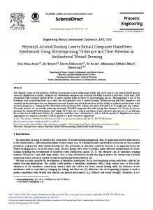
3. Results and discussions Change of colour of the reaction medium from pale yellow to light purple is the first physical indication about formation of AuNPs due to excitation of electrons in gold nanoparticles. As the reaction progresses with time, more gold ions are converted into gold nanoparticles and colour of the gold solution becomes more dense and intense. UV-vis spectrophotometer was used to record absorbance after 30 minutes and λmax of reaction mixture in the 700 nm to 400 nm range. λ max was observed at 540 nm with no other peaks in this region indicating formation of monodispersed gold nanoparticles in the reaction medium. 3.1 Effect of volume of extract on synthesis of AuNPs Different volumes of extracts ranging from 0.20 ml to 2 ml were used to observe their effect on AuNP synthesis by dropwise addition into 1 ml of 2.28 mM gold solution diluted with 5 ml distilled water. It was observed that increase of extract volume results in increase of absorbance of the AuNP solution measured by UV-vis spectrophotometer (Fig. 1). Change in colour of solution was appeared after 27, 21, 17, 14, 13 and 10 minutes using 0.20, 0.40, 0.60, 0.80, 1 and 2 ml of extract respectively. Colour of the solution also changed from ruby purple to deep purple with increase in volume of extract. Similar observations were reported for AuNP synthesis using Sesbania grandiflora leaf extract [20].
Fig 1: UV spectra using different volumes of extract
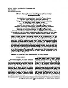
Fig 2: Change of λmax using different volumes of Extract
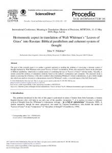
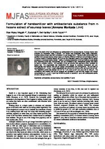
Absorbance of reaction medium increased from 0.86 A to 1.34 A up to addition of 1 ml of extract and became constant thereafter when higher volume of extract (up to 2 ml) was added to the reaction medium (Fig. 1). SPR band became sharper and λmax was blue shifted from 546 nm to 539 nm when volume of extract was increased from 0.20 ml to 0.60 ml (Fig. 2). This blue shifting of λmax indicating more formation of small nanoparticles [21]. Any further addition in extract quantity resulted in red shift of SPR band up to 557 nm showing an increase in particle size [20, 22] due to changes in dielectric properties of surrounding layer covers the surface of nanoparticles that was also observed during bio synthesis with olive leaf extract [21]. 3.2 TEM analysis of AuNPs Morphology of gold nanoparticles was studied using TEM image analysis which revealed that the majority of the synthesized AuNPs were spherical in shape with narrow size distribution. Trigonal, pentagonal and some irregular shaped particles were also observed. OPL extract was also prepared in distilled water without addition of any Emim chloride and was used to synthesize gold nanoparticles to evaluate the effect of ionic liquid on size of AuNPs. TEM image analysis confirmed that Emim chloride reduced particles size from 39.60 nm to 20.09 nm indicating its tendency to work as capping agent (Fig. 3). Particle size synthesized using Emim chloride was in range of 7.2 nm to 39.36 nm with an average particle diameter of 20.09 nm. On higher magnification it was observed that particles have a thin layer of ionic liquids on their surfaces and thus played its role to increase stability (Fig. 3). This layer of biomoieties, mostly flavonoids, were originally present in oil palm leaves extract [20]. AuNPs synthesized by OPL extract using distilled water only showed wide range of distribution with average diameters of 39.60 nm (Fig. 4).
571
Muhammad Irfan et al. / Procedia Engineering 148 (2016) 568 – 572
Fig 3: TEM image analysis and Histogram of AuNPs using distilled water
Fig 4: TEM image analysis and Histogram of AuNPs using aq. EMIM Cl
3.3 DLS & Zeta potential Particle size distribution was determined by DLS technique and hydrodynamic particle diameter of gold nanoparticles was measured as 55.76±0.35 nm (Fig. 5a). Particles size measured by DLS is higher than TEM image analysis might be due to capping of ionic liquids along with other organic moieties. Surface charge of AuNPs prepared in presence of Emin chloride was 14.9±0.15 mV indicating stability of gold solution [23] as shown in Fig. 5b.
(a)
(b)
Fig 5: DLS analysis of AuNPs; (a) size distribution (b) zeta potential
3.4 FTIR analysis FTIR analysis of OPL extracts before and after reaction with gold precursor indicated involvement of active biomolecules in reduction of gold ions (AuCl4-) to gold nanoparticles. FTIR spectrum showed significant stretching-frequency shifting around 3435 cm-1 indicating presence of OH group that was derived from polyphenolic compounds. Reduction in band intensity observed in the region of 698 cm-1 and 1639 cm-1 indicated presence of aromatic C-H bending, amide of polypeptides and stretching of carboxylate groups in amino acids groups of protein molecules (Fig. 6). These protein molecules are reported to cap the surface of AuNPs providing long term stability and reduce agglomeration of particles [24].
572
Muhammad Irfan et al. / Procedia Engineering 148 (2016) 568 – 572 Fig 6: FTIR analysis for OPL extract and AuNPs solution
4. Conclusion Stable gold nanoparticles were synthesized using oil palm leaves extract in presence of Emim chloride. Using Emim chloride average AuNP particle size was 20.06 nm and these were smaller than AuNPs prepared with aqueous OPL extract. Emim chloride capped the surface of gold nanoparticles and increased stability at room temperature. High value of zeta potential (-14.9 mV) confirms stability of AuNPs in presence of Emin chloride. Volume of extract showed significant impact on rate of reduction and surface plasmon resonance band moved to lower wavelength (blue shifted) up to 0.6 ml added extract and thereafter red shifted on increase in volume of extract. FTIR analysis confirms presence of polyphenolic and amide groups that are involved in reduction of gold precursor. Acknowledgements The author would like to express his gratitude towards project supervisor, AP Dr. Sekhar Bhattacharjee and grateful to Universiti Teknologi Petronas for financial support through FRGS-0153AB-I96. Author would also graciously acknowledge the financial assistance through Graduate Assistance (GA) scheme by Universiti Teknologi Petronas, Malaysia for pursuing PhD studies. References [1] T.S. Santra, F.-G. Tseng, T.K. Barik, Green biosynthesis of gold nanoparticles and biomedical applications, American Journal of Nano Research and Application 2 (2014) 5-12. [2] P.P. Gan, S.H. Ng, Y. Huang, S.F.Y. Li, Green synthesis of gold nanoparticles using palm oil mill effluent (POME): A low-cost and eco-friendly viable approach, Bioresource technol. 113 (2012) 132-135. [3] P. Ghosh, G. Han, M. De, C.K. Kim, V.M. Rotello, Gold nanoparticles in delivery applications, Adv. Drug Deliv. Rev. 60 (2008) 1307-1315. [4] A. Khan, R. Rashid, G. Murtaza, A. Zahra, Gold Nanoparticles: Synthesis and Applications in Drug Delivery, Trop. J. Pharm. Res. 13 (2014) 1169-1177. [5] Z. Khan, O. Bashir, J.I. Hussain, S. Kumar, R. Ahmad, Effects of ionic surfactants on the morphology of silver nanoparticles using Paan (Piper betel) leaf petiole extract, Colloids Surf B Biointerfaces. 98 (2012) 85-90. [6] P. Khademi-Azandehi, J. Moghaddam, Green synthesis, characterization and physiological stability of gold nanoparticles from Stachys lavandulifolia Vahl extract, Particuology, 19 (2015) 22-26. [7] P. Kuppusamy, M.M. Yusoff, G.P. Maniam, N. Govindan, Biosynthesis of metallic nanoparticles using plant derivatives and their new avenues in pharmacological applications–An updated report, Saudi Pharm J. (2014). [8] O.V. Kharissova, H.R. Dias, B.I. Kharisov, B.O. Pérez, V.M.J. Pérez, The greener synthesis of nanoparticles, Trends Biotechnol. 31 (2013) 240-248. [9] A.I. Lukman, B. Gong, C.E. Marjo, U. Roessner, A.T. Harris, Facile synthesis, stabilization, and anti-bacterial performance of discrete Ag nanoparticles using Medicago sativa seed exudates, J. Colloid Interface Sci. 353 (2011) 433-444. [10] L. Ren, L. Meng, Q. Lu, Z. Fei, P.J. Dyson, Fabrication of gold nano-and microstructures in ionic liquids—a remarkable anion effect, J. Colloid Interface Sci. 323 (2008) 260-266. [11] A. Safavi, S. Zeinali, M. Yazdani, Synthesis of biologically stable gold nanoparticles using imidazolium-based amino acid ionic liquids, J Amino Acids. 43 (2012) 1323-1330. [12] X. Zhang, Z. Sun, Z. Cui, H. Li, Ionic liquid functionalized gold nanoparticles: Synthesis, rapid colorimetric detection of imidacloprid, Sens Actuators B Chem. 191 (2014) 313-319. [13] P. Singh, K. Kumari, A. Katyal, R. Kalra, R. Chandra, Synthesis and characterization of silver and gold nanoparticles in ionic liquid, Spectrochim. Acta A Mol. Biomol. Spectrosc. 73 (2009) 218-220. [14] H. Passos, M.G. Freire, J.A. Coutinho, Ionic liquid solutions as extractive solvents for value-added compounds from biomass, Green Chem, 16 (2014) 47864815. [15] J. Anuradha, T. Abbasi, S. Abbasi, An eco-friendly method of synthesizing gold nanoparticles using an otherwise worthless weed pistia (Pistia stratiotes L.), Journal of Advanced Research 6 (2015) 711-720. [16] A.F.M. Cláudio, A.M. Ferreira, M.G. Freire, J.A. Coutinho, Enhanced extraction of caffeine from guaraná seeds using aqueous solutions of ionic liquids, Green Chemistry 15 (2013) 2002-2010. [17] Y. Sun, W. Li, J. Wang, Ionic liquid based ultrasonic assisted extraction of isoflavones from Iris tectorum Maxim and subsequently separation and purification by high-speed counter-current chromatography, J Chromatogr B. 879 (2011) 975-980. [18] S. Mohamed, Oil palm leaf: a new functional food ingredient for health and disease prevention, J. Food Process Eng Tech 5 (2014). [19] C. Ibegbulem, P. Chikezie, Serum lipid profile of rats (Rattus norvegicus) fed with palm oil and palm kernel oil-containing diets, Asian. J. Biochem 7 (2012) 46-53. [20] J. Das, P. Velusamy, Catalytic reduction of methylene blue using biogenic gold nanoparticles from Sesbania grandiflora L, J Taiwan Inst Chem E 45 (2014) 2280-2285. [21] M. M. H. Khalil, E.H. Ismail, F. El-Magdoub, Biosynthesis of Au nanoparticles using olive leaf extract. Arab J Chem. 5 (2012) 431-437. [22] B. Sharma, D.D. Purkayastha, S. Hazra, L. Gogoi, C.R. Bhattacharjee, N.N. Ghosh, J. Rout, Biosynthesis of gold nanoparticles using a freshwater green alga, Prasiola crispa, Mater Lett. 116 (2014) 94-97. [23] K. Anand, R. Gengan, A. Phulukdaree, A. Chuturgoon, Agroforestry waste Moringa oleifera petals mediated green synthesis of gold nanoparticles and their anti-cancer and catalytic activity, J. Ind. Eng. Chem. 21 (2015) 1105-1111. [24] R.K. Das, B.B. Borthakur, U. Bora, Green synthesis of gold nanoparticles using ethanolic leaf extract of Centella asiatica, Mater Lett. 64 (2010) 1445-1447.