¤ 2010 Zoological Society of Japan
ZOOLOGICAL SCIENCE 27: 931–938 (2010)
Ontogenetic Changes in the Morphology and Morphometry of Cuban Gar (Atractosteus tristoechus) Yamilé Comabella1, Andrés Hurtado2 and Tsai García-Galano1 * 1
Centro de Investigaciones Marinas, Universidad de la Habana, Calle 16 #114 e/ 1ra y 3ra, Playa, La Habana, Cuba 11300 2 Centro de Reproducción de la Ictiofauna Indígena, Ciénaga de Zapata, Cuba
Detailed examination of early development and growth of Cuban gar (Atractosteus tristoechus) was conducted using morphologic and morphometric characters. Larvae were reared at a constant water temperature (28 ± 1°C) from hatching to 18 days after hatching (DAH). Observation of the disappearance, reduction, or appearance of external structures, pigment characteristics, and behavior, identified three developmental stages (attached 0–3 DAH; transitional 4–10 DAH; and freeswimming 11–18 DAH). For the 18 day trial, the average growth rate was 1.30 mm/d and the specific growth rate averaged 10.2%/d. The slowest growth rates (0.02 mm/d and 2.8%/d, 7–11 DAH) coincided with the exhaustion of yolk reserves and the transition from endogenous to exogenous feeding. In addition to the slowest growth rates, the most dramatic morphological changes in A. tristoechus were observed during the transition from endogenous to exogenous feeding. Five total length (TL) groupings were established using 25 morphologic and morphometic characters. The characters snout length, pelvic fin length, snout width at nares, head length, and head width best identified length groupings. As the larvae developed, the snout and head lengthened and narrowed. Proportionally, the snout was narrower and the head longer in Cuban gar larvae than in other lepisosteid larvae. Key words:
Cuban gar, larvae, morphology, morphometry, lepisosteids
INTRODUCTION The Cuban gar (Atractosteus tristoechus) is an endemic freshwater fish (family: Lepisosteidae) that inhabits western Cuba, primarily in Ciénaga de Zapata. In conjunction with its restricted range, other factors such as habitat loss and ecological alteration, have contributed to the decline of natural populations. For these reasons, the species was listed as vulnerable in 1999 (Pérez et al., 1999) and interest in aquaculture production increased in order to preserve and restore natural populations through the stocking of cultured individuals. Research contributing to the development of management plans and strategies to protect the existence and survival of this species in Cuba are needed. Morphological descriptions of adult Cuban gar (Parra, 1787; Bloch and Schneider, 1801; Poey, 1854) and the ontogeny of digestive enzymes in larvae (Comabella et al., 2006) have been published, although the morphology of larval development has not yet been described. However, morphological characteristics of early development have been described for the longnose gar Lepisosteus osseus, shortnose gar L. platostomus, spotted gar L. oculatus, alligator gar Atractosteus spatula and tropical gar Atractosteus tropicus (Netch and Witt, 1962; Pearson et al., 1979; Yeager * Corresponding author. Phone: +53-7-203-0617; Fax : +53-7-204-3275; E-mail:
[email protected] doi:10.2108/zsj.27.931
and Bryant, 1983; Simon and Wallus, 1989; Simon and Tyberghein, 1991; Long and Ballard, 2001; Aguilera et al., 2002). The goals of this study were to qualitatively and quantitatively describe the development of Cuban gar larvae under laboratory conditions, to identify distinguishable morphological stages, and to determine growth rates during early development. MATERIALS AND METHODS Cuban gar (Atractosteus tristoechus) eggs were obtained from the induced spawning of one female and three males maintained in captivity at the Center for Native Ichthyofauna Reproduction located in Ciénaga de Zapata, Cuba. Breeding adults were placed in a 3 × 2.5 m concrete pond containing 50 cm of water (24–27°C). Artificial branches were spread throughout the pond to provide spawning substrate (León et al., 1978; Dean, 1895; Simon and Wallus, 1989). An initial injection of luteinizing hormone-releasing hormone analog (LHRH-A; 25 μg/ml) was administered to the broodstock, with a second injection given 16 h later. Courtship and spawning occurred 9 h after the second injection. Fifteen minutes after release from the female, spawned adhesive eggs were removed from the pond and placed in a 100 L circular fiberglass tank until hatching (68–100 h). The eggs taken from broodstock pond and transferred to experimental tanks were gradually adapted (4 h) to test temperature (28 ± 1°C). Experimental design, sampling and measurements After hatching, 300 larvae were distributed among three 15 L circular fiberglass tanks (6.7 larvae/l) containing submerged
932
Y. Comabella et al.
branches to facilitate larval adherence. Larvae were reared at a constant water temperature (28 ± 1°C), under a light regime of 08:00 to 20:00 h, and oxygen levels were maintained above 6 ppm. Each morning, 50% of the water was exchanged in each tank after bottom cleaning. Larvae were fed ad libitum with live Moina spp. three times a day (09:00, 14:00, and 19:00 h). Each day, three larvae per tank were randomly selected, anaesthetized with tricaine methanesulphonate (MS 222), individually weighed on an Ohaus balance (± 0.1 mg) and fixed in 70% ethanol for later examination. Identification of early developmental stages was carried out through observation (10, 20 and 40 × magnification) of the disappearance, reduction, or appearance of external structures and in situ observation of pigmentation and larval behavior. A Kodak Z 760 camera mounted on an Omano binocular dissecting microscope, using semi-dark field or total illumination, was used to photograph preserved larvae. Morphometric characters were measured (± 0.01 mm) using an ocular micrometer and digital calipers. Twenty morphometric characters (standard length, snout length, eye diameter, head length, predorsal length, preanal length, yolk sac length, pectoral and pelvic fin length, head depth at eyes, head depth at pectorals, preanal depth, mid-postanal depth, caudal peduncle depth, yolk sac depth, snout width at nares, mid-snout width, snout width at anterior margin of eyes, head width, and suctorial disk width) and three meristic characters (number of fin rays in dorsal, anal and caudal fins) were measured or counted for each specimen based on the criteria defined and illustrated by Simon and Wallus (1989) for lepisosteid larvae. Growth rate (GR; mm/d) was calculated as: [TL2 – TL1] / (t2 – t1) where TL2 and TL1 are the total length at times t2 and t1, respectively. The specific growth rate (SGR; %/day) was calculated as: 100 [ln W2 – ln W1] / (t2 – t1) where W2 and W1 are wet body weights at times t2 and t1, respectively. Statistical analyses Data were analyzed by one-way analysis of variance (ANOVA) to detect differences (p < 0.05) and significant ANOVA were followed by a post hoc multiple comparison test (Tukey test). Prior to ANOVA analysis, data expressed as percentages were arcsine square-root-transformed. Regression analysis was used to describe growth of larvae. The analyses were conducted using Statistica for Windows (StatSoft, Tulsa, Okla.).
RESULTS Larval morphology Three stages of larval development were established for this species (Table 1). Stage 1: Attached 0 DAH–3 DAH (28°C) Newly hatched larvae had an unformed mouth cavity (stomodeum) without snout extension, and possessed an adhesive papillose suctorial disk which allowed larvae to remain adhered to vegetation until the yolk sac was absorbed. During this stage the suctorial disk shrank (90%) and the mouth opened and communicated with the digestive system (Fig. 1A, B).
Table 1.
The head was was deflected over the pale-yellow yolk sac and an unpigmented optic cup covered the spherical eyes (Fig. 1B). The yolk sac was large, spherical to ovoid, often with a pointed posterior tip. Yolk sac depth gradually decreased (75%) (Fig. 1C). The first distinguishable fin, the transparent median fin-
Fig. 1. Developmental Stage 1 (attached) of Atractosteus tristoechus. sd- suctorial disc, s- stomodeum, e- eye, mf- median finfold, ys- yolk sac, o- operculum, pf- pectoral fins. Scale bar = 1 mm.
Description of developmental stages of Cuban gar larvae from 0–18 DAH. Stage 1 Attached
Stage 2 Transitional
Stage 3 Free Swimming
DAH (28°C) Total Length Weigth Suctorial disc Mouth
0–3 10.5–17.7 mm 20–37 mg Evident Unformed mouth cavity- mouth opens Yolk sac Present Median finfold Evident
4–10 16.2–24.9 mm 32–52 mg Almost absorbed Elongated snout. Teeth barely visible. Reduced Reduced
Pelvic fin Caudal fin
Absent Without fin rays Attached, vertical position Behaviour Rhythmic movements jaw-operculum Pigmentation Sparsely pigmented (head, lateral dark line from yolk sac to anus and dorsal brown line)
Initiate Fin rays initiated Detached, horizontal position Beginning of exogenous feeding Brown ventral sides, pale-yellow-brown stripe with irregular white margins on dorsal region. Dorsal brown line disappears.
11–18 22.9–35.2 mm 43–179 mg Absent Snout length increased and teeth present. Absent Almost absent (only between anus-anal fin) Fin rays defined Fin rays defined Free swimming
Phase
Lecithoexotrophic GR = 1.30 mm/d SGR = 10.2%/d.
Lecithotrophic
Conspicuous lateral brown line between dorsal yellow-brown and ventral gray side, from snout to caudal fin with an upper unpigmented stripe. Exotrophic
Cuban Gar Larvae Morphology and Morphometry
933
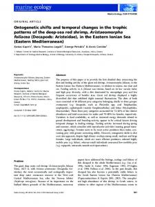
fold, was evident posterior to the yolk sac, with aggregated pigmentation, indicating the location of the future dorsal, caudal and anal fins (Fig. 1C). The pelvic fins were absent and the pectoral fins were located above the yolk sac. No fin rays were visible at this stage. The large operculum covered the anterior sides of the yolk sac to the base of the pectoral fins (Fig. 1D). Gill filaments and their circulatory network were visible. Larvae remained adhered to vegetation Fig. 2. Developmental Stage 2 (transitional) of Atractosteus tristoechus. sd- suctorial or the water surface by the suctorial disk in disc, no- nasal openings mf- median finfold, ys- yolk sac, o- operculum, pf- pectoral fins, vertical or semi-vertical orientation. Rhythpvf- pelvic fins. Scale bar = 1 mm. mic movements of the jaw and fleshy opercula began. Recently hatched larvae were only sparsely pigmented, mainly on the head. Scattered melanophores were present on the mid-dorsal yolk sac and along the junction of the yolk sac and body. Melanophores were densely concentrated on the upper surface of the gut from the yolk sac to anus, forming dark lines at the locations of future anal and caudal fins. No pigmentation was present on the dorsal surface of the body until a brown line formed extending from the snout to the future dorsal fin at 2 DAH. During this stage, yolk sac melanophores increased in number (Fig. 1E). Stage 2: Transitional Fig. 3. Developmental Stage 3 (free swimming) of Atractosteus 4 DAH–10 DAH (28°C) tristoechus. t- teeth, cr-caudal rays. Scale bar = 1 mm. The mouth formed and the majority of the adhesive suctorial disk was absorbed, leaving only a small remnant at the tip of the snout. The snout began to adopt its characteristic Stage 3: Free-swimming elongated shape. The maxilla was longer than the mandible, 11 DAH–18 DAH (28°C) and tooth primordia were barely visible on the jaws (Fig. 2A, Larvae were free-swimming and developed the characB). The nasal apparatus was evident with incurrent and teristic elongated adult shape. The suctorial disc and yolk excurrent openings. The eyes became spherical and pigsac were absent. Snout length increased and the jaws were mented (Fig. 2C). studded with sharp teeth (Fig. 3A). The median finfold was Pelvic fin buds were visible at the posterior end of the observable only between the anus and anal fin with a tenyolk sac, which was considerably reduced (Fig. 2D). Remdency to disappear. Rays began to form in the dorsal, anal nants of the median fin were observed between the develand pelvic fins. Pectoral and caudal fin ray formation was oping dorsal, caudal, and anal fins, but a more conspicuous complete and the paired fins were pigmented (Fig. 3B). finfold remained between the anal and pelvic fin buds. The The body was gray ventrally and ventro-laterally. A dark median finfold was gradually lost as the other fins develbrown longitudinal line between the yellow-brown dorsal and oped. Rudiments of fin rays became visible in unpaired fins, ventral sides became increasingly conspicuous, initiating mainly in the caudal fin as basal pterygiophores. Pectoral just behind the posterior end of the snout, continuing fins increased in length and developed black pigmentation through the eyes and ending at the caudal fin (Fig. 3C). basally with unpigmented margins. Fin rays began to form Numerous scattered melanophores covered the entire body in the pectoral fins. The opercula began to shrink and except for an unpigmented lateral stripe from snout to anus. develop a solid structure. At this time, the larvae detached from vegetation Growth remained, near the water surface and were capable of movLarvae increased 3.1 times in total length and 6.3 times ing in a horizontal direction, by means of undulations of the in weight from 0 to 18 DAH (Fig. 4). Increase in total length caudal part of the body. Exogenous feeding began, and was was defined by the following equation: TL = 12.884 + 1.2167 observable by lateral movement of the head followed by × DAH, and the increase in total weight was described the bites to catch live food. Digestive tracts full of food items equation TW = 23.887 × exp(0.091 × DAH). The growth rate were observed, as were the first fecal pellets. Pigmentation was 1.30 mm/d and the specific growth rate averaged consisted mostly of brown areas on the ventral side of the 10.2%/d. Three larval growth periods were identified during body, and the dorsal region developed a pale-yellow-brown the first 18 days after hatching: 0–6 DAH, 7–11 DAH and stripe with irregular white margins extending from the head 12–18 DAH. Rapid growth was observed from 0–6 DAH and to the posterior region. The distinctive light-colored dorsal 12–18 DAH, however, growth was slowest (0.02 mm/d and stripe seen in stage 1 was no longer visible. By the end of 2.8%/d) from 7–11 DAH. this stage, all the fins were black with unpigmented margins (Fig. 2D).
934
Y. Comabella et al.
Larval morphometry Five length intervals were determined (Fig. 5). The first interval (10.5–14.9 mm) occurred during the attached stage, stage 1 of development, from hatching until 2 DAH at 28°C. The second interval (16–22.1 mm) included larvae passing from first to second stage of development at 3 to 5 DAH.
Fig. 4. Growth in weight (left scale) and total length (right scale) of Cuban gar larvae from hatching until 18 days after hatching (DAH). Each point represents the mean of nine individuals ± SE. Discontinuous vertical lines define the three growth periods (0–6 DAH, 7– 11 DAH, 12–18 DAH). Growth rates (GR-mm/d) and specific growth rates (SGR-%/d) are presented for each growth period.
The next interval (22.4–24.9 mm) represented by larvae from 6 to 12 DAH, had the slowest growth of all intervals in length (GR = 0.4 mm/d) and weight (SGR = 2.14%/d) and
Fig. 5. Relationships among total length, age and developmental stage of Cuban gar larvae from hatching until 18 days after hatching (DAH). Circles represent total length intervals.
Table 2. Morphometric characters (mean ± SD; Minimum-Maximum) of Cuban gar larvae grouped by selected total length intervals (N = simple size). Characters are expressed as percentages of total length (TL) or head length (HL). Fin ray counts are also listed for each interval. Different superscripts indicate significant differences between intervals (p < 0.05). Total length (TL) intervals (mm) Measure
10.5–14.9 (N = 27)
Mean ± SD Min-Max Length, % of TL Snout length 4.1 ± 0.94e (2.5–7.5) Eye diameter 6.4 ± 0.53a (5.3–7.6) Head length (HL) 21.9 ± 2.33d (18.5–27.3) Predorsal length 72.5 ± 2.39a (68.5–77.4) Preanal length 69.4 ± 2.02a (65.8–73.6) Standard length 98.4 ± 0.70a (97.0–99.2) Yolk sac length 36.9 ± 2.29a (31.5–41.1) Fin length, % of TL Pectoral 4.3 ± 1.27c (2.5–6.7) Pelvic Body Depth, % of TL Head at eyes 14.9 ± 1.64a (12.4–18.9) Head at Pectoral 22.6 ± 2.60a (18.8–28.3) Preanal depth 7.3 ± 1.32b (4.4–9.8) Postanal depth 5.5 ± 0.80b (3.5–6.9) Caudal Peduncle 5.2 ± 0.69ab (4.5–6.1) Yolk sac depth 16.8 ± 2.98a (12.1–22.6) Body width, % of HL Snout at nares Mid-snout Snout anterior eyes 41.4 ± 5.82a (32.3–52.2) Head width 63.8 ± 7.42a (48.6–83.3) Disc width 29.0 ± 6.70a (17.6–40.0) Fin ray counts Dorsal 0 Anal 0 Caudal 0
16–22.1 (N = 27)
22.4–24.9 (N = 46)
26.2–31 (N = 12)
31.8–35.2 (N = 11)
Mean ± SD
Min-Max
Mean ± SD
Min-Max
Mean ± SD
Min-Max
Mean ± SD
Min-Max
6.9 ± 1.34d 5.7 ± 0.41b 26.1 ± 1.47c 66.7 ± 1.22c 63.1 ± 1.30c 96.0 ± 1.34c 31.9 ± 3.04b
(5.2–9.5) (4.9–6.8) (23.6–29.1) (64.7–69.6) (61.1–65.7) (94.2–99.3) (27.2–37.5)
11.2 ± 1.10c 5.5 ± 0.29bc 28.6 ± 1.60b 67.4 ± 1.26c 63.7 ± 1.68c 95.4 ± 1.04c
(8.5–13.3) (4.9–6.2) (25.5–33.6) (65.0–69.7) (59.2–67.2) (93.2–97.7)
13.7 ± 1.30b 5.5 ± 0.33bc 31.2 ± 2.07a 69.7 ± 2.61b 66.5 ± 2.53b 97.3 ± 1.34b
(12–16.4) (4.8–6) (27.9–34.5) (66.0–74.9) (63.2–71.5) (94.6–98.9)
14.8 ± 0.88a 5.3 ± 0.28c 31.5 ± 1.12a 69.5 ± 1.20b 66.0 ± 1.34b 96.9 ± 1.08b
(13.4–16.2) (4.7–5.6) (29.4–33.0) (67.9–71.8) (64.0–69.1) (94.9–98.7)
6.0 ± 0.54b 3.2 ± 0.42c
(4.4–7.1) (2.4–3.9)
5.5 ± 0.59b 4.2 ± 0.71b
(4.5–6.3) (3.4–5.8)
5.7 ± 0.36b (5.2–6.5) 4.9 ± 0.42a (4.3–5.5)
8.9 ± 0.88c 9.7 ± 0.67c 7.7 ± 0.71b 5.2 ± 0.36b 4.9 ± 0.33b
(6.9–10.5) (8.3–10.4) (6.2–8.5) (4.6–5.7) (4.4–5.3)
8.5 ± 0.38c 9.6 ± 0.31c 7.7 ± 0.25b 5.2 ± 0.28b 5.0 ± 0.29b
(7.9–9.4) (9.1–10.1) (7.3–8.1) (4.8–5.8) (4.6–5.4)
11.0 ± 1.01c (9.1–12.8) 9.9 ± 0.44d 21.6 ± 1.91b (19.2–24.4) 20.2 ± 1.33b 29.2 ± 2.04b (25.9–32.9) 27.0 ± 1.32b 33.9 ± 1.70d (30.3–36.7) 32.2 ± 1.02d
(9.3–10.9) (18.7–22.3) (24.8–29.1) (30.6–33.7)
6.9 ± 0.68a (5.8–8.1) 2.1 ± 0.59d (1.0–2.8) 10.9 ± 1.34b 12.6 ± 3.01b 8.5 ± 0.86a 6.2 ± 0.56a 5.7 ± 0.46a 9.8 ± 1.40b
(8.8–14.1) (1.4–17.5) (6.8–10.6) (5.2–7.7) (4.7–6.7) (6.9–12.7)
8.8 ± 0.51c 9.5 ± 0.47c 7.0 ± 0.50b 5.2 ± 0.42b 4.9 ± 0.43b
(7.6–10.1) (8.6–10.7) (5.7–7.7) (3.8–5.9) (3.8–5.8)
15.8 ± 1.53a 32.0 ± 4.89a 41.2 ± 4.61a 50.1 ± 5.31b 11.7 ± 1.09b
(14–19.1) (25.8–48.7) (33.9–51.3) (41.9–60.0) (10.6–12.8)
13.5 ± 1.13b 23.7 ± 1.71b 31.4 ± 2.70b 38.5 ± 2.37c
(10.8–15.9) (20.3–27.1) (23.2–36.8) (33.3–42.4)
0 4 6–10
4–6 4–7 9–12
6–8 5–7 11–13
6–8 6–8 12–14
Cuban Gar Larvae Morphology and Morphometry
contained larvae from stages 2 and 3. The last two length intervals (26.2–31 mm; 31.8–35.2 mm) were characterized by the greatest increase in weight (17.2%/d) and length (1.5 mm/d). Selected morphometric and meristic characters (Table 2) were grouped by the five total length intervals described above. The characters that differed statistically among all the intervals were: snout length, pelvic fin length and snout width at nares. Snout length represented 4.1% of TL in recently hatched larvae (ranging from 10.5–14.9 mm TL) and increased to 14.8% of TL when larvae reached 31.8– 35.2 mm TL (18 DAH). Pelvic fin length increased from 2.1% of TL when these fins first appeared (16–22.1 mm) to 4.9% of TL at 18 DAH. However, snout width at nares significantly shrank from 15.8% to 9.9% of HL from the second to fifth length interval. Characters such as head length and head width differed statistically in all length intervals with exception of the last two. In newly hatched larvae, the head length was 22% of
Table 3. Morphometric characters of lepisosteid larvae using Cuban gar total length intervals (expressed in percentages using proportionality described in Table 2). Bold number- A. tristoechus (present study), a- L. osseus (Simon and Wallus, 1989), b- L. oculatus (Simon and Tyberghein, 1991), c- A. spatula (Aguilera et al., 2002), d- A. tropicus (Aguilera et al., 2002), e- L. platostomus (Simon and Wallus, 1989). Intervals
Snout length
Head length
Fin Pelvic length
Snout width
Head width
4.1 5.1–5.7 4–6.2 6–7.4 6.8–10.6
21.9 20.4–24.6 18.4–20.8 25.6–26 27.6–29.4
– – – – 0.7–1.5
– –20.1 –15.3 22.3–22.7 18.8–24.8
63.8 64.4–52.1 52.5–60.9 30.7–38.6 43.8–44.2
6.9 26.1 8.9–10.5 26.9–26.2 6.2–12.2 20.8–26.6 7.4–9.9 26–27.8 10.6–13.8 27.6–29.4
2.1 2.8–3.2 –2.7 1.6–3.6 1.5–4.3
15.8 18.5–16.3 15.3–14.9 22.7–18.6 18.8–14.4
50.1 42.7–35 52.5–35.8 38.6–36.2 44.2–36.6
11.2 28.6 10.5–11.5 26.2 12.2–11 26.6–25.4 9.9–14.2 27.8–29.5 13.8 29.4
3.2 3.2–3.3 2.7–3.1 3.6–5.1 4.3
13.5 38.5 16.3–13.9 35–32.4 14.9–15.4 35.8–35.4 18.6–14.8 36.2–33.4 14.4 36.6
a b c d e
13.7 31.2 11.5–12.4 26.2–26.2 11–14.3 25.4–27.2 14.2 29.5 13.8 29.4 11.9 29.2 14.8 31.5
4.2 3.3–3.5 3.1–3.4 5.1 4.3 3.8 4.9
11 33.9 13.9–12.9 32.4–33.9 15.4–12.2 35.4–27.6 14.8 33.4 14.4 36.6 10.2 32.6 9.9 32.2
a b c d e
14.3 27.2 14.2–15.3 29.5–30.7 13.8 29.4
3.4 5.1–5.8 4.3
12.2 27.6 14.8–16.9 33.4–33.2 14.4 36.6
I a b c d e II a b c d e III a b c d e IV
V
935
TL, and increased 31.5% of TL by 15–18 DAH. Head width decreased from 63.8% to 32.2% of HL in the studied period. A qualitative comparison with other lepisosteid larvae such as L. osseus, L. oculatus, A. spatula, A. tropicus and L. platostomus (Simon and Wallus, 1989; Simon and Tyberghein, 1991; Aguilera et al., 2002), using Cuban gar total length intervals appears in Table 3. DISCUSSION During larval development, many fish undergo rapid growth and dramatic changes in morphology, metabolism, and behavior in preparation for the metamorphosis into juvenile fish period (Gisbert et al., 2002; Verhaegen et al., 2007; Darias et al., 2008). In the present study, three periods of larval development were identified. The first stage was characterized by a lecithotrophic phase when larvae remained attached and vertically oriented to vegetation and obtained energy exclusively from the yolk sac. The defining event of this stage was the switch from cutaneous to branchial respiration, manifested by rhythmic movements of the mandible and opercula and the development of gill filaments. In early larvae, the gills were relatively underdeveloped, although gill growth was rapid (Oikawa and Itazawa, 1985), thus supporting increased oxygen consumption required for changes in morphology, metabolism, swimming ability, and behavior. Similar development was described in 6 DAH sturgeon larvae (Gisbert et al., 2001; Gisbert and Doroshov, 2006) and 18–19 DAH California halibut larvae (Gisbert et al., 2002). This stage may incur a relatively high risk of predation; however, gar species have evolved cardiotoxic substances that are found in the eggs and yolk sac and may protect against predation (Netch and Witt, 1962; Burns et al., 1981). The most drastic morphological changes observed in A. tristoechus occurred during the second developmental stage. During this lecithoexotrophic stage, larvae began to feed exogenously but continued to utilize yolk reserves that meet the energetic demands of prey capture (Moteki et al., 2001; Williams et al., 2004). This transitional period was defined by internal, external, and behavioral changes. Also, survival of the larvae depends on the quantity of endogenous food supply, the rate at which yolk is used after hatching (Jobling, 1995; Betti et al., 2009), incubation temperature (Mendiola et al., 2007), the ability to capture, ingest, and digest food, and the availability of food at first feeding. This is a critical stage in larval life due to competition for food and predation (Balon, 1985; Coughlin, 1991). Propulsion both for food capture and predator avoidance is critical and depends on the development of organs necessary for feeding (Porter and Theilacker, 1999; Makrakis et al., 2005) and swimming (Murphy et al., 2007; Huysentruyt et al., 2009). The concurrent development of organs associated with feeding and predator avoidance and must occur in mutual balance (Osse et al., 1997). During this stage, larval behavior was characterized by periods of resting and swimming activity. At 10 DAH most larvae remained relatively immobile maintaining position in the water column although employed active swimming for feeding. Studies of larval swimming show that in general, early fish larvae use anguilliform swimming with a large amplitude over a substantial part of the body (Webb and Weihs, 1986; Osse and van den Boogaart, 1995), which gradually develops into the characteristic adult swimming
936
Y. Comabella et al.
pattern (Russo et al., 2007). The pectoral and caudal fins developed more rapidly than did the other fins. This rapid development may be associated with standard maneuvering and “start movements” usually observed in fishes in this stage (Barros and Higuchi, 2007). According to Walker (2004), maneuvering and “start movements” are commonly associated with predation strikes, which involve both caudal fin movements to generate the impulse and pectoral fins for maneuvering. Larval Cuban gars were observed to initiate feeding 5–7 DAH, when the pectoral and caudal fins predictably developed faster than the other fins. Additionally, the functionality of the swimbladder at this age (unpublished data) may allow a higher incidence of prey capture, as a result of efficient buoyancy regulation. After the critical stage of transition, larvae are exotrophs capable of detecting and predating zooplankton in the water column. During the third stage, snout lengthening was most evident, sharp teeth were visible ,and the development of fin rays improved their carnivorous habits. With active feeding, larval growth and survival increased and remnants of the finfold were resorbed. Most teleost fishes possess a larval finfold during early development (Kendall et al., 1984). The timing of finfold growth, differentiation and disappearance varies greatly among different fish groups (Balon, 1985; Fukahara, 1985). The function of the larval finfold is still speculative; however, van Snik et al. (1997) considered it a possible adaptation for locomotion providing an enlarged surface for cutaneous larval respiration. At the end of the third stage, Cuban gar larvae gradually transformed into juveniles. This transformation was not reflected solely by fin growth, but included a drastic change in total body form from larvae to juvenile. The growth and ossification of fin rays in the caudal fin mark the transition to the adult swimming pattern. The general pigmentation pattern observed for A. tristoechus larvae was similar to descriptions of pigmentation in other gar species (Suttkus, 1963; May and Echelle, 1968; Echelle and Riggs, 1972; Moore et al., 1973; Simon and Wallus, 1989, Aguilera et al., 2002). The unpigmented lateral stripe extending from head to the caudal fin appeared during the second stage (16.2–24.9 mm) of development of Cuban gar larvae, similar to the case in L. oculatus (22– 26 mm), L. osseus (15.4 mm) and A. spatula (19.2–22.5 mm; Simon and Wallus, 1989; Aguilera et al., 2002). In these species, the lateral stripes have irregular margins, whereas in tropical gar, the lateral stripes between the dark areas of the flank were yellow, had well-defined margins and appeared earlier (12.5–13.8 mm; Aguilera et al., 2002). Nevertheless the impact of feeding and culture conditions on pigmentation may result in different pigmentation patterns between reared and wild larvae. Different patterns of larval growth with respect to weight and length were observed during the experiment. Up to 6 DAH, length increased significantly by 2.01 mm/d and weight increased 11.6%/d although this increase was not stastically significant. This period coincides with lecithotrophic stage 1 when larvae may only increase in length due to the utilization of yolk sac elements while weight remains stable because there is no uptake of external nutrients. This was confirmed by a previous study (Comabella et al., 2006)
where larval protein concentration increased significantly starting on 9 DAH, which coincided with the input of new protein sources from exogenous feeding. From 7 to 11 DAH, neither length nor weight increased significantly, coinciding with the transition from endogenous and exogenous feeding, the most important and vulnerable period during the ontogeny of fishes (Kamler, 1992; Makrakis et al., 2005). According to Williams et al. (2004), the greatest reduction in yolk sac volume corresponds to the ‘intensity’ of development, and is reflected in the period of the greatest change in larval appearance, and organ and structure development. This transition is a complex process involving critical physiological changes associated with yolk depletion. Similar patterns of growth reduction have been reported for several teleost species (Blaxter, 1969; Yada and Furukawa, 1999; Gisbert et al., 2002; Geerinckx et al., 2008). The exotrophic, free-swimming stage 3 was characterized by a pronounced daily increase in weight due to effective assimilation of external nutrients. Our results are in agreement with the growth pattern reported for the first 15 DAH for A. spatula (Mendoza et al., 2002) at the same temperature; however, growth and development rates probably differ at different temperatures. The growth rates of Cuban gar larvae were the second fastest reported for lepisosteid larvae (Table 4). Our experimental conditions and temperature (28 ± 1°C) were similar to experiments conducted with other Atractosteus spp. Species, thus allowing for comparison of larval growth rates among the three members of the genus. The growth rates of Cuban gar larvae were intermediate between those of alligator gar and tropical gar larvae. The first total length interval of Cuban gar larvae was characterized by a lecithotrophic phase and was comparable to the first and second intervals described for A. spatula and A. tropicus by Aguilera et al. (2002). The second total length interval of Cuban gar was analogous to intervals III and IV described by Aguilera et al. (2002) for other the Atractosteus species. The second total length interval was a transitional, lecithoexotrophic stage. However, in the third total length interval, growth was slowest, due to the change from endogenous to exogenous feeding that concludes with the exhaustion of yolk reserve. After this critical period of transition, larvae in the last two total length intervals were Table 4. species.
Published growth rates for larval and juvenile lepisosteid
Specie
Growth rates Stage (mm/d)
Lepisosteus osseus
0.8 larvae 2.33–4.5 juvenile
Lepisosteus oculatus
0.83 1.3–1.7 1.55 5.06 0.86–1 2.5 1.30
Atractosteus spatula Atractosteus tropicus Atractosteus tristoechus
Reference
Pearson et al. (1979) Netch and Witt (1962) Echelle and Riggs (1972) larvae Simon and Tyberghein (1991) juvenile Simon and Wallus (1989) 0–10 DAH Aguilera et al. (2002) 10–15 DAH 0–15 DAH Aguilera et al. (2002) 15–30 DAH 0–18 DAH Present study
Cuban Gar Larvae Morphology and Morphometry
exotrophs, with the largest incremental increase in weight and length. Intervals IV and V of Cuban gar were similar to intervals V and VI reported for A. spatula and A. tropicus by Aguilera et al. (2002). In addition to early ontogenetic changes in Cuban gar growth rates, changes in body proportions were also observed. The characters snout length, pelvic fin length, snout width at nares, head length and head width were used to determine morphometric differences among total length intervals of Cuban gar larvae. Changes in most of the characters used to differentiate total length intervals were related to cephalic development. During the development of Cuban gar larvae, the snout narrowed and the head lengthened, similar to other lepisosteids, reflecting the ichthiophagous habits of this family. A comparison of the four differential characters related to head development, among A. tristoechus, L. osseus, L. oculatus, L. platostomus, A. spatula and A. tropicus larvae, yielded an interesting proportionality relationship. For the first total length intervals of larval development (10.5–22 mm), the Cuban gar possessed proportionally the shortest snout and the widest head, in comparison to the other lepisosteids mentioned above. For last total length intervals (26.2– 35.2 mm), the head of Cuban gar was longer and the snout narrower than the other species. Based on research into the relationships between morphological characters and feeding ecology in other fish species (Luczkovich et al., 1995; Clifton and Motta, 1998; Luboschek and McCormick, 2001; Wesneat and Marshall, 2008) the observed differences in snout width and head length in lepisosteids may be due to variation in habitats and food resources among the species. Unfortunately, there are no observations of spawning areas or diet of Cuban gar larvae in natural habitats, thus leaving many questions unanswered. ACKNOWLEDGMENTS This study was supported by Centro de Investigaciones Marinas (CIM) and Centro de Reproducción de la Ictiofauna Indígena from Cuba. The authors wish to thank Dr. J. Canabal for his excellent technical assistance during the experiment. Special thank to Dr. Allyse Ferrara (Nicholls State University, USA) for her many helpful comments on the manuscript and English corrections. We acknowledge the two anonymous referees, who greatly contributed to the improvement of this work by providing corrections, suggestions, and comments.
REFERENCES Aguilera C, Mendoza R, Rodríguez G, Márquez G (2002) Morphological description of alligator gar and tropical gar larvae, with an emphasis on growth indicators. Trans Am Fish Soc 131: 899–909 Balon EK (1985) The theory of saltatory ontogeny and life history models revisited. In “Early Life History of Fish” Ed by EK Balon, Dr W Junk Publishers, Boston, Massachusetts, pp 13–30 Barros B, Higuchi H (2007) Notes on morphological characters in early developed amazonian leaffish, Monocirrhus polyacanthus (Polycentridae, Perciformes). Kempffiana 3: 18–22 Betti P, Machinandiarena L, Ehrlich MD (2009) Larval development of Argentine hake Merluccius hubbsi. J Fish Biol 74: 235–249 Blaxter JHS (1969) Development: eggs and larvae. In “Fish Physiology Vol 3” Ed by WS Hoar, DJ Randall, Academic Press, New York, pp 177–252 Burns TA, Stalling DT, Goodger W (1981) Gar ichthyotoxin
937
(Lepisosteus sp.): its effect on crayfish, with notes on bluegill sun-fish. Southwest Nat 25: 513–515 Case JE, Westneat MW, Marshall CD (2008) Feeding biomechanics of juvenile red snapper (Lutjanus campechanus) from the northwestern Gulf of Mexico. J Exp Biol 211: 3826–3835 Clifton K, Motta PJ (1998) Feeding morphology, diet and ecomorphological relationship among five Caribbean Labrids (Teleostei, Labridae). Copeia 1998: 953–966 Comabella Y, Mendoza R, Aguilera C, Carrillo O, Hurtado A, García-Galano T (2006) Digestive enzyme activity during early larval development of the Cuban gar Atractosteus tristoechus. Fish Physiol Biochem 32: 147–157 Coughlin DJ (1991) Ontogeny of feeding behaviour of first-feeding Atlantic salmon (Salmo salar). Can J Fish Aquat Sci 48: 1896– 1904 Darias MJ, Zambonino-Infante JL, Hugot K, Cahu CL, Mazurais D (2008) Gene Expression Patterns During the Larval Development of European Sea Bass (Dicentrarchus labrax) by Microarray Analysis. Mar Biotechnol 10: 416–428 Dean B (1895) The early development of gar-pike and sturgeon. J Morphol 11: 1–55 Echelle AA, Riggs CD (1972) Aspects of the early life history of gars (Lepisosteus) in Lake Texoma. Trans Am Fish Soc 101: 106– 112 Fukahara O (1985) Functional morphology and behaviour of early life stages of red sea bream. Bull Jap Soc Sci Fish 51: 731–743 Geerinckx T, Verhaegen Y, Adriaens D (2008) Ontogenetic allometries and shape changes in the suckermouth armoured catfish Ancistrus cf. triradiatus Eigenmann (Loricariidae, Siluriformes), related to suckermouth attachment and yolk-sac size. J Fish Biol 72: 803–814 Gisbert E, Doroshov SI (2006) Allometric growth in green sturgeon larvae. J Appl Ichthyol 22 (Suppl 1): 202–207 Gisbert E, Cech JJ, Doroshov SI (2001) Routine metabolism of larval green sturgeon (Acipenser medirostris Ayres). Fish Physiol Biochem 25: 195–200 Gisbert E, Merino G, Muguet JB, Bush D, Piedrahita RH, Conklin DE (2002) Morphological development and allometric growth patterns in hatchery-reared California halibut larvae. J Fish Biol 61: 1217–1229 Huysentruyt F, Moerkerke B, Devaere S, Adriaens D (2009) Early development and allometric growth in the armoured catfish Corydoras aeneus (Gill, 1858). Hydrobiologia 627: 45–54 Jobling M (1995) Development of eggs and larvae. In “Environmental Biology of Fishes” Ed by M Jobling, Chapman & Hall, London, pp 357–390 Kamler A (1992) Early life history of fish: an energetics approach. Chapman & Hall, London Kendall AW, Ahlstrom EH, Moser HG (1984) Early life history stages of fishes and their characters. In “Ontogeny and Systematics of Fishes Vol 1” Ed by Special Publication of the American Society of Ichthyology and Herpetology, pp 11–22 León R, Aguiar R, Hernández I (1978) Estudio sobre la biología y el cultivo artificial del manjuarí (Atractosteus tristoechus) Blosh y Schneider, Dirección Ramal de Acuicultura, La Habana Long W, Ballard WW (2001) Normal embryonic stages of the longnose gar, Lepisosteus osseus. BMC Dev Biol: 1–6 Luczkovich JJ, Norton SR, Gilmore RG (1995) The influence of oral anatomy on prey selection during the ontogeny of two percoid fishes, Lagodon rhomboides and Centropomus undecimalis. Environ Biol Fish 44: 79–95 Lukoschek V, McCormick MI (2001) Ontogeny of diet changes in a tropical benthic canivorous fish, Parupenaeus barberinus (Mullidae): relationship between foraging, boehaviour, habitat use, jaw size and prey selection. Mar Biol 138: 1099–1113 Makrakis MC, Nakatani K, Bialetzki A, Sanches PV, Baumgartnera G, Gomes LC (2005) Ontogenetic shifts in digestive tract mor-
938
Y. Comabella et al.
phology and diet of fish larvae of the Itaipu Reservoir, Brazil. Environ Biol Fish 72: 99–107 May EB, Echelle A (1968) Young-of-year alligator gar in Lake Texoma, Oklahoma. Copeia 3: 629–630 Mendiola D, Alvarez P, Cotano U, Murguía AM (2007) Early development and growth of the laboratory reared north-east Atlantic mackerel Scomber scombrus L. J Fish Biol 70: 911–933 Mendoza R, Aguilera C, Rodríguez G, González M, Castro R (2002) Morphophysiological studies on alligator gar (Atractosteus spatula) larval development as a basis for their culture and repopulation of their natural habitats. Rev Fish Biol Fish 12: 133–142 Moore G, Trautman M, Curd M (1973) Description of postlarval gar (Lepisosteus spatula Lacepede, Lepisosteidae), with a list of associated species from the Red River, Choctaw County, Oklahoma. Southwest Nat 18: 343–344 Moteki M, Yoseda K, Sahin T, Ustundag C, Kohno H (2001) Transition from endogenous to exogenous nutritional sources in larval Black Sea turbot Psetta maxima. Fish Sci 67: 571–578 Murphy BF, Leis JM, Kavanagh KD (2007) Larval development of the Ambon damselfish Pomacentrus amboinensis, with a summary of pomacentrid development. J Fish Biol 71: 569–584 Netch NF, Witt A (1962) Contributions to the life history of the longnose gar (Lepisosteus osseus) in Missouri. Trans Am Fish Soc 91: 251–262 Oikawa S, Itazawa Y (1985) Gill and body surface areas of the carp in relation to body mass, with special reference to the metabolism–size relationship. J Exp Biol 117: 1–14 Osse JWM, van den Boogaart JGM (1995) Fish larvae, allometric growth, and the aquatic environment. ICES Marine Sciences Symposium 201: 21–34 Osse JWM, van den Boogaart JGM, van Snik GMJ, van der Sluys L (1997) Priorities during early growth of fish larvae. Aquaculture 155: 249–258 Pearson WD, Thomas GA, Clark AL (1979) Early piscivory and timing of the critical period in postlarval longnose gar at mile 571 of the Ohio River. Trans Ky Acad Sci 40: 122–128 Pérez E, Matamoros Y, Ellis S (1999) Taller para el análisis de la conservación y manejo planificado de una selección de especies cubanas (CAMP). Sección IV, Peces, Habana Porter S, Theilacker G (1999) The development of the digestive tract
and eye in larval walleye pollock, Theragra chalcogramma. Fish Bull 97: 722–729 Russo T, Costa C, Cataudella S (2007) Correspondence between shape and feeding habit changes throughout ontogeny of gilthead sea bream Sparus aurata L., 1758. J Fish Biol 71: 629– 656 Simon T, Wallus R (1989) Contributions to the early life histories of gar (Actinopterygii: Lepisosteidae) in the Ohio and Tennesse River Basins with emphasis on larval development. Trans Ky Acad Sci 50: 59–74 Simon TP, Tyberghein EJ (1991) Contributions to the early life history of the spotted gar, Lepisosteus oculatus Winchell, from Hatchet Creek, Alabama. Trans Ky Acad Sci 52: 124–131 Suttkus RD (1963) Order lepisostei. In “Fishes of the Western North Atlantic” Ed by HB Bigelow, WC Schroeder, Memoirs of the Sears Foundation for Marine Research I, Part 3, New Haven, Connecticut van Snik GMJ, van den Boogaart JGM, Osse JWM (1997) Larval growth patterns in Cyprinus carpio and Clarias gariepinus with attention to the finfold. J Fish Biol 50: 1339–1352 Verhaegen Y, Adriaens D, Wolf TD, Dhert P, Sorgeloos P (2007) Deformities in larval gilthead sea bream (Sparus aurata): A qualitative and quantitative analysis using geometric morphometrics. Aquaculture 268: 156–168 Walker JA (2004) Kinematics and performance of maneuvering control surfaces in teleost fishes. IEEE J Ocean Eng 29: 572–584 Webb PW, Weihs D (1986) Functional morphology of early life history stages of fishes. Trans Am Fish Soc 115: 115–127 Williams K, Papanikos N, Phelps RP, Shardo JD (2004) Development, growth, and yolk utilization of hatchery-reared red snapper Lutjanus campechanus larvae. Mar Ecol Prog Ser 275: 231–239 Yada O, Furukawa A (1999) Relationship between external and internal morphological changes and feeding habits in the fry stage of Japanese catfish Silurus asotus. UJNR Tech Rep 28: 157–162 Yeager BL, Bryant RT (1983) Larvae of the longnose gar, Lepisosteus osseus, from the little river in Tennessee. J Tenn Acad Sci 58: 20–22 (Received December 22, 2009 / Accepted May 10, 2010)