Dec 10, 2013 - 2009). Aquatic leeches that cause parasitic infestation in humans and animals include Limnatis nilotica, Myxobdella africana, Dinobdella ferox, ...
ttp://www.bsava.com
CASE REPORT
Oral infestation with leech Limnatis nilotica in two mixed-breed dogs S. M. Rajaei*, H. Khorram*, M. Ansari Mood*, S. Mashhadi Rafie* and D. L. Williams† *Department of Clinical Sciences, Faculty of Specialized Veterinary Sciences, Science and Research Branch, Islamic Azad University, Tehran, Iran †Department of Veterinary Medicine, University of Cambridge, Cambridge CB3 0ES
Leeches are bloodsucking hermaphroditic parasites that attach to tissues using two muscular suckers, ingest large amounts of blood and may cause severe anaemia in the host. Two four-month-old mixed-breed dogs (one bitch and one male) were referred with anorexia, retching, hypersalivation and bleeding from the oral cavity. On physical examination, two live leeches were detected on the ventral aspect of the tongue of the bitch and one in a similar position in the male. The leeches were gently detached and removed using Adson tissue forceps after applying vinegar over the area. Microcytic hypochromic anaemia was detected in the bitch and mild leukocytosis in the dog. One month after treatment both animals were re-examined and a complete blood count was normal. Given that infestation with leeches as described here is associated with contaminated water, the use of clean and safe drinking water is recommended to avoid such diseases. Journal of Small Animal Practice (2014) 55, 648–651 DOI: 10.1111/jsap.12166 Accepted: 9 October 2013; Published online: 10 December 2013
INTRODUCTION The word leech is derived from the Anglo-Saxon word loece, meaning to heal, in reference to the early blood-letting practices of Western medicine. Worldwide, over 600 species have been described, which occur in freshwater, marine, estuarine and moist-terrestrial ecosystems. In freshwater systems, leeches are an integral benthic component functioning as predators or ectoparasites. (Moser et al. 2009). Leeches have a cylindrical body with anterior and posterior suction discs. The anterior suction disc is circular and may be very prominent or just a minor expansion of the lips of the mouth. The posterior suction disc is circular, directed ventrally, and can be either larger or smaller than the body of the leech (Moser et al. 2009). Aquatic leeches that cause parasitic infestation in humans and animals include Limnatis nilotica, Myxobdella africana, Dinobdella ferox, Phytobdella catenifera and Teromyzon tessulatom (Gholami-Ahangaran et al. 2012). The Nile leech Limnatis nilotica is found in countries around the Mediterranean and in Bulgaria and Romania, reaching as far east as Iran (Orevi et al. 2000, Grosser and Pesic 2006) and Tadjikistan, and as far west as the Azores. It is the most widely distributed species of leech in Egypt and Lebanon (Orevi et al. 2000). The typical leech life cycle consists of the egg, which is deposited inside a cocoon, the juvenile and finally the reproductive 648
hermaphroditic adult. Freshwater leeches typically breed in the spring, lay eggs into a secreted cocoon in the summer, and overwinter in the benthos or on a host. Juveniles of blood sucking species require a minimum of 3 to 5 blood meals to reach sexual maturity. The typical life span for leeches is 1 to 3 years (Moser et al. 2009). Leech infestation primarily occurs in tropical areas, such as in Mediterranean countries, Africa and Asia (Uygur et al. 2003). Leech infestations have been reported in the veterinary literature in cats (Chang et al. 2006) and dogs (Hatherill 1967, Gothe et al. 1991, Bahmani et al. 2011). To the authors’ knowledge, this is the first report in English of naturally occurring leech infestation of the oral cavity of dogs.
CASE HISTORY Two four-month-old mixed-breed dogs (one bitch and one male) with anorexia, retching, hypersalivation and bleeding from the oral cavity were referred to the Islamic Azad University specialised veterinary clinic (Saadat Abad, Tehran, Iran). Vaccination and parasite control were adequate and up to date for both dogs. According to the owner, during a week-long trip to a rural area in the Fars province of Iran, bleeding was observed from the dogs’ mouths. The dogs resisted opening their mouth by the owner,
Journal of Small Animal Practice
•
Vol 55
•
December 2014
•
© 2013 British Small Animal Veterinary Association
Oral infestation with leech in two mixed-breed dogs
but veterinary assistance was not available at that time. Two days after returning home, 7 to 9 days after possible exposure to contaminated water, the two dogs were anorexic and were brought for veterinary attention. Both dogs had good body condition scores (5 and 4·5 of a 9-point body condition score system) and weighed 14·5 and 13·6 kg each. On physical examination, the heart and respiratory rates of both dogs were within reference limits. Both animals showed pallor of the oral mucous membranes and pain on opening the mouth especially in the bitch. Two live leeches were detected on the ventral aspect of the tongue of the bitch (Fig. 1) and one in a similar position in the male (Fig. 2). The leech species isolated from the two dogs was dark brown in colour with two lateral yellow-orange and dark green bands on either side. Strong jaws and muscular suckers at the anterior and posterior ends were prominent. The three invertebrates measured 32, 31 and 32 mm, respectively, in length and were identified to be a Limnatis nilotica species based on morphological characteristics (Gholami-Ahangaran et al. 2012). Both animals were sedated by using combination of 10 mg/kg ketamine (Ketamine; Alfasan Co.) and 0·3 mg/kg of midazolam (Midamax; Tehran Chemie Pharmaceutical Co.) for complete
examination. Two millilitres of vinegar was applied over the area where the leeches were attached to the oral mucosa, and the leeches were gently detached using a pair of Adson tissue forceps. The haemorrhagic sites on the oral mucosa were initially rinsed with saline (Sodium Chloride 0·9%; Shahid Ghazi Pharmaceutical Co.) and subsequently with 0·2% chlorhexidine oral solution (Hexodine®, Shahr Darou Co.). In order to examine for presence of other leeches in the oesophagus and upper airways, plain radiographs of the thorax in right lateral and ventrodorsal projection were obtained. No abnormalities were visible. Endoscopy was not available. Complete blood counts (CBC) and serum biochemistry profiles were performed. The CBC abnormalities in the bitch included decreased packed cell volume (PCV), haemoglobin (Hb), red blood cell (RBC) count, mean corpuscular volume (MCV) and mean corpuscular haemoglobin (MCH) (Table 1). Microcytic hypochromic anaemia was diagnosed. No other abnormalities were found in the leukogram and serum biochemistry profiles. The bitch was treated with oral iron supplementation (Feriron®; Darou Pakhsh), multivitamin drop (Mr Vit®; Shahr Darou Co.) and prophylactic oral antibiotics; 20 mg/kg amoxicillin/clavulanic acid (Farmentin®; Farabi Co.) three times daily for 3 days. In the male dog only a mild leukocytosis with left shift was noted (data not shown) and the animal received 20 mg/kg amoxicillin/ clavulanic acid (Farmentin®) three times daily orally for 7 days. One week later, the owner reported that treatment was effective and the dogs were bright, responsive and eating solid food. In a follow-up visit 1 month later, both dogs were re-examined and no clinical abnormalities were detected. The complete blood counts and serum biochemistry profiles had returned to their respective reference intervals (Table 1).
DISCUSSION
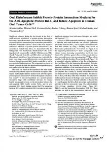
FIG 1. Two live leeches (arrowed) adhering to the mucous membrane of the ventral tongue in the bitch. The rostral leech is blood engorged
FIG 2. Limnatis nilotica leeches in sublingual mucosa of male dog Journal of Small Animal Practice
•
Vol 55
•
December 2014
•
The leech Limnatis nilotica normally is attracted to animals or humans when they are exposed to water. It enters the mouth or nostrils from water holes, ponds or streams and attaches itself to the mucous membranes of the pharynx, larynx or nostrils. Occasionally, it invades the eye, urethra or vagina. (Orevi et al. 2000). Leech infestations have been reported in humans and animals in several countries. Infestation with Limnatis nilotica has been reported several times in Iran and its neighbouring countries especially Iraq and Turkey, Iran’s western neighbours (Almallah 1968, Alcelik et al. 1997, Shirzadeh 2005, Agin et al. 2008, Bulent et al. 2010, Shirzadeh and Golmohammadi 2012). Three reports of leech infestation in dogs have been documented, one in the oral cavity (Bahmani et al. 2011, in Farsi) and one in the nasal cavity (Gothe et al. 1991). No data was available from another report (Hatherill 1967). This report appears to be the first in the English veterinary literature of oral cavity infestation with leeches in two dogs. A general assumption is that all leeches are sanguivorous (blood-feeding). However, leeches exhibit a diverse range of feeding strategies, with approximately half of the leech species being
© 2013 British Small Animal Veterinary Association
649
S. M. Rajaei et al.
Table 1. Complete blood count and biochemistry profile of bitch (initial and after 1 months) Test
Result
Reference interval
First examination
1 months later
30% 53 g/L 3×10⁶/µL 53 fL 17·6 pg 14×103/µL 8·1×103/µL 0·22×103/µL 4·3×103/µL 0·4×103/µL 0·98×103/µL 285
38% 124 g/L 5·9×106/µL 65 fL 20·6 pg 13×103/µL 9·2×103/µL 0·1×103/µL 2·9×103/µL 0·6×103/µL 0·2×103/µL 322
37 to 55% 120 to 180 g/L 5·5 to 8·5×106/µL 60 to 77 fL 19·5 to 24·5 pg 6 to 17×103/µL 3 to 11·5×103/µL 0 to 0·3×103/µL 1 to 4·8×103/µL 0·15 to 1·25×103/µL 0·1 to 1·25×103/µL 210 to 450×103/µL
57 g/L 28 g/L 4.95 mmol/L 6.78 mmol/L 70.72 mmol/L 30 U/L 23 U/L 103 U/L 11 U/L 0.0011 mmol/L 0.009 mmol/L
62 g/L 30 g/L 4.67 mmol/L 5.35 mmol/L 44.2 mmol/L 27 U/L 28 U/L 98 U/L 9 U/L 0.0026 mmol/L 0.007 mmol/L
54 to 71 g/L 26 to 33 g/L 3.57 to 6.49 mmol/L 3.57 to 9.996 mmol/L 44.2 to 132.6 mmol/L 23 to 66 U/L 21 to 102 U/L 20 to 156 U/L 1·15 to 28·4 U/L 0.0008 to 0.005 mmol/L 0.004 to 0.012 mmol/L
Haematology Packed cell volume Haemoglobin Red blood cell Mean corpuscular volume Mean corpuscular haemoglobin White blood cell Neutrophils (Segmented) Neutrophils (Band) Lymphocytes Monocytes Eosinophils Platelets Serum Biochemistry Total protein Albumin Glucose BUN Creatinine AST ALT ALP CK Fe TIBC
BUN Blood urea nitrogen, AST Aspartate aminotransferase, ALT Alanine aminotransferase, ALP Alkaline phosphatase, CK Creatine kinase, TIBC Total iron binding capacity
predatory on invertebrates (such as earthworms, gastropods, crustaceans and insect larvae) the others being temporary bloodfeeding ectoparasites (Moser et al. 2009). Anaemia is a worldwide clinical problem and usually associated with an underlying disease. Whilst these are often associated with chronic diseases such as renal failure, immune-mediated diseases, infections, toxicosis and deficiencies (Tyler and Cowell 1996), in tropical climates anaemia is often associated with parasitic diseases including internal or external parasites, such as fleas, ticks, lice or more rarely leeches. In this respect, leeches are well known in tropical areas for their ability to cause severe anaemia (Taskesen et al. 2009) to such a degree that it can lead to the death of the patient (Cundall et al. 1986). This anaemia can occur due to the anticoagulatory salivary gland secretion of blood-feeding leeches and/or the ability of leeches to ingest large amounts of blood. The number of leeches that infest humans or animals is also important, although as shown here one or two leeches may cause sufficient blood loss to result in anaemia. Leeches can ingest large amounts of blood into their stomach, even up to over ten times their body weight (Moser et al. 2009, Sarathi 2011). Various anticoagulant agents are known to be injected by the leech; theromyzon tessulatum, cytin (chymotrypsin inhibitors), tessulin (a trypsin and chymotrypsin inhibitor), therin (a trypsin inhibitor), theromin (a thrombin inhibitor) and therostatin (a factor Xa inhibitor) have been identified (Salzet 2001). These agents can cause bleeding and subsequent severe prolonged blood loss in infested humans or animals. In this report, the bitch was diagnosed with anaemia associated with leech infestation. This has been reported in several human studies (Cundall et al. 1986, Estambale et al. 1992, Krüger et al. 650
2004, Kabiri et al. 2009, Kose et al. 2008, Montazeri et al. 2009, Taskesen et al. 2009, Çoban et al. 2011, Senthilkumaran et al. 2012) but less commonly in the canine species. Diagnosis of leech infestation in the oral cavity is relatively easy. Various diagnostic methods have been used in humans for diagnosis of leeches in the oesophagus, nasopharynx and respiratory system. Radiography has been used for evaluation of the gastrointestinal and respiratory systems in some humans with leech infestation (Krüger et al. 2004, Mekonnen 2013, Moini and Farbod-Ara 2013) but has some limitations. Leeches can cause severe problems, especially in the lower airways, where they can cause life-threatening acute bleeding and/or obstruction and must be removed immediately by bronchoscopy (Krüger et al. 2004). Endoscopy appears to be the best diagnostic technique to diagnose the presence of leeches in the oesophagus, and other visually inaccessible parts of the gastrointestinal tract. Various ways of removing the leech from the mucous membranes have been described including the use of an insecticide spray (Pryor et al. 1970), cocaine drops (White 1998), topical lidocaine (Bilgen et al. 2002) or distilled water (Chang et al. 2006). In the authors’ experience, vinegar renders it easy to detach leeches. Additionally, there are no reports of vinegar toxicity in the dog. In humans, various techniques have been used to detach leeches from mucous membranes, including suction (Adhikari 2005), normal saline (Hannan and Hoque 2012), lidocaine (Ghimire and Acharya 2008), forceps with or without bipolar electro-cautery (Chen et al. 2010) and vinegar (Bilgen et al. 2002, Ghazzawi et al. 2005, Garca et al. 2011). Removal of the leech should be done gently; forceful removal may cause the invertebrate’s mouthparts to remain in the tissue inciting infection (Kose et al. 2008).
Journal of Small Animal Practice
•
Vol 55
•
December 2014
•
© 2013 British Small Animal Veterinary Association
Oral infestation with leech in two mixed-breed dogs
Contact with water infested with leeches has been documented to be the most important risk factor for oropharynx and nasopharynx infestation of aquatic leeches in humans (Chen et al. 2010, Çoban et al. 2011) and animals (Chang et al. 2006). In this report access to contaminated drinking water was associated with leech infestation in these dogs. To prevent this type of disease in companion animals, use of clean and safe water especially in suburban areas with rivers, ponds, canals and swamps is an important consideration. Conclusion This case report details the infestation of oral leeches in two dogs with oral bleeding and mucous membrane pallor associated with anaemia in one animal. Leeches should be included in the differential diagnoses for unexplained anaemia in animals which may have been exposed to contaminated water in tropical or subtropical areas. Conflict of interest None of the authors of this article has a financial or personal relationship with other people or organisations that could inappropriately influence or bias the content of the paper. References Adhikari, P. (2005) Nasal leech infestation in children: comparison of two different innovative techniques. International Journal of Pediatrics and Otorhinolaryngology 73, 853-855 Agin, H., Ayhan, F. Y., Gülfidan, G., et al. (2008) Severe anemia due to the pharyngeal leech Limnatis nilotica in a child. Türkiye Parazitoloji Dergisi 32, 247-248 Alcelik, T., Cekic, O. & Totan, Y. (1997) Ocular leech infestation in a child. American Journal of Ophthalmology 124,110-112 Almallah, Z. (1968) Internal hirudiniasis as an unusual cause of haemoptysis. British Journal of Diseases of the Chest 62, 215-218 Bahmani, M., Nekouei, S. H., Parsaei, P., et al. (2011) Case report infestation with Limnatis nilotica in a Persian male dog from Shahrekord. Journal of Modern Veterinary Researches 2, 29-32 (in Farsi) Bilgen, C., Karci, B. & Ulouz, Ü. (2002) Case report: a nasopharyngeal mass: leech in the nasopharynx. International Journal of Pediatric Otorhinolaryngology 64, 73-76 Bulent, A., Ilknur, O., Beray, S., et al. (2010) an unusual cause of hemoptysis in a child: live leech in the posterior pharynx. Tropical biomedicine 27, 208-210 Chang, S. C., Cheng, F. P., Tung, K. C., et al. (2006) Nasal infestation with the leech Dinobdella ferox in a domestic shorthair cat. Veterinary Record 21, 99-100 Chen, W. C, Chien, C. Y., Yang, C. H., Li, et al. (2010) Nasal leech infestation: report of seven leeches and literature review. European Archives of Oto-RhinoLaryngology 267, 1225-1229 Çoban, S., Tutal, E., Alpay, D., et al. (2011) An unexpected cause of severe anemia in an adult patient: a pharyngeal leech (with video). Gastrointestinal Endoscopy 73, 360-361 Cundall, D. B., Whitehead, S. M. & Hechtel, F. O. (1986) Severe anaemia and death due to the pharyngeal leech Myxobdella africana. Transactions of the Royal Society of Tropical Medicine and Hygien 80, 940-944 Estambale. B. B., Knight, R.& Chunge, R. (1992) Haematemesis and severe anaemia due to a pharyngeal leech (Myxobdella africana) in a Kenyan child: a case
Journal of Small Animal Practice
•
Vol 55
•
December 2014
•
report. Transactions of the Royal Society of Tropical Medicine and Hygien 86, 458 Garca, M. F., Yelken, M. K. & Okur, M. H. (2011) Leech infestation of the nasopharynx; a rare cause of epistaxis and hemorrhage. European Journal of General Medicine 8, 141-143 Ghazzawi, I., Zuraiqat, F., Burqan, O., et al. (2005) Leech infestation causing upper gastrointestinal bleeding. Jordanian Royal Medical Services 12, 47-49 Ghimire, A. & Acharya, A. (2008) Unusual cause of unilateral epistaxis: nasal leech infestation. Journal of Nepal Medical Association 47, 38-40 Gholami-Ahangaran, M., Bahmani, M.& Zia-Jahromi N. (2012) Comparative and evaluation of anti-leech (Limnatis nilotica) effect of olive (Olea europaea L.) with Levamisol and Tiabendazole. Asian Pacific Journal of Tropical Disease 2, S101-S103 Gothe, R., Barutzki, D., Schöl, H., et al. (1991) Imported infestations of nasopharyngeal parasites in dogs. Tierärztliche Praxis 19, 84-7. (In German) Grosser, C. & Pesic, V. (2006) On the diversity of Iranian leeches (Annelida: Hirudinea). Archives of Biological Science Belgrade 58, 21-24 Hannan, M. J. & Hoque, M. M. (2012) Leech infestation in children through body orifices: experience in a hospital in Bangladesh. World Journal of Surgery 36, 2090-2092 Hatherill, C. W. (1967) Diestecostoma mexicanum infestation of dogs. Veterinary Record 81, 262 Kabiri, M., Hassani, A., Barkat, A. et al. (2009) Severe anemia by epistaxis caused by leech infestation. Archive of Pediatrics 16, 384-385 (in French) Kose, A., Zengin, S., Kose, B., et al. (2008) Leech bites: massive bleeding, coagulation profile disorders, and severe anaemia. American Journal of Emergency Medicine 26, 1067e3-1067e6 Krüger, C., Malleyeck, I.Olsen, O. H. (2004) Aquatic leech infestation: a rare cause of severe anaemia in an adolescent Tanzanian girl. European Journal of Pediatrics 163, 297-299 Mekonnen, D. (2013) Leech infestation: the unusual cause of upper airway obstruction. Ethiopian Journal of Health Sciences 23, 65-68 Moini, L. & Farbod-Ara. T. (2013) Hemoptysis caused by leech infestation: a unique case. Zahedan Journal of Research in Medical Sciences 15, 85-87 Montazeri, F., Bedayat, A., Jamali, L., et al. (2009) Leech endoparasitism: report of a case and review of the literature. European Journal of Pediatrics 168, 39-42 Moser, W. E., Govedich, F. R. & Klemm, D. J. (2009) Annelida, Hirudinida (Leeches). In: Encyclopedia of Inland Waters, Three-Volume Set. Vol. 1, 1st edn. Ed G. E. Likens. Academic Press, Waltham, MA, USA. pp 116-123 Orevi, M., Eldor, A., Giguzin, I., et al. (2000) Jaw anatomy of the blood-sucking leeches, Hirudinea Limnatis nilotica and Hirudo medicinalis, and its relationship to their feeding habits. Journal of Zoology 250, 121-127 Pryor, W. H., Bergner, J. F. & Raulston, G. L. (1970) Leech (Dinobdella ferox) infection of a Taiwan monkey (Macaca cyclopis). Journal of the American Veterinary Medical Association 157, 1926-1927 Salzet, M. (2001) Anticoagulants and inhibitors of platelet aggregation derived from leeches. FEBS Letters 492, 187-192 Sarathi, K. (2011) Nasal leech infestation causing persistent epistaxis. Journal of Emergencies, Trauma and Shock 4, 413-414 Senthilkumaran, S., Menezes, R. G., Pant, S., et al. (2012) Unexpected reach of a leech. Journal of Parasitic Diseases 6, 1-3 Shirzadeh, E. (2005) Red eye due to leech in the eye. Iranian Journal of Medical Sciences 30, 197-198 Shirzadeh, E. & Golmohammadi, R. (2012) Eye bleeding due to leech infestation in an ischemic heart-disease patient. Iranian Red Crescent Medical Journal 14, 245-247 Taskesen, M., Katar, S. & Basçik, H. (2009) an unusual cause of gastrointestinal bleeding and severe anaemia in a child: leech infestation. Journal of Tropical Pediatrics 55, 338-339 Tyler, R. D. & Cowell, R. L. (1996) Classification and diagnosis of anaemia. Comparative Hematology International 6, 1-16 Uygur, K., Yasan, H., Yavuz, L., et al. (2003) Removal of laryngeal leech: a safe and effective method. American Journal of Otolaryngology 24, 338-340 White, G. B. (1998) Leeches and leech infestation. In: Manson’s Tropical Diseases. 20th edn. Ed G. C. Cook.W. B. Saunders, London. pp 1523-1525
© 2013 British Small Animal Veterinary Association
651