The MOC-25 tumour arose spontaneously in a female nude mouse and was established as a continuous line intraperitoneally in nude mice, where it reproduces ...
CLIN. EXPL. METASTASIS, 1991, VOL. 9, NO. 5, 4 8 5 - 4 9 7
Organ-specific growth of a murine lymphoma of spontaneous origin in nude mice ANTONELLA TOMASONI, EUGENIO SCANZIANI*, GABRIELLA MASSAZZA, GIOVANNI GIUDICI?, GIUSEPPE SIRONI*, CORRADO CASLINI, ALESSANDRO RAMBALDI and R A F F A E L L A G I A V A Z Z I ~ : Istituto di Ricerche Farmacologiche Mario Negri, Via Gavazzeni 11, 24100 Bergamo, *Istituto di Anatomia Patologica Veterinaria e Patologia Aviare, Universit~t degli Studi di Milano, Via Celoria 10, 20133 Milano, and ?Clinica Pediatrica, Universith de Milano, Ospedale S. Gerardo, 20052 Monza, Italy
(Received 11 June 1991; accepted 13 August 1991) The MOC-25 tumour arose spontaneously in a female nude mouse and was established as a continuous line intraperitoneally in nude mice, where it reproduces the topological features of its origin, growing preferentially in the uterus, ovaries and liver. Karyotype analysis showed that MOC-25 cells are hyperdiploid. Tumorigenicity and malignant behaviour were studied by transplanting tumour cells into different sites in nude mice. The comparison of tumour take after i.p. and s.c. injections of scaled concentrations of MOC-25 cell suspension showed preferential growth in the peritoneum. Regardless of the route of implantation (s.c., i.v., i.p.), this tumour rapidly and preferentially disseminated to the liver, uterus, ovaries, spleen and bone marrow. No significant differences in tumour growth and metastatic behaviour were observed when MOC-25 was injected in ovariectomized nude mice or in male nude mice. Morphology studies using light and electron microscopy, immunophenotyping and molecular analysis indicated a B-lymphoid origin of the MOC-25 tumour.
Introduction The capacity of tumour cells to disseminate to and grow at different anatomical sites is a particular characteristic of malignancy. The locations of distant secondary tumours in m a n y clinical cancers and animal tumours are not r a n d o m and their distribution cannot be explained by simple anatomical or mechanical hypotheses [22, 32]. The mechanism(s) that regulate the homing of metastases to specific sites remain largely undefined. A p p r o p r i a t e models of cancer cell dissemination and metastasis are indispensable if progress is to be m a d e in this area [8, 19]. The majority of spontaneous and induced animal tumours currently used in investigations produce metastases to the lung; few tumour lines spontaneously metastastize to the liver and the peritoneal organs in general or localize in bone marrow; this pattern of dissemination has often been described for tumours of the haematopoietic system [3, 14, 19, 29]. T u m o u r lines that metastasize to specific organs have been obtained by sequential injection of metastases from organ to organ [7, 23]. Artificial metastases, e.g. in the liver or b o n e - b o n e m a r r o w have been obtained by injecting ~To whom correspondence should be addressed. (~) 1991 Rapid Communications of Oxford Ltd.
486
A. Tomasoni et al.
tumour cells directly or indirectly into the bloodstream draining the target organ [1, 13, 16]. More recently the injection of tumour cells into pertinent organs has provided a way by which dissemination of tumour cells can more closely resemble the clinical pattern of tumour dissemination [8, ll]. However, these methods are often technically complicated and are not always reproducible. The ideal model for studying organ specificity of tumour cells is one where tumour cells preferentially or only grow in certain sites without selective pressure, independently from the route of injection. We describe here a murine lymphoma of spontaneous origin that appeared in the peritoneal organs of a nude mouse. This tumour line, maintained in nude mice, repeatedly and selectively metastasizes to the organs from which it was isolated.
Materials and methods
Animals Nude (Ncr nu/nu) mice were obtained from the National Cancer Institute, Animals Program, Frederick, MD, and used when 6-8 weeks old. Throughout the experiments mice were housed in laminar flow cabinets under specific pathogen-free conditions. Mice received proper care and maintenance in accordance with institutional guidelines. Tumour line maintenance The MOC-25 line, derived from a spontaneous murine tumour that arose in the peritoneal cavity of a female nude mouse, was established and maintained for serial passage i.p. in nude mice by injecting tumour cells obtained from enzymatic digestion of solid tumours, as previously described [12]. Briefly, tumour tissue fragments, aseptically removed from the infiltrated peritoneal organs (mainly the uterus and ovaries) were subjected to 20 rain sequential digestion at 37 ~ "in Eagle's minimum essential medium (MEM) containing collagenase type I (200 units/ml) and DNase (270 kunits/ml) (Sigma, St. Louis, MA). The cell suspension was filtered through 250/xm sterile mesh, washed three times and resuspended in Ca 2+, Mg2+-free Hank's balanced salt solution (HBSS). Tumour cells (1 x 10⁶), shown to be viable by Trypan Blue exclusion, were injected i.p. into nude mice in 0.2 ml HBSS.
Flow cytometric analysis A cell suspension of MOC-25 was obtained as described above. The surface immunophenotype was determined by indirect immunofluorescence using a FACScan analyser (Becton Dickinson). The following monoclonal antibodies were employed: rat anti-mouse CD45 (clone M1/89.18.7.HK): mouse anti-mouse I-A d (clone MK-D6) and rat anti-mouse lymphocyte surface receptor for endothelium (MEL-14), obtained from the American Type Culture Collection (ATCC). The mouse anti-mouse C D l l b (clone MAC-l) was kindly provided by Dr T. Springer, Dana Farber Cancer Institute, Boston, MA. The mouse anti-mouse CD3 (clone 2Cll) and anti-mouse CD5 (clone 53.7.3); anti-IgM, anti-K and anti-A chains were obtained courtesy of Dr S. Alberti (Mario Negri Institute, Milano, Italy). Non-immune rat ascites were used as negative control.
Metastatic murine lymphoma
487
The secondary reagents were an affinity-purified FITC-labelled goat anti-mouse (Tecno Genetics, Recordati, Italy) and rabbit anti-rat (Bio-Yeda, Rehovot, Israel) immunoglobulin antiserum.
Southern blot analysis DNA was prepared by cell lysis, proteinase K digestion, extraction with phenol and precipitation with ethanol. Ten micrograms of DNA were digested with the appropriate restriction endonuclease, size-fractionated by electrophoresis through 0.8% agar gel, denatured, neutralized and transferred to synthetic membranes (Genescreen plus, Boston, MO). The immunoglobulin genes arrangement was evaluated with a 1.9 kb BamHI-EcoRI cDNA fragment that recognizes the J region of the mouse IgH gene (clone J 11, kindly provided by Dr E. D'Andrea, Instituto di Oncologia, University of Padua, Italy) [17]. This probe was labelled to a high specific activity with 32p using the random primer method. Membranes were hybridized and washed as described [6]. Karyotype A single-cell suspension of the MOC-25 tumour was prepared as described above. Cells were incubated in MEM containing 30% FCS, supplemented with 0.1/zg/ml colcemid (Gibco) for 2 h. Chromosome spreads were prepared by standard techniques following hypotonic treatment with KC1 75 M and fixation in methanol:acetic acid (3:1). Spreads were stained by QFQ banding and 50 metaphases were examined. Histopathological studies For histological examination samples of peritoneal organs infiltrated by tumours were fixed in 10% buffered formalin, processed for paraffin embedding and sectioned according to standard techniques. Sections 5/xm thick were stained with haematoxylin and eosin. For metastasis development, lung, spleen, liver, kidney, uterus, ovary, spine, femur, and brain were processed for histological examination. Selected samples were immunohistochemically tested by the avidin-biotin peroxidase complex (ABC) procedure (Vecstain Elite, Vector Laboratories, Burlingame, CA) for the presence of cytokeratins using a commercially available polyclonal antibody (Dakopatts, Golstrup, Denmark). Histological sections from snap-frozen tumour samples were examined for the enzyme c~-naphthyl acetate esterase using a commercially available kit (Sigma). Transmission electrort microscopy was performed on multiple fresh fragments of tumour tissue fixed overnight in 2.5% glutaraldehyde in Milloning phosphate buffer at 4 ~ washed in buffer and post-fixed in 1% osmium tetroxide. These were then dehydrated in graded ethanols and embedded in Araldite. The sections were cut at 60-90 nm, stained with uranyl acetate and lead citrate and examined in a Zeiss EM 109 electron microscope. Tumour growth For intraperitoneal growth tumour cell suspensions obtained as described above were injected i.p. into nude mice at the concentrations Specified in the Results. Mice were checked twice a week. Criteria for growing tumours were abdominal distension, palpable tumour masses in the peritoneal cavity and visible swelling of ovary and uterus. Tumour growth was confirmed at autopsy.
488
A. Tomasoni et al.
Subcutaneous tumours were induced by a s.c. injection of 0.1 ml tumour cell suspension in the dorsal scapular region. Mice were observed twice a week. Diameters of growing tumours were measured in millimetres with calipers and the tumour weight in mg was calculated from the formula length • width 2 2 T u m o u r weights were plotted against days after inoculation and tumour doubling time was calculated from the resulting graph.
Metastasis formation For spontaneous metastasis studies, mice were given a s.c. injection of 10 • 106 viable tumour cells in 0 . 1 m l HBSS. When the tumour reached 1> 1.5 cm or mice became moribund they were killed and autopsied. The ability of tumour cells to disseminate after i.p. injection was studied in all mice bearing a growing i.p. tumour. These animals were killed and autopsied when they became moribund or showed a heavy tumour burden. For artificial metastasis studies 1 • 105 viable tumour cells suspended in 0.2 ml HBSS were injected into the lateral tail vein of nude mice. The animals were killed and autopsied 21-28 days later (by which time they were paraplegic)i Lung, lymph nodes, spleen, liver, kidneys, uterus, and ovary were routinely examined for metastases or tumour infiltration. Paraplegia was considered a sign of spine involvement. Metastasis development was confirmed by histological analysis as described above. Results
Origin of MOC-25 tumour A tumour of unknown origin was discovered in a 13-month-old female nude mouse. The mouse presented marked abdominal distension and at autopsy the ovary and uterus were found to be replaced by tumour. The liver contained tumour nodules with 50% of parenchymal infiltration. No ascitic effusion was observed. No gross tumours were evident in other organs. The tumour, called MOC-25, now at its 15th passage, was serially transplanted i.p. to other nude mice. The transplanted tumour reproduced the topological and histological features found in the mouse of origin.
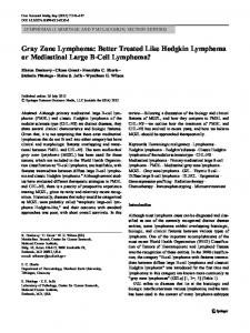
Characterization of MOC-25 tumour Morphological analysis. Histologically the MOC-25 tumour tissue consisted of rounded or polygonal pleomorphic cells with round-to-oval voluminous nuclei with scant chromatin (Figure 1). T u m o u r cells were negative for cytokeratins and cr-naphthyl acetate esterase. Electron microscope analysis showed irregular round-to-oval cells with large ovoid, occasionally invaginated or lobulated nuclear outlines (Figure 2). The scanty cytoplasm was limited and contained numerous free ribosomes, some cisternae of rough endoplasmic reticulum and mitochondria. No other structures were seen in the cytoplasm, and no cell junctions joined opposed cell membranes. On the basis of these findings the MOC-25 tumour was initially classified as of lymphoid origin.
Metastatic murine lymphoma
489
Immunophenotype and molecular analysis. Positive staining with the anti-CD45 monoclonal antibody confirmed the haematopoietic origin of the neoplastic cells (Figure 3). These cells were consistently I-A d positive, but myeloid (antiC D l l b ) , lymphoid-T (anti-CD3 and CD5), and B (anti-IgM, anti-K, anti-Z) cell surface antigens were never expressed by MOC-25 (Figure 3). Staining with MEL-14, the anti-lymphocyte surface receptor for endothelium, was also negative. The lack of a clear relationship of these haematopoietic cells with a specific cell differentiation pathway prompted us to investigate the genomic organization of the genes encoding for the heavy chain of immunoglobulins, which undergoes
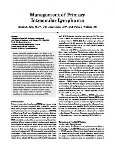
Figure 1. Photomicrographs of MOC-25 secondary growth in nude mice. (A) MOC-25 tumour cells infiltrating the spleen. A remnant of the splenic tissue is still recognizable (H&E, x200). (B) Liver with neoplastic nodules (H&E, x300).
490
A. Tomasoni et al.
Figure 1. (cont.) (C) Involvement of the bone marrow showing the medullary cavity of the proximal epiphysis of the tibia completely filled by tumour cells. The bone cortex is lysed and the muscular tissue is infiltrated. Bone marrow on the left is unaffected (H&E, x80).
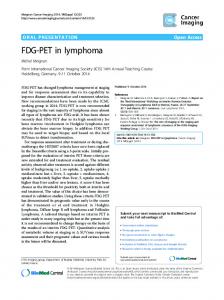
early rearrangement during B cell ontogeny [17]. Tumour cell D N A was digested with specific restriction endonucleases and hybridized to an IgH c D N A probe. As shown in Figure 4 (lane 1), rearranged bands were evident in D N A from MOC-25, thus indicating the cells' B-lymphoid origin.
Figure 2. Electron micrograph of MOC-25 cells infiltrating the liver tissue. Cells have large oval or invaginated nuclei and scanty cytoplasm containing many ribosomes, mitochrondria and some cisternae of rough endoplasmic reticulum, x3000.
Metastatic murine lymphoma
W'r~ -7z I