Jan 15, 1997 - (Miles, Elkhart, IN), snap-frozen in liquid nitrogen, and stored at -700C. ...... Alternatively, it could bea response to tissue injury or ischemia that ...
Anerica n Joturnal of Patbology, Vol. 150, No. 5, May 1997 Copyright © Amercan Society for Investigative Pathology
Expression of Fibronectin Splicing Variants in
Organ Transplantation A Differential Pattern between Rat Cardiac Allografts and Isografts
Ana J. Coito,*t Lawrence F. Brown,* John H. Peters,t Jerzy W. Kupiec-Weglinski,t and Livingston Van De Water*§ From the Departments ofPathology,* Beth Israel Hospital
and Harvard Medical School, the Surgical Research Laboratory,t Harvard Medical School, and Department of Surgery, Brigham and Women's Hospital, and the Departments of Surgery,5 Massachusetts General Hospital and Harvard Medical School, Boston, Massachusetts, and the Division of Pulmonary Medicine,' Cedars-Sinai Medical Center, Los Angeles, California
AUograft rejection is associated with infiltration of inflammatory ceUls and deposition of extracellular matrix proteins. The extent to which diversity in the extracellular matrix regulates inflammatory ceU function in transplants remains unclear. One group of extracellular matrix proteins, termedfibronectins (FNs), exhibits inherent diversity as a consequence of alternative splicing in three segments: EIIIA, EIIIB, or V. Although the EIIIA segment has documented functions in mesenchymal ceUl differentiation, neither this segment nor the EIIIB segment have been testedfor effects specific to leukocytefunctions. By contrast, the V region can include the CS-I segment to which leukocytes may adhere through a481 integrins. In this study, we demonstrate that EIIIA +, EIIIB+, and V+ FN variants are synthesized, primarily by macrophages in distinct temporal and spatial patterns in two rat cardiac transplant models: either with antigenic chaUlenge, allografts, or without chaUenge, isografts. The ratio of EIIIA inclusion into FN increases by day I in allografts and isografts and remains high until allografts are rejected (-7 days) but faUs to normal levels in tolerated isografts (day 6). EIIIB+ FN ratios in allografts peak later than do EHIJA + FNs (day 4). EIIIB+ FN
ratios remain relatively low in isografts. Interestingly, EHIA + and EIIIB+ FNs are deposited prominently in the myocardium of rejecting allografts in close association with infiltrating leukocytes, and FN expression and deposition are prominent at sites of infarction. By contrast, these FNs are largely restricted to the epicardium and to a lesser degree in the immediately adjacent myocardium in isografts. CS-I + FNs increase in allografts and isografts at 3 hours after transplantation but are particularly prominent in alografts 1 to 3 days before rejection. Our data suggest that FN splicing variants have a differential role in the effector functions of leukocytes in allografts and isografts and provide a foundation for testing their function on leukocytes and a rationale for FNbased therapeutics to modulate allograft reyection in transplant recipients. (Am J Pathol 1997,
150:1757-1772)
There is a growing body of evidence that extracellular matrix molecules play important roles in the cellular migration and positioning that occur during embryogenesis, antigenic responses, and tissue repair.1'2 Both in vitro and in vivo data have been brought to bear on the central question: do variations in extracellular matrix composition govern the types of cells recruited to a specific site? To address this question, it is necessary to determine the extent to Supported by National Institutes of Health grants R01 A123847 (to J. W. Kupiec-Weglinski) and GM-36812 (to L. Van De Water) and by grants from the American Heart Association (grant GIA 92-758 to L. Van De Water), the B. I. Pathology Foundation, Inc. (to L. Van De Water), and the Charles B. See Foundation (to J. H. Peters). Accepted for publication January 15, 1997. Address reprint requests to Dr. Livingston Van De Water, Shriners Burns Institute, Building 1400W, One Kendall Square, Cambridge, MA 02139.
1757
1758 Coito et al AJP May 1997, Vol. 150, No. 5
SH
SH t
NQ2
OOCH
EIIIB
In
EIIIA""
CR
-2W
ss /V
/V
I
~~~~0_0V 95 out V120
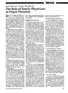
Figure 1. Stnrcture offibroniectin (F). A diagramnmatic represenltatioto of EN shouwing the three regions of alternative splicing cesignlated EIIIB, EIIIA, and V in rats ( modifiedfronm figiu?se in Rcf 62). Note that the cell adhesive peptide CS- I is included in V120.
which variations in extracellular matrix composition are characteristic of a particular inflammatory reaction and whether or not this has a role in specific cell recruitment or retention by tissues. One system in which direct comparisons can be made is in the analysis of extracellular matrix composition in the transplanted organ. In the case of allograft rejection, recirculating lymphocytes contact alloantigen on graft vascular endothelium and then migrate to host lymphoid organs to interact with other as yet uncommitted cells. Various extracellular matrix components may have a crucial role during lymphocyte traffic and positioning3'through their interactions with cell surface antigens, such as integrins.6'7 Several surface receptors, mostly belonging to the (31, ,B2, and (37 integrin subfamilies, have been characterized in T as well as in B lymphocytes.2'6'8 Lymphocyte interactions with extracellular matrix components have also been shown to exert synergistic effects on T cell activation in general9'10 and in specific functions such as secretion of cytokines.10'11 Fibronectin (FN) is likely a key extracellular matrix component involved in these events as it can act as a co-stimulator in both CD4+ and CD8+ T cell activation through the T cell receptor (TcR).9'12 For example, the density of immobilized anti-CD3 or anti-TcR required to induce degranulation and tyrosine phosphorylation of cellular proteins by CD8 T cells is approximately 10-fold lower in the presence of FN.12 Lymphocytes interact with FN mainly via two receptors that belong to the (31 integrin family: the aC4g1 and a5(1 integrins. 13'14 The FNs constitute a group of extracellular matrix proteins known to mediate cellular functions such as activation, proliferation, and migration. 15.16 Structural diversity in the FNs arise by regulated alternative splicing of a single gene transcript in three segments termed EIIIA, EIIIB, and V in rats (Figure 1) and ED-A, ED-B, and IIICS, respectively, in humans.17 The EIIIA and EIIIB domains are each either included or excluded as intact type Ill homology repeats. The V (or IIICS) may be excluded, partially included, or fully included. The latter form contains
the CS-1 region, a sequence recognized by a4(31 integrins and known to mediate cell adhesion. 13,14.18 The EIIIA and EIIIB segments are prominent in FNs produced during embryogenesis, and their expression is regulated in a cell-specific manner. However, FN variants that include the EIIIA and EIIIB segments are also expressed in a highly restricted pattern in some normal adult tissues.19-21 For example, adult liver synthesizes the plasma form of FN, and this form normally excludes the EIIIA and EIIIB domains. In specific pathological conditions, such as cutaneous wound healing,22'23 neointimal hyperplasia,24 27 and glomerular nephritis,28'29 marked increased expression of the EIIIA and EIIIB segments is observed. Although little is known about the function of these domains, recently the EIIIA segment has been reported to regulate cell adhesion30' 31 and differentiation.32 The importance of these FN variants in fundamental processes such as cell migration and differentiation is underscored by the recent observation that mice made null for FN die at a time during embryonic development when the expression of these FNs is prominent.33 Despite growing evidence of important functions for FN variants, the pattern of FN splicing in immunological settings (ie, allografts) has not been analyzed. An organ allograft, unlike soluble antigen, represents a useful experimental system in which to examine the role of the extracellular matrix constituents in the immune response. 3 The effector arm is dependent on the migration of alloreactive cells into the foreign tissue. In our previous studies in rat cardiac allografts, we have shown a markedly increased intragraft deposition of laminin (LN) and FN in the early post-transplant period that precedes the massive cellular infiltration.35 Interestingly, leukocytes infiltrating the rejecting cardiac allograft were consistently detected in close proximity to FN, rather than LN, in the interstitial and perivascular areas.35 Furthermore, the treatment of the cardiac recipients with anti-tumor-necrosis-factor (TNF)-a antibody prolonged the survival of allografts and was associated with an intragraft decrease in FN deposition and a diminution in the magnitude of the leukocyte infiltrate.36 In cholesterol-fed rabbits receiving cardiac allografts, soluble TNF-a receptor decreased intimal thickening, reduced the vascular inflammatory reaction, and decreased the accumulation of FN in coronary arteries.37 In this model, TNF-a blockade had specific effects on the vascular wall, without modulation of myocardial rejection. In piglets, the up-regulation of FN synthesis by interleukin (IL)-1 in donor coronary arteries was associated with the development of post-cardiac transplant arteriopathy.38 Recently, it
Fibronectins in Allografts and Isografts 1759 AJP May 1997, Vol. 150, No. 5
was shown that the treatment of rabbit cardiac allograft recipients with a synthetic CS-1 FN peptide decreased the incidence and severity of donor coronary artery intimal thickening by blocking the binding of a431 (VLA-4) to FN.39 Together, these results suggest potentially important links between FN expression and leukocyte infiltration. This work establishes the presence of specific FN variants at sites of tissue damage. We seek here to determine the spatial and temporal regulation of FN variants in a model in which pronounced leukocyte infiltration occurs. We demonstrate by RNAse protection assay, in situ hybridization, and immunofluorescence that 1) there is molecular heterogeneity in the FNs found in cardiac allografts and isografts as a result of alternative splicing of the primary gene transcript of FN and 2) the incorporation of the EIIIA, EIIIB, and V domains in the FN during allograft rejection has a distinct temporal and spatial pattern of distribution. These findings provide the basis for the analysis of functional differences in specific FN variants as they occur in allografts as compared with isografts. They also provide the rationale for FNbased therapeutics to modulate the allograft rejection cascade in the transplantation setting.
Materials and Methods Animals and Allografting Techniques Male inbred adult rats (Harlan Sprague-Dawley, Indianapolis, IN) weighing 200 to 250 g were used. Hearts from Lewis x Brown Norway F1 hybrids (LBNF1; RT1"ln) or from Lewis (LEW; RT11) were transplanted to the abdominal great vessels of LEW recipients using standard microvascular techniques. Briefly, the animals were anesthetized with ether, the donor vena cava and pulmonary veins were ligated, and the heart was removed. The ascending aorta was anastomosed end-to-side to the infrarenal aorta of the host. The pulmonary artery of the donor heart was anastomosed to the inferior vena cava of the recipient animals. Graft function was assessed by daily palpation through the abdominal wall, and rejection was taken as the time of cessation of myocardial contractions. LEW isografts contracted in a normal physiological manner for well over 100 days; LBNF1 hearts were rejected at 7.5 days in LEW hosts.
RNA Isolation and Northern Blot Analysis
transplantation. The cellular RNA was prepared by the guanidinium thiocyanate/cesium chloride method.40 RNA (10 ,ug/lane) was subjected to electrophoresis through 1% agarose containing 2 mmol/L MOPC and 0.03% formaldehyde; RNA loading was monitored by ethidium bromide staining. RNAs were transferred by positive pressure (Stratagene Corp., La Jolla, CA) onto nylon membranes (ICN Biotrans, Aurora, OH). After 16 hours, the filters were washed, ultraviolet cross-linked, and prehybridized at 65°C for 4 to 5 hours in a solution containing 5% sodium dodecyl sulfate (SDS), 50 mmol/L Pipes (pH 6.5), 50 mmol/L sodium phosphate (pH 7.0), 100 mmol/L NaCL, 1 mmol/L EDTA (pH 8.0), and 100 jig/ml denatured salmon sperm DNA. Membranes were hybridized (20 hours at 65°C) with either a rat FN cDNA probe (prlf-141) or a probe for glyceraldehyde 3-phosphate dehydrogenase (Clontech, Palo Alto, CA) that had been radiolabeled with [a-32P]dCTP (Multiprime DNA labeling system, Amersham, Arlington Heights, IL). The filters were washed in 5% SDS, 1X standard saline citrate (SSC) once at room temperature and twice at 55 to 650C for 15 minutes and then exposed to x-ray film (Kodak X-OMAT AR) between two intensifying screens for 12 hours to 3 days at -80°C. Laser densitometry (Molecular Dynamics model 300A, Sunnyvale, CA) was used to quantify the relative signal intensity of the FN bands.
RNAse Protection Assays Assays were carried out as described previously.22,25 Probes for the EIIIA and EIIIB segments were transcribed from the prFN-A-280 and prFN-B-350 constructs.42 Probes for the V region were transcribed from the pV-527 construct.25 In brief, probes were synthesized using a commercial transcription kit (Promega, Madison, WI) with [32P]UTP and _106 cpm were hybridized (42 to 45°C) overnight to a mixture of purified cardiac RNAs (5 or 10 ,tg/sample) and yeast tRNA (10 jig/sample). Samples were digested, phenol extracted, ethanol precipitated, and analyzed on a denaturing 6% polyacrylamide gel.
Probes for in Situ Hybridization Probes and procedures for transcription of rat FN and murine lysozyme have been described.22'23'25 Single-stranded RNA probes with sense or antisense orientation were synthesized to a specific activity of 1 o8 cpm/,ug with 35S-labeled UTP using a commercial transcription kit (Promega) and purified on denaturing polyacrylamide gels. The DNA inserts were _
Cardiac allografts (LBNF1) or isografts (LEW) were harvested at 0, 3, 6, 24, 96, and 144 hours after
1760
Coito et al
AJP May 1997, Vol. 150, No. 5
270, 213, 255, and 438 nucleotides for total FN, EIIIA+ FN, EIIIB+ FN, and lysozyme, respectively. The antisense mouse lysozyme probe reacts with virtually all rat peritoneal macrophages but not with lymphocytes, neutrophils, or mast cells; no peritoneal cells were labeled with the sense probes.22
In Situ Hybridization Cardiac specimens were fixed in 4% paraformaldehyde, transferred to 30% sucrose, allowed to sediment overnight at 40C, frozen in OCT compound, and stored at -700C. In situ hybridization was carried out as described.23 Briefly, the slides were passed through 0.2 mol/L HCL, 1 gg/ml proteinase K, 0.2% glycine, 4% paraformaldehyde, and 1/200 (v/v) acetic anhydride in 0.1 mol/L triethanolamine (pH 8.0) before incubation in the hybridization mixture. The buffer (50% deionized formamide, 10% dextran sulfate, 0.3 mol/L NaCI, 10 mmol/L Tris, 5 mmol/L EDTA, 0.2% Ficoll 400, 0.02% polyvinylpyrollidone, 0.02% bovine serum albumin (BSA), 10 mmol/L dithiothreitol, 100 jig/ml yeast tRNA, and 500 ,tmol/L nonradiolabeled thio-UTP) containing the appropriate probe (0.3 ,ug/ml per kb probe complexity) was placed on the sections and incubated overnight at 500C. The slides were rinsed sequentially in 50% formamide, 2X SSC, and 10 mmol/L dithiothreitol at 500C, digested with 10 jig/ml RNAse A (Sigma Chemical Co., St. Louis, MO), and washed in 50% formamide at 65°C. Sections were viewed on a Zeiss Axiophot microscope. Black and white and color photographs were taken with transmitted light.
Antibodies for Immunohistochemical Analysis Antibodies to rat monocytes/macrophages (ED1), T helper/inducer (CD4; W3/25), and T cytotoxic/suppressor cells (CD8; OX8) were purchased from Bioproducts for Science (Indianapolis, IN). Rabbit and goat antisera to rat FN (Chemicon International, Temecula, CA, and Calbiochem, La Jolla, CA, respectively) were solid-phase absorbed to remove crossreactivity with other rat plasma proteins. A monoclonal antibody specific to the EIIIA domain of FN (IST-9) (Accurate Chemical, Westbury, NY)43 was used, and an immunopurified polyclonal antibody to the EIIIB domain of FN, as previously described,21 was also used. Swine anti-rabbit Ig conjugated with
peroxidase, fluorescein-isothiocyanate-conjugated rabbit anti-goat Ig, rabbit anti-mouse Ig, alkaline phosphatase anti-alkaline phosphatase (APAAP)
complexes (Dako, Carpinteria, CA), and fluoresceinconjugated goat anti-mouse IgG and donkey antirabbit IgG (Pierce, Rockford, IL) were also used.
Immunohistochemical Analysis Cardiac allografts (LBNF1) and isografts (LEW) were harvested at 0, 3, 6, 24, 96, and 144 hours after transplantation. They were then examined in parallel by immunohistochemistry for cellular infiltration as well as for FN splicing variant expression.
APAAP Staining To evaluate the graft-infiltrating cells, cardiac tissues were embedded in Tissue Tek OCT compound (Miles, Elkhart, IN), snap-frozen in liquid nitrogen, and stored at -700C. Cryostat sections (4 ,gm) were dried at room temperature, fixed in acetone, and incubated with optimally diluted primary antibody (0.05% BSA in 0.1 mol/L Tris-buffered saline (TBS), pH 8.2) for 1 to 24 hours at 40C, followed by incubation with bridging antibody complexes and APAAP complexes.35 The two last steps were repeated, if necessary. The control sections were performed by replacing the primary monoclonal antibody with either 0.05% BSA in TBS or normal mouse serum. The phosphatase reaction was developed with naphthol As-Mx phosphate in TBS, 2% dimethylformamide, 10 mmol/L levamisole, and Fast Red TR Salt (Sigma). Labeled cells within 20 high-power fields per section were counted with the aid of an ocular grid micrometer.
Immunoperoxidase Staining To detect the expression of total FN, cryostat sections were air dried, fixed in acetone, and then incubated with primary antibody at room temperature for 30 minutes (anti-FN antibody, 1:100 dilution), followed by incubation with secondary antibody conjugated with peroxidase.35 The primary antibody was substituted by 0.1% BSA in TBS or normal rabbit serum in the control sections. The peroxidase reaction was developed with 3,3-diaminobenzidine tetrahydrochloride (Sigma) in a 50 ,umol/L NaN3/0.05 mol/L Tris/HCI buffer (pH 7.6) with 0.01% H202 for 10 minutes at room temperature. The slides were counterstained with hematoxylin and mounted. The staining was assessed semiquantitatively, due to the diffusion of reaction products around individual cells and because of extensive, often continuous labeling patterns. Labeling of 20 adjacent high-power-field sections were evaluated and the results were judged
Fibronectins in Allografts and Isografts 1761 A/P May 1997, Vol. 150, No. 5
Table 1. Sequential Immunohistochernical Analysis qf Intramnyocardial
Hours after transplantation
0 3-6 24 96 144
ED1+ macrophages Isografts Allografts 8 8 35 305 256
The results of immunostaining ND, not detected.
+ + ±
+ ±
3 3 10 85 73
are
expressed
Immunostaining (mean + SD) OX8+ T cells Isografts Allografts ND ND
8 -+ 3 7 ±+ 3 28 8 94 ± 22 21 +6
6 3 90 ± 13 66+ 10
±
as mean
+
Iqfiltrating Cells
ND ND 5+2 6 3 2+2
W3/25+ T cells Allografts Isografts 10 + 4 9 -+- 4 11 + 4 15 ± 6 8+3
10 + 4 10 + 3 12 + 4 12 + 4 6+3
SD of data from 20 high-power fields per section per rat and three to six rats
per group.
as +,
weakly positive; + +, clearly positive; and
+++, strongly positive.
Immunofluorescence EIIlA+ FN Detection Cryostat sections were air dried, fixed in acetone, and then incubated (at room temperature for 1 hour) with a blocking solution (PBS with 1% nonfat milk, 0.05% Tween 20, 0.02% sodium azide, and 10% heat-inactivated goat serum). Monoclonal antibody (IST-9) was diluted 1:5 in PBS containing 1% nonfat milk, 0.05% Tween 20, 0.02% sodium azide. Sections were incubated (2 hours at room temperature), washed, and incubated with the secondary fluorescein-conjugated antibody; control sections were performed by substituting the primary monoclonal antibody with normal mouse serum. Cryostat sections of rat embryo were also used as positive control.
EIIIB+ FN Detection Hearts were fixed (1 hour at 4°C) in 3.7% paraformaldehyde, transferred to 0.1 mol/L glycine (1 hour), kept overnight in 0.6 mol/L sucrose at 40C, frozen in OCT compound, and stored at -70°C. Cryostat sections were treated (8 hours at 370C) with peptide N-glycosidase F at 25,000 U/ml in 0.05 mol/L sodium phosphate buffer (or 0.05 mol/L sodium phosphate buffer alone).21 After washing, sections were incubated (1 hour) with a blocking solution consisting of PBS with 1% nonfat milk, 0.05% Tween 20, 0.02% sodium azide, and 10% heat-inactivated donkey serum. Polyclonal antibody anti-EIIIB+ FN diluted 1:20 in PBS containing 1% nonfat milk, 0.05% Tween 20, 0.02% sodium azide was then applied to the section (for 9.5 hours at 37°C). After washing, the sections were then incubated with fluorescein-conjugated secondary antibody.
Results Intramyocardial Leukocyte Infiltration and FN Deposition Our earlier immunohistochemical and laser scanning confocal microscopy studies in rats demonstrated that infiltrating cells localize selectively in FN-rich interstitial and perivascular areas of cardiac allografts.35 In the present study, we performed a comparative evaluation by immunostaining of both intramyocardial cellular infiltration and FN deposition in allografts and isografts at different time points after transplantation. We chose a central area of the myocardium for much of our analysis because this is a primary tissue for cardiac allograft rejection and because of the difficulty in enumerating cells in the epicardial region of cardiac allografts at the later time points of the rejection process. Mononuclear cells begin to infiltrate the central myocardial region between 6 and 24 hours after transplant in both cardiac allografts and isografts. However, we noted the presence of an increase in mononuclear cells in the epicardium and adjacent myocardium at 3 hours after transplantation. In the myocardium of allografts, the infiltrating cells consisted predominantly of ED1 + macrophages and OX8+ (CD8+) T cells (Table 1). The number of CD8+ and ED1 + cells increased significantly during the later time points of the rejection process; the number of macrophages and CD8+ T cells counted from 20 high-power fields per section increased from 35 10 and 6 + 3, respectively, in cardiac allografts at 24 hours to 305 + 85 and 90 + 13, respectively, in those at 4 days after transplantation. The number of macrophages and CD8+ T cells remained high until graft rejection occurred at -7 days. In the myocardium of isografts, we again observed an increase in ED1 + cells between 6 and 24 hours (7 + 3 to 28 + 8), but a more modest addi-
1762
Coito et al
AJP May 1997, Vol. 150, No. 5
Northern Blot Analysis of Cardiac Graft FN
Table 2. Sequential Immunobistochemical Analysis of Intramyocardial Depositio)n qf FN
Hours after transplantation
0 3-6 24 96 144
mRNA
Staining intensity Isografts Allografts
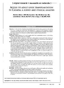
To determine whether or not the observed differences in FN deposition in the allografts and in the isografts resulted from intragraft FN expression, cardiac allografts and isografts were subjected to total RNA isolation and FN mRNA analysis by Northern blotting (Figure 2). We observed an increase in the expression of FN mRNA in both isografts and allografts (lanes 2 and 3) at 3 hours after transplantation as compared with naive hearts (lane 1). These levels increased further and remained comparable in both groups through 24 hours after transplantation (lanes 5 and 6). By 4 days, allograft FN mRNA levels remained consistently high (lanes 8 and 10), whereas isograft FN mRNA levels varied between individual rats (two examples shown, lanes 7 and 9). However, by day 6 (lane 1 1) in isografts, these levels fell to nearly those observed in naive hearts. By contrast, the cardiac allografts consistently maintained very high FN mRNA levels through days 4 and 6 after transplantation (lane 12).
+ ++
++ ++I+
+I++ ++
Staining intensity was evaluated in a semiquantitative fashion on a scale from + (weak) to +++ (strong).
tional increase was observed between 24 and 96 hours (28 + 8 to 94 + 22) as compared with allografts at this interval (Table 1). Rather than remaining high, as observed in rejecting allografts, the number of ED1] macrophages in well functioning cardiac isografts decreased to 21 + 6 by day 6. The number of CD8+ cells observed in the myocardium of the isografts remained low throughout the observation period. The total FN deposition, detected by immunohistochemistry in the grafts, was increased by 3 to 6 hours after transplantation. This occurred at a time when the cell number in the epicardial region began to increase but preceded the massive intramyocardial cellular infiltration (Table 2). In the myocardium of the cardiac allografts, the FN deposition was very strong at 4 and 6 days and correlated temporally with the pronounced cellular infiltration. In the isografts, the levels of FN deposited in the myocardium increased during the first 24 hours and by day 6 decreased sharply to levels comparable with those of a normal heart.
0mI13 i
a a
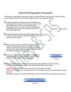
We performed ribonuclease protection assays to determine whether or not changes in the pattern of expression of specific FN variants occurred in cardiac grafts after transplantation. The probes used detected either inclusion (+) or exclusion (-) of the EIIIA, EIIIB, V25 (CS-1) segments (Figure 3). RNAse protection assays were quantitated with a phosphoimager, permitting analysis of ratios even at early intervals after transplantation when bands were faint on autoradiographs. In cardiac allografts and isografts alike, no changes in
r
1 44 hrs.
96
24
6 I
Ribonuclease Protection Assay
i1. a i a i 1
i
FN
a
i
=
I
a
_I
GAPDH 1
2 3 4 5
6 7 8
9 10 11 12
Figure 2. Northern blot analysis of cardiac grafts. Northern blots of total RNA (10 pug/lane) extracted from naive hearts (lane 1 ), cardiac allografis (a) at 3, 6, 24, 96, anzd 144 hours after transplantation (lanes 3, 4, 6, 8, 10, anzd 12, respectively) or cardiac isografts ( i) at 3, 24, 96, anid 144 houirs after tranisplantation (lanes 2, 5, 7, 9. 11, respectively).Blots were hvbridized uith cDNA probes for either total FN or glyceraldebyde 3-phosphate dehydrogenase (GAPDI-!). Conmparable levels ojfFN mRNA aere observed 3 houirs after transiplantation in the cardiac allografts and isografts. These levels increasedfurther but remained conmparable in both groups u.ntil 24 hours after- transplantation. In cardiac isografts, some variability in the levels of FN mRNA uwas obsenred itn inidividuial rats by 4 days. blt by 6 days these levelsfrill to those obsenred in naive hearts. The cardiac allografts continuied to express very high levels of the EN mtRANA unltil the tioie of rejection ( 6 to 7 days). n =3 to 4 sainplesltine poinlt.
Fibronectins in Allografts and Isografts
1763
AJP May 1997, Vol. 150, No. 5
_
_
_
_
_
_
.
hrs. +
EIIIA
-
EIIIB
-
remained relatively low (0.3) in naive hearts and in cardiac allografts (Figure 3) and isografts alike through 24 hours. The ratio increased from 0.3 to 0.7 at day 4 and 0.8 at day 6 in allografts but did not change appreciably in isografts. However, the overall level of FN increased in these isografts (Figure 2), and therefore more EIIIB+ FNs are present in isografts than in naive hearts. The V region has a more complex pattern of alternative splicing than EIIIA or EIIIB and can be found in three different forms: completely included (V120), partially included (V95), and completely excluded (VO). The two larger forms differ in the presence (V120) or absence (V95) of the 25-amino-acid, celladhesive sequence termed CS-1 or V25.15 Because little to no VO was observed, only the V120 (+ in Figure 3) and V95 (- in Figure 3) were subjected to densitometry. The ratio of inclusion of the CS-1 sequence remained at 0.8 in naive hearts for up to 24 hours after transplantation (Figure 3C, allografts). However, the ratio increased in the allograft from 0.8 to 1.3 (day 4) and 1.5 (day 6) after transplantation. In cardiac isografts, the ratio was 0.8 through 24 hours, rising to 1.0 by day 4 and day 6 after transplantation.
In Situ Hybridization
V
nl
1
2
3
4
5
6
Figure 3. RNAse protection assay analysis of cardiac allografts. Representative ribonuclease protection experimenit examining the inclusion of EIMA, EIIB, and CS-1 (V region) into FN mnRNA from rat cardiac allograft dunng rejection. Total RNAs ( 5 or 10 jig) uere hybridized with a -2P-labeled probes for either EIIIA, EIIIB, or Vprobes, digested with ribonucleases and anialyzed ont denaturing 6% polyacrylamide gels to assess segment inclusion ( +) or cexclusion (-). During rejection, the EIIIA+EIHIA- ratio increased at day I and remained high through 6 days as compared with naive hearts. In contrast, an increased ratio of EllIB+EIIIB- uwas nioted at later time points (days 4 anid 6). CS-1+ FN incluisioni was noted as ear4ly as 3 hours after transplantation, and the ratio increased gradually thereafter. A similar pattern u'as seen in
tu'o additional experimenits.
the ratio (0.7) of EIIIA inclusion to exclusion were observed between 3 and 6 hours after transplantation (Figure 3). At 24 hours, however, an increase in the ratio from 0.7 to 3.0 (allografts, Figure 3) and from 0.7 to 2.7 (isografts, not shown) was observed. In allografts, the ratio remained high through day 6 with variability evident in individual rats, with ratios ranging from 1.9 to 3.2 at day 4 and 1.5 to 4.6 at day 6. In isografts, the ratios remained high through 4 days (1.5 to 2.6) but decreased consistently (to 1.1) by day 6. Cardiac transplants were also analyzed for inclusion of the EIIIB segment. In contrast to the results observed with EIIIA, the ratio of inclusion of EIIIB
To identify the sources of total FN mRNA, EIIIA+ FN mRNA, EIIIB+ FN mRNA, and V25+ FN mRNA in the cardiac grafts, we performed in situ hybridization studies with segment-specific 35S-labeled RNA probes. Using a probe that reacts with all forms of FN, we observed FN mRNA in arterioles and labeling of scattered macrophages in naive heart. In the first 3 hours after transplantation, both arteriole labeling and macrophage labeling were observed (Figure 4, A and B). There was an increase in the numbers of macrophages positive for FN mRNA compared with naive heart. In the arterioles, both endothelial cells and medial cells were labeled for FN mRNA (Figure 4A). The presence of active macrophages within early transplants was confirmed by in situ hybridization with a probe to lysozyme, but neither small nor large arterioles were labeled for lysozyme (Figure 4B). When adjacent sections of the myocardium of allografts at 4 days were analyzed, we consistently observed that the distribution of cells labeled for FN mRNA (Figure 4D) coincided with the distribution of cells positive for lysozyme mRNA (Figure 4E). Those cells usually had the convoluted nuclear morphology typical of macrophages, best seen in sense controls in which nuclear detail was not obscured by silver grains (Figure 4, C and F). By day 4 and day 6, a high proportion of the infiltrating cells in the cardiac
1764
Coito et al
AJPMav 1997, Vol. 150, No. 5
aw
.VA
A
4k jk
Figure 4. In situL hybridization detection of FN and lysozyme mRNA. Sections of cardiac allografts at 3 hours (A to C) or 4 days (D to F) after transplantation were hybridized with 35S-labeled probes for FN (A and D) or lysozyme (B and E) or with a control probe transcribed in the sense orientation for FN(C and F). FN mRNA uas observed in the intimal and medial layers of arterioles (A) and adjacent macrophages (B) at 3 houns. Hoivever, lVsozyme mRNA was not observed in either small or large arteroles (B, curved arrows). Heavy labeling was observed u'ith probes for FN (D) and lysozyme (E) in the mnocardium at 4 days. Cotntrol probes did not label cells with tissue sections (C and F). Not all macrophages labeled for both lysozyme and FN mRNAs (D and E). Magnification, X330.
allografts had features of macrophages and were heavily labeled for FN and lysozyme mRNAs, confirming our previous results that the macrophages are the main source of FN mRNAs in the rejection process.36 However, in some instances, macrophages labeling for lysozyme mRNA were not all positive for FN mRNAs (Figure 4). We also performed a comparative study, by in situ hybridization, to analyze the distribution of FN mRNAs in allografts and isografts at intervals after transplantation. For intervals of up to 3 hours after transplantation of either allografts or isografts, arterioles and macrophages expressed FN mRNA. The accumulation of ElIIA+ FN mRNA was noted as early as 3 hours after transplantation in both isografts and allografts, and was synthesized by arterioles and
mononuclear cells infiltrating the epicardium and myocardium. At intervals up to 4 days, the predominant cell type expressing FN also expressed lysozyme mRNA and had the characteristic nuclear morphology of macrophages. By day 4 after transplantation, we detected obvious differences in the distribution pattern of FN mRNA between allografts (Figure 5, A and B) and isografts (Figure 5, C and D). In the cardiac allografts, large areas of myocardial infarction were noted and the expression of FN mRNA could be primarily detected within the epicardium and deeper myocardium and endocardium (Figure 5, A, B, E, F, and G). These cells were predominately macrophages (see Figure 5L and Figure 4) and were intensely labeled for FN (Figure 5, G and K) at the
Fibronectins in Allografts and Isografts 1765 AJPMav 1997, Vol. 150, No. 5
11:4,~ ~ ~
g
jal
II ?. --,
f .1
M
.''
IC q,
" ..IL .
-
--t. p ,,
--A
It
;.7
i °
-
.
-t
-
* l ~
-'
.
O .. e-
9-
~ ~
I,
-., Ip. ~
~
~
v~
J&I. t f. ~
~
, 1;
Figure 5. In situ hybridizationi detection ofFN mRNA anid EIIIA+ EN rtRNA in cardiac allografis anid isografts. In situ hybridization detection of EN mRNA expression is shown at lown magnification otn cardiac sections at day 4 after transplantation in allograf?s (A and B) and isografts (C and D) in dark-field (A and C) and in bright-field (B and D). FN ?nRNA was localized primarily in the mnyocardium of allografis, uwhereas in isografts, labelinzg was largely restricted to the epicardium. In situL hybridization onsections at day 6 after tranisplantation is shoun at higher magnijication itsing probes that react either with allform,s of FN(E, F, G, M, and N), uith EIIIA+ FN(I, J, K, 0, and P), or u.ith lysozyme (L) in cardiac allografts (E to L) and cardiac isografts (M to P). In allografts, a high proportion of infiltrating inflammatory cells (idenltified as macrophages in L) wvere strongly labeledfor total FN mRRNA (E to G) in the epicardium (arrow in E), myocardium (arrows in F), and adjacent to areas ofinifarction (arrows in G) andforEUll4 plus FN mRRNA (I to K). EHIMA FN mRNA in allografts had a similar distribution to total FN mRNA but was less intensely labeled (arrows in to K). In isografts (M to P), total FN mRNA was restricted to smaller numbers of infiltrating cells localized predominantly in the epicardium (M, arrow) anid adjacent myocardium. In isografts, fewer cells were noted in the deeper nyocardium adjacent to the enidocardium (N). EUIL4 FN mRNA labeling appeared to be in infiltrating cells predominantly in the epicardium (0) bhot not in the myocardium (P). In situ hybridizationfor either lysozyme niRNA, a specific markerfor macrophages (L), or its control, a sense probe (H) illnstrates the large number of macrophages in cardiac allografts 6 days after transplantation. Magnification, X 22.3 (A to D) and X 142 (E to P). n = 2 to 4 samples/time point/exeimental group.
1766 Coito et al AJPMav 1997, Vol. 150, No. 5
Table 3. Immuinofluorescence Staining for EIILA+ FN and EHIB+ FN in the Cardiac Grafts FN isoforms
Hours after transplantation
0
EIIIA
3-6 24 96 144
0
EIIIB
3-6 24 96 144 Staining intensity
was
Type of graft Allo Iso Allo Iso Allo Iso Allo Iso Allo Iso
Endocardium
+__
+ +-F
+ +4
Allo Iso Allo Iso Allo Iso Allo Iso Allo Iso
evaluated in a semiquantitative fashion
Staining intensity Myocardium Epicardium
Arteries
+
++
+
++
on a
edges of infarcted areas. EIIIA+ FN mRNA had a similar cellular distribution pattern to that of total FN mRNA (Figure 5, I-K). The expression of EIIIB+ FN mRNA and V25+ FN mRNAs in the cardiac allografts at 4 and 6 days after transplantation co-localized with the ElIIA+ FN, but in general the level of labeling for EIIIB+ FN was less pronounced than for EIIIA+ FN (data not shown). Unlike cardiac allografts, areas of myocardial infarction were not seen in isografts. In contrast to widespread expression of FN mRNA in allografts, in cardiac isografts (days 4 and 6), the expression of total FN mRNA was largely restricted to leukocytes in the epicardium and in the myocardium adjacent to the epicardium (Figure 5, C, D, M, and N). Expression of the ElIlA+ FN (Figure 5, 0 and P), V25 and EIIIB (not shown) mRNAs coincided with expression of total FN mRNA.
Immunofluorescence Staining We next determined the extent to which specific FN variants were deposited in the transplant matrix. This was done by immunohistochemistry with segmentspecific antibodies with the results summarized in Table 3. In naive hearts and in the early interval (3 and 6 hours) after transplantation of both allografts and isografts, low levels of EIIIA staining were observed, which was restricted to arterioles (Figure 6A). At 24 hours after transplantation, in the cardiac allografts, EIIIA+ FN was deposited in the epicar-
scale from
+
++ ++ ++ ++
++ ++
(very weak) to + + + (strong).
dium (Figure 6B) and to a lesser extent in the myocardium (Figure 6C); in isografts at this time, EIIIA+ FNs were detected primarily in the epicardium (Figure 6D). This apparent difference in staining was more pronounced in allografts by 4 and 6 days after transplantation with EIIIA+ FNs observed in the endocardium, myocardium, and epicardium (Figure 6, E and F). By contrast, EIIIA+ FNs were restricted to the epicardium of isografts (Figure 6G). Consistent with data obtained by Northern analysis (Figure 2) and in situ hybridization (Figure 5), EIIIB+ FNs were detected at the later time points (4 and 6 days) of the rejection process (Figure 7). The intensity of staining for ElIIB+ FN was always considerably less intense than staining for EIIIA+ FN on replicate sections of either allografts or isografts. In allografts, EIIIB+ FN was deposited in the endocardium, myocardium, and epicardium (Figure 7A), whereas in the isografts, deposition of this FN isoform was confined to the epicardium (Figure 7C).
Discussion We report here our experiments on the expression of total FN and the splicing pattern of the primary gene transcript for FN in rat cardiac allografts and isografts. The principal findings of this work are as follows. First, the local synthesis of FN, in both allografts and isografts, is increased as early as 3 hours after transplantation. Second, the sources of
Fibronectins in
Allografts and Isografts
1767
AJP May 1997, Vol. 150, No. 5
Figure 6. Immunofluorescence analysis of EIIIA+ FN in cardiac grafts. Immunofluorescence localization of EIIIA+ FN is shown in naive heart (A), in cardiac allografts at day 1 (B and C), day 4 (E), and day 6 (F) after transplantation, and in cardiac isografts at day 1 (D) and day 6(G). In the naive heart, EIIIA+ FN was restricted to the arteries, whereas in the cardiac allografts, the EIIIA+ FN was detected in moderate amounts in the epicardium (B) and myocardium (C) at day 1 and increasing significantly by day 4 (E) and day 6(F). In the cardiac isografts, the EIIIA+ FN was restricted to the epicardium but was absent from the myocardium (D and G). No staining was detected in the sections incubated with control IgG (H). Magnification, X217(A and F) and x109 (B to E and G to H); n = 3 animals/group.
this FN at 3 hours include macrophages as well as arterioles, most likely from endothelial and smooth muscle cells. At intervals beyond 24 hours, infiltrating macrophages play a predominant role in both isografts and allografts. Third, the expression of FN in cardiac allografts remains high during later intervals to the point of allograft rejection, whereas the expression of FN in isografts approaches those observed in naive hearts. Fourth, the newly synthesized FNs in the cardiac grafts include EIIIA+, EIIIB+, and V25+ variants that are generated by alternative splicing of the FN pre-mRNA. Fifth, the temporal expression and spatial distribution patterns of FN variants differs in cardiac allografts and isografts. This study extends our earlier findings that local synthesis of total FN mRNA occurs in cardiac allografts during acute cardiac rejection and that macrophages are the primary source of FN during rejection.36 Interestingly, as shown in the present study, FN synthesis also occurs in macrophages in cardiac isografts, albeit with a different pattern of expression
and alternative splicing, particularly in the interval between 4 and 6 days. Although macrophages are an early and continuing source of FN in both allografts and isografts, they were not the only cellular sources of FN. We also found that arterioles that expressed FN mRNAs in naive hearts also did so at the earliest time points (3 to 6 hours) in both isografts and allografts. At intervals up to -6 days, macrophages were a predominant source; we expect that other cell types (eg, fibroblasts) also serve as sources of FN. These findings suggest that the cellular mechanisms responsible for regulating individual patterns of FN expression are important in governing the outcome. These data support earlier reports demonstrating that FN expression is a ubiquitous feature of the host response to injury or in disease. Indeed, in all settings examined, including cutaneous wounds,22'23 vascular intimal proliferations, myocardial infarctions, and hypertensive arteries24-27,44,45 and during glomerulonephritis29,46 and liver fibrosis,32 FN expression is a prominent feature.
1768
Coito et al
AJP May 1997, Vol 150, No 5
5
I
w
Figure 7. Immunofluorescence analysis ofEIIIB+ FN in cardiac grafts. Immunofluorescence localization ofEIIIB+ FN in cardiac sections 6 days after transplantation in cardiac allografts (A and B) and isografts (C and D). Sections were deglycosylated by pretreatment with peptide N -glycosidase F (A and C). Those pretreated with buffer alone (B and D) showed little specific staining. Controls in which anti-EIIIB antibodies were substituted with normal IgG or in which the peptide N-glycosidase F step was omitted before anti-EIIIB antibody, yielded background staining. Magnification, X 21 7; n = 3 animals/group.
Despite the constancy of this pattern, there does appear to be a tissue-specific pattern to the cell types responsible for this synthesis. Thus, in healing cutaneous wounds, during glomerular nephritis, and in cardiac allografts, macrophages are an important source of FN.3646 In vascular injury, such as after balloon catheterization or during atherogenesis, medial smooth muscle cells produce FN.2425 In some instances, an orderly progression of synthesis occurs. Thus, during cutaneous wound healing and in glomerulonephritis, macrophages are an early source, whereas mesenchymal cells (such as fibroblasts in cutaneous wounds) express FN at later intervals in the process.22 46 Myocardial infarctions are a prominent feature of the pathophysiology of cardiac allograft rejection. We observe infarcts at day 4 through day 6 in our model. Infiltrating T cells appear earlier, rising some 15-fold between day 1 and day 4 (Table 1). In earlier work, we observed close association between FN and lymphocytes.35 A similar increase in T-cell infiltration is not observed in isografts, nor are infarcts (Table 1). We also observed that EIIIA+ FNs rise to maximal levels in both isografts and allografts by day 4 (Figure 3). Interestingly, the percent inclusion of EIIIB in FN rises in allografts and isografts and occurs in distinct compartments. Taken together, our data demonstrate important differences in the splicing patterns of FN in allografts and isografts and suggest a role for specific FN variants in leukocyte recruitment and infarction. The mechanisms regulating FN expression are likely to vary between individual cell types. While transforming growth factor-4l is well known to increase FN expression by fibroblasts, its role in mod-
ulating FN expression in other cell types remains unclear.4748 Our recent data suggest a potential role for TNF-a in modulating macrophage appearance, and potentially FN expression, during cardiac allograft rejection.36 Other data also indicate that, in coronary arteriopathy after transplantation, endothelial and smooth muscle cells produced increased FN under the regulation of IL-1,B and TNF-a.3849 Moreover, soluble TNF-a receptor reduces the expression of FN and leukocyte infiltration within these intimal thickenings.37 Local synthesis of FN by cells in the transplanted organ is not likely the only FN present. The extravasation of plasma FN along with other plasma proteins, such as fibrinogen, appears to be a ubiquitous feature of all inflammatory sites examined.50 Extravasated plasma proteins constitute a provisional matrix that is a prominent feature of early healing wounds and cutaneous delayed hypersensitivity reactions.i152 In both healing wounds and cutaneous delayed hypersensitivity reactions, a permeabilityenhancing factor and endothelial growth factor, termed VPFNEGF, is likely responsible for the marked increase in protein extravasation.5354 An important consequence of FN alternative splicing that occurs during inflammation is the gradual replacement of plasma FN, which largely lacks the EIIIA and EIIIB segments, with locally produced FNs that contain these segments. Our observation that CS-1 inclusion increases in cardiac allografts is particularly interesting in this regard because this segment of FN has a known cell adhesive function for lymphocytes. 13,14 Emerging evidence suggests function(s) for EIIIA+ FNs in mesenchymal cell activation. However,
Fibronectins in Allografts and Isografts 1769 AJP May 1997, Vol. 150, No. 5
no specific biological functions have been ascribed to either the EIIIA or the EIIIB segments during transplantation. We do observe marked differences in the temporal appearance, relative levels, and spatial deposition of the two segments in allografts and isografts. For example, the cells expressing FN mRNA, and the deposition of FN into the matrix, were predominantly in the epicardium and adjacent myocardium in isografts. By contrast, the pattern of staining in allografts was much stronger in the myocardium. The common expression of FN in the epicardium in both isografts and allografts may reflect a common pathway of leukocyte migration or activation. Alternatively, it could be a response to tissue injury or ischemia that may occur during the brief intervals (-45 minutes) that the hearts are undergoing transplantation. Interestingly, the temporal pattern in both allografts and isografts, first ElIIIA FNs and then E111B+ FNs, is similar to the pattern observed during liver fibrosis32 but somewhat different than that in cutaneous wounds in which both appear together but disappear at different times (Reimer CL, Plantefaber L, Peters JH, Hynes RO, Van De Water L, submitted). The regulatory mechanisms governing the temporal and spatial patterns are of some interest. This apparent regulation supports our hypothesis that these forms exert important effects in the repair of tissue damaged during surgery. Moreover, by comparing the patterns in allografts and isografts, our data support the hypothesis that these FN variants are involved in lymphocyte adhesion, migration, or differentiation in allografts. Interestingly, it has been shown that the EIIIA segment mediates adhesion of fibroblastic cells and promotes the differentiation of lipocytes in the liver to become myofibroblasts.2728 Because the EIIIA segment exerts effects on cellular adhesion and differentiation, we think that similar processes in lymphocytes may be affected. For example, CD8+ T-lymphocyte-mediated lysis of target cells requires the formation of tight conjugates between these cytotoxic T lymphocytes and the target cell. Both adhesion and de-adhesion events very likely involve specific signals generated by the activated T cell.55 The presence of specific FN variants may provide a spatial address at which appropriate co-stimulatory signals could initiate or augment critical T cell signaling events. In that regard, it is of interest that the forms of FN variants present in normal adult paratracheal lymph nodes are predominantly EIIIB- and EIIIA-.21 Taken together, these data suggest that the EIIIB and EIIIA segments subserve functions important to the activation of lymphocytes at sites of immunological reactions.
The initiation of FN synthesis in the early interval after transplantation is also a novel and intriguing observation coming from this work, especially from the point of view of lymphocyte recruitment during graft rejection. Mounting evidence indicates that FN plays important roles in the early recruitment of lymphocytes in various pathological states. CS-1 and RGD peptides block a4fB1 and a5f31 integrins on lymphocytes, and these may inhibit the extravasation of lymphocytes from post-capillary venules. Indeed, FN peptides modeled after either the heparin-binding region or CS-1 segment interrupt multi-organ cellular infiltration, inflammatory lesion and the development of the lethal wasting syndrome in transforming-growth-factor-,B1-deficient mice56 and in a model of experimental arthritis.57 FN is synthesized by transplant arterioles during cardiac arteriopathy that accompanies transplant rejection, and soluble TNF-a receptor reduces inflammatory cell infiltration and FN deposition at these sites.37 Although factors other than FN participate in the recruitment of T cells, our data and that of other workers underscores an important role for these matrix proteins. Indeed, CS-1 and RGD peptides inhibit lymphocyte transendothelial migration in a porcine endothelial/smooth muscle cell co-culture system.58 Moreover, administration of CS-1 peptides to rabbit cardiac allograft recipients was associated with a significant reduction of T cell infiltration and subendothelial FN accumulation in coronary arteries, presumably by blocking the binding of a4f1 to FN.39 Although other roles for FN are possible, indeed even likely, our data taken with the work of others provide a basis for a model in which early FN deposition, resulting from both extravasated plasma FN and locally expressed cellular FN, establishes an environment that promotes early leukocyte infiltration, including the infiltration by lymphocytes and macrophages within the first 24 hours. Consistent with a role in lymphocyte accumulation at the graft site, we also observed during the later phase of the rejection process (4 to 6 days) that CS-1 was preferentially expressed in the allografts compared with the isografts. In the cardiac isografts, the predominant leukocytes are the macrophages; a modest increase in T cells was noted. By contrast, in cardiac allografts, the relative increase in T cells present in myocardium after 24 hours of transplantation was significantly higher (10-fold). These data are consistent with evidence gathered in vitro, which show that the RGD and CS-1 domains of FN have a role in lymphocyte activation. Indeed, interactions of a4/31 and a5j31 integrins with their specific ligands are known to act as co-stimulatory signals with CD3/ TcR pathways to induce T cell proliferation and IL-2
1770 Coito et al AJP May 1997, Vol. 150, No. 5
induction.59'60 Stimulation of IL-2 production by ligation of FN via a5p1 on CD4+ T cells is associated with induction of AP-1 transcription factors. The adhesion of natural killer cells, through a4X31 and asp1 receptors to FN, stimulates tyrosine phosphorylation of natural killer cell substrates.61 FN can enhance degranulation of CD8+ cytotoxic T lymphocytes in response to substimulatory amounts of anti-TcR and anti-CD3.9'61 These data strongly imply a role for CS-1 in regulating the appearance and activation of lymphocytes in cardiac allografts. Indeed, in our preliminary studies, treatment of rat recipients with CS-1 peptides alone abrogated acute rejection at 7 days and doubled the length of survival of cardiac allografts (A. J. Coito, unpublished data). Hence, our studies support the contention that distinct FNs play a functional role in the host immune cascade triggered by organ transplantation and offer potential new means of immunosuppression in organ transplantation.
Acknowledgments We are grateful to Dr. Maria de Sousa for invaluable discussions, to Drs. Jochen Binder and Kazuhiko Onodera for excellent assistance in providing us with the cardiac grafts, and to Carol Foss for care in helping us prepare the manuscript.
References 1. Hynes RO, Lander AD: Contact and adhesive specificities in the association, migrations, and targeting of cells and axons. Cell 1992, 68:1-20 2. Shimizu Y, Shaw S: Lymphocyte interactions with extracellular matrix. FASEB J 1991, 5:2292-2298 3. De Sousa M, Tilney NL, Kupiec-Weglinski JW: Recognition of self within self: specific lymphocyte positioning and the extracellular matrix. Immunol Today 1991, 12: 262-266 4. Coito AJ, de Sousa M, Kupiec-Weglinski JW: The role of cellular and extracellular matrix adhesion proteins in organ transplantation. Cell Adhes Commun 1994, 2:249-255 5. Kupiec-Weglinski JW, de Sousa M: Lymphocyte traffic is modified in vivo by anti-laminin antibody. Immunology 1991, 72:312-313 6. Lobb RR, Hemler ME: The pathophysiological role of a4 integrins in vivo. J Clin Invest 1994, 94:1722-1728 7. Springer TA: Traffic signals for lymphocyte recirculation leukocyte emigration: the multistep paradigm. Cell 1994, 76:301-314 8. Springer TA: Adhesion receptors of the immune system. Nature 1990, 346:425-434 9. Matsuyama T, Yamada A, Kay J, Yamada K, Akiyama
S, Schlossman S: Activation of CD4 cells by fibronectin and anti-CD3 antibody: a synergistic effect mediated by the VLA-5 fibronectin receptor complex. J Exp Med 1989, 170:1133-1148 10. Hershkoviz R, Gilat D, Miron S, Mekori Y, Aderka D, Wallach D, Vlodavsky I, Cohen I, Lider 0: Extracellular matrix induces tumor necrosis factor-a sections by an interaction between resting rat CD4+ T cells and macrophages. Immunology 1993, 78:50-57 11. Hershkoviz R, Cahalon L, Gilat D, Miron S, Miller A, Lider 0: Physically damaged extracellular matrix induces TNF-a secretion by interacting resting CD4+ T cells and macrophages. Scand J Immunol 1993, 37: 111-120 12. Ostergaard HL, Ma EA: Fibronectin induces phosphorylation of a 120 kDa protein and synergizes with the T cell receptor to activate cytotoxic T cell clones. Eur J Immunol 1995, 25:252-256 13. Guan J, Hynes R: Lymphoid cells recognize an alternatively spliced segment of fibronectin via the integrin receptor a4g1. Cell 1990, 60:53-61 14. Wayner E, Garcia-Pardo A, Humphries M, McDonald J, Carter W: Identification and characterization of the T lymphocyte adhesion receptor for an alternative cell attachment domain (CS-1) in plasma fibronectin. J Cell Biol 1989, 109:1321-1330 15. Hynes R: Fibronectins: Springer Series in Molecular Biology. Edited by A Rich. New York, Springer-Verlag, 1990 16. Mosher DF (Ed): Fibronectin. New York, Academic Press, 1989 17. Schwarzbauer JE: Alternative splicing of fibronectin: three variants, three functions. BioEssays 1991, 13: 527-533 18. Mould AP, Wheldon LA, Komoriya A, Wayner EA, Yamada KM, Humphries MJ: Affinity chromatographic isolation of the melanoma adhesion receptor for the IIICS region of fibronectin and its identification as the integrin a4f31. J Biol Chem 1990, 265:4020-4024 19. Carnemolla B, Balza E, Siri A, Zardi L, Nicotra MR, Bigotti A, Natali PG: A tumor-associated fibronectin isoform generated by alternative splicing of messenger RNA precursors. J Cell Biol 1989, 108:1139-1148 20. Vartio T, Laitinen L, Narvanen 0, Cutolo M, Thornell LE, Zardi L, Virtanen I: Differential expression of the ED sequence-containing form of cellular fibronectin in embryonic and adult human tissues. J Cell Sci 1987, 88: 419-430 21. Peters JH, Chen G, Hynes RO: Fibronectin isoform distribution in the mouse: II. Differential distribution of the alternatively spliced EIIIB, EIIIA and V segments in the adult mouse. Cell Adhes Commun 1996, 4:127-148 22. Brown LF, Dubin D, Lavigne L, Logan B, Dvorak HF, Van De Water L: Macrophages and fibroblasts express alternatively spliced fibronectins during cutaneous wound healing. Am J Pathol 1993, 142:793-801 23. ffrench-Constant C, Van De Water L, Dvorak HF, Hynes RO: Reappearance of an embryonic pattern of fi-
Fibronectins in Allografts and Isografts 1771 AJP May 1997, Vol. 150, No. 5
24.
25.
26.
27.
28.
29.
30.
31.
bronectin splicing during wound healing in the adult rat. J Cell Biol 1989, 109:903-914 Glukhova M, Frid M, Shekhonin B, Vasilevskaya T, Grunwald J, Saginati M, Koteliansky V: Expression of extra domain A fibronectin sequence in vascular smooth muscle cells is phenotype dependent. J Cell Biol 1989, 109:357-366 Dubin D, Peters JH, Brown LF, Logan B, Kent KC, Berse B, Berven S, Cercek B, Sharifi BG, Pratt RE, Dzau VJ, Van De Water L: Balloon catheterization induces arterial expression of embryonic fibronectins. Arterioscler Thromb Vasc Biol 1995, 15:1958-1967 Knowlton AA, Connelly CM, Romo GM, Mamuya W, Apstein CS, Brecher P: Rapid expression of fibronectin in the rabbit heart after myocardial infarction with and without reperfusion. J Clin Invest 1992, 89:1060-1068 Mamuya WS, Brecher P: Fibronectin expression in the normal and hypertrophic rat heart. J Clin Invest 1992, 89:392-401 Barnes JL, Torres ES, Mitchell RJ: Expression of alternatively spliced fibronectin variants during remodeling in proliferative glomerulonephritis. Am J Pathol 1995, 147:1361-1371 Neckeleit V, Zagachin L, Nishikawa K, Peters JH, Hynes RO, Colvin RB: Embryonic fibronectin isoforms are synthesized in crescents in autoimmune glomerular nephritis in the rat. Am J Pathol 1995, 147:965-978 Xia P, Culp LA: Adhesion activity in fibronectins alternatively spliced domain EDa (F. IIIA) and its neighboring type IlIl repeats: oncogene-dependent regulation. Exp Cell Res 1994, 213:253-265 Xia P, Culp LA: Adhesion activity in fibronectin's alternatively spliced domain EDa (EIIIA): complementarity to plasma fibronectin functions. Exp Cell Res 1995, 217:517-527
32. Jarnagin WR, Rockey DC, Koteliansky VE, Wang S-S, Bissell DM: Expression of variant fibronectins in wound-healing: cellular source and biological activity of the EIIIA segment in rat hepatic fibrogenesis. J Cell Biol 1994, 127:2037-2048 33. George EL, Georges-Labouesse EN, Patel-King RS, Rayburn H, Hynes RO: Defects in mesoderm, neural tube and vascular development in mouse embryos lacking fibronectin. Development 1993, 119:10791091 34. Kupiec-Weglinski JW, Coito AJ, Gorni A, de Sousa M: Lymphocyte migration and tissue positioning in allograft recipients: the role played by extracellular matrix proteins. Transplant Rev 1995, 9:29-40 35. Coito AJ, Binder J, de Sousa M, Kupiec-Weglinski JW: The expression of extracellular matrix proteins during accelerated allografts in sensitized rats. Transplantation 1994, 57:599-605 36. Coito AJ, Binder J, Brown LF, de Sousa M, Van De Water L, Kupiec-Weglinski JW: Anti-TNF-a treatment downregulates the expression of fibronectin and decreases cellular infiltration of cardiac allografts in rats. J Immunol 1995, 154:2949-2958
37. Clausell N, Molossi S, Sett S, Rabinovitch M: In vivo blockade of tumor necrosis factor-a in cholesterol-fed rabbits after cardiac transplant inhibits acute coronary artery neointimal formation. Circulation 1994, 89:27682779 38. Clausell N, Molossi S, Rabinovitch M: Increased interleukin-1l3 and fibronectin expression are early features of the development of the postcardiac transplant coronoary arteriopathy in piglets. Am J Pathol 1993, 142: 1772-1786 39. Molossi S, Elices M, Arrhenius T, Diaz R, Coulber C, Rabinovitch M: Blockade of very late antigen-4 integrin binding to fibronectin with connecting segment-1 peptide reduces accelerated coronary arteriopathy in rabbit cardiac allografts. J Clin Invest 1995, 95:2601-2610 40. Chirgwin J, Przybyla A, MacDonald R, Rutter W: Isolation of biologically active ribonucleic acid from sources enriched in ribonuclease. Biochemistry 1979, 18: 5294-5299 41. Schwarzbauer JE, Tamkun JW, Lemischka IR, Hynes RO: Three different fibronectin mRNAs arise by alternative splicing within the coding region. Cell 1983, 35:421-431 42. Schwarzbauer J, Patel R, Fonda D, Hynes R: Multiple sites of alternative splicing of the rat fibronectin gene transcript. EMBO J 1987, 6:2573-2580 43. Borsi L, Carnemolla B, Castellani P, Rosellini C, Vecchio D, Allemanni G, Chang SE, Taylor-Papadimitriou J, Pande H, Zardi L: Monoclonal antibodies in the analysis of fibronectin isoforms generated by alternative splicing of mRNA precursors in normal and transformed human cells. J Cell Biol 1987, 104:595-600 44. Takasaki I, Chobanian AV, Mamuya WS, Brecher P: Hypertension induces alternatively spliced forms of fibronectin in rat aorta. Hypertension 1992, 20:20-25 45. Samuel JL, Barrieux A, Dufour S, Dubus I, Contard F, Koteliansky V, Farhadian F, Marotte F, Thiery J-P, Rappaport L: Accumulation of fetal fibronectin mRNAs during the development of rat cardiac hypertrophy induced by pressure overload. J Clin Invest 1991, 88: 1737-1746 46. Barnes JL, Hastings RR, De La Garza MA: Sequential expression of cellular fibronectin by platelets, macrophages, and mesangial cells in proliferative glomerulonephritis. Am J Pathol 1994, 145:585-597 47. Roberts C, Birkenmeir T, McQuillan J, Akiyama S, Yamada S, Chen W-T, Yamada K, McDonald J: Transforming growth factor ,B stimulates the expression of fibronectin and of both subunits of the human fibronectin receptor by cultured human lung fibroblasts. J Biol Chem 1988, 263:4586-4592 48. Ignotz R, Endo T, Massague J: Regulation of fibronectin and type 1 collagen mRNA levels by transforming growth factor-p. J Biol Chem 1987, 262:6443-6446 49. Clausell N, Molossi S, Rabinovitch M: Tumor necrosis factor-a induction of smooth muscle cell fibronectin synthesis via interleukin-/3 is associated with the devel-
1772
Coito et al
AJP May 1997, Vol. 150, No. 5
50.
51.
52.
53.
54.
55.
56.
opment of graft atherosclerosis. Circulation 1993, 88: 1419A Dvorak HF: Tumors: wounds that do not heal. Similarities between tumor stroma generation and wound healing. N EngI J Med 1986, 315:1650-1659 Clark RAF, Dvorak HF, Colvin RB: Fibronectin in delayed-type hypersensitivity skin reactions: associations with vessel permeability and endothelial cell activation. J Immunol 1981, 126:787-793 Grinnell F, Billingham R, Burgess L: Distribution of fibronectin during wound healing in vivo. J Invest Dermatol 1981, 76:181-189 Brown LF, Berse B, Yeo K-T, Yeo T-K, Senger DR, Dvorak HF, Van De Water L: Expression of vascular permeability factor/vascular endothelial growth factor (VPFNEGF) by epidermal keratinocytes during wound healing. J Exp Med 1992, 176:1375-1379 Brown LF, Olbricht SM, Berse B, Jackman RW, Matsueda G, Tognazzi KA, Dvorak HF, Van De Water L: Overexpression of vascular permeability factor (VPF/ VEGF) and its endothelial cell receptors in delayed hypersensitivity skin reactions. J Immunol 1995, 154: 2801-2807 Mescher MF, O'Rourke AM, Champoux P, Kane KP: Equilibrium binding of cytotoxic T lymphocytes to class antigen. J Immunol 1991, 147:36-41 Hines KL, Kulkarni AB, McCarthy JB, Tian H, Ward JM, Christ M, McCartney-Francis NL, Furcht LT, Karlsson S, Wahl SM: Synthetic fibronectin peptides interrupt inflammatory cell infiltration in transforming growth factor
57.
58.
59.
60.
61.
62.
,31 knockout mice. Proc Natl Acad Sci USA 1994, 91: 5187-5191 Wahl SM, Allen JB, Hines KL, Imamichi T, Wahl AM, Furcht LT, McCarthy JB: Synthetic fibronectin peptides suppress arthritis in rats by interrupting leukocyte adhesion and recruitment. J Clin Invest 1994, 94:655-662 Molossi S, Elices M, Arrhenius T, Rabinovitch M: Lymphocyte transendothelial migration toward smooth muscle cells in interleukin-1lf-stimulated co-cultures is related to fibronectin interactions with a4f1 and a5f1 integrins. J Cell Physiol 1995, 164:620-633 Shimizu Y, van Seventer GA, Horgan KJ, Shaw S: Roles of adhesion molecules in T cell recognition: fundamental similarities between four integrins on resting human T cells (LFA-1, VLA-4, VLA-5, VLA-6) in expression, binding and costimulation. Immunology 1990, 114: 109-143 Yamada A, Nikaido T, Nojima Y, Schlossman SF, Morimoto C: Activation of human CD4 T lymphocytes: interaction of fibronectin with VLA-5 receptor on CD4 cells induces the AP-1 transcription factor. J Immunol 1991, 146:53-56 Gismondi A, Milella M, Palmieri G, Piccoli M, Frati L, Santoni A: Stimulation of protein tyrosine phosphorylation by interaction of NK cells with fibronectin via a4X1 and a5X31. J Immunol 1995, 154:3128-3137 ffrench-Constant C, Hynes RO: Alternative splicing of fibronectin is temporally and spatially regulated in the chicken embryo. Development 1989, 106:375-388