The lymphatic system performs several vital func tions in the human body, including returning water, proteins, and other macromolecules to the blood,.
ISSN 0362�1197, Human Physiology, 2014, Vol. 40, No. 1, pp. 52–57. © Pleiades Publishing, Inc., 2014. Original Russian Text © A.I. Krupatkin, 2014, published in Fiziologiya Cheloveka, 2014, Vol. 40, No. 1, pp. 62–67.
Oscillatory Processes in Lymph Microcirculation in the Human Skin A. I. Krupatkin Priorov Central Institute of Traumatology and Orthopedics, Ministry of Health and Social Development, Moscow, Russia Received November 26, 2012
Abstract—This study was the first to use laser Doppler flowmetry followed by wavelet analysis in order to esti� mate oscillations in lymph microcirculation in 30 subjects with (n = 13) or without (n = 17) edema of the dis� tal part of the upper limb. Lymph flow in the human skin exhibited clear dominance of pacemaker phase oscillations in the frequency ranges of 0.021–0.042 and 0.016–0.035 Hz in the skin of the palm surface of the finger nail bone and in the skin of the forearm, respectively. Edema was associated with an increase in the peak frequencies and normalized maximum amplitudes (Al/Ml, where Al is the mean value of the maximum amplitude of phase oscillations, and Ml is the value of the averaged lymph flow expressed in perfusion units). Low�amplitude oscillations were recorded rarer in the myogenic, endothelial, and respiratory ranges. We did not find any cardiac pulse rhythm in the wavelet spectrum of the lymph flow. We did not find any interaction between the Al/Ml value and the skin temperature. In the group of subjects without edema, under physiolog� ical conditions only, we found a negative correlation between the Al/Ml value and the amplitudes of myoge� nous proper blood flow oscillations, which reflected the number of functional capillaries and activity of oxi� dative metabolism in the tissue. In the group with edema, we did not find any correlations between the indices of lymph flow and blood flow. The values of normalized amplitude and frequency of phase oscillations may be used as efficient diagnostic tools in the studies on lymph microcirculation. Keywords: lymph microcirculation, phase oscillations, edema DOI: 10.1134/S0362119713040087
lation at the level of the roots of the lymphatic system is the functional activity of an organ and accumulation of the interstitial liquid related to this activity and the growing interstitial pressure. For example, lymph for� mation in an inactive, motionless paw is very low. A pressure gradient of 0.12 mmH2O between the inter� stitial space and the lymphatic capillaries is considered sufficient; physiological measurements show this gra� dient to be no less than 0.2–0.8 mmH2O, which is higher than the lower threshold [4]. According to Orlov [5], there are two groups of smooth muscle cells in the lymphangions of the lymphatic vessels: the first group exhibits spontaneous excitability due to pace� maker activity generating phase rhythmic contraction, and the second group of less excitable cells that are responsible for induced tonic contractions [5]. Each lymphangion has its own pacemaker located close to the valve. Actually, the activity of the pacemakers is the main driving force of the lymph, i.e., the internal mechanism of lymph transportation; the lymphatic pulse is the result of phase contractions and pace� maker activity. The main mechanism triggering the functioning of the pacemaker is an increase in the intravascular pressure and stretching of the vascular smooth muscles. There are few data on in vivo measurement of the indices of lymph microcirculation in humans. In the 1990s, a group of Swiss researchers studied lymph microcirculation in the skin of the distal region of the
The lymphatic system performs several vital func� tions in the human body, including returning water, proteins, and other macromolecules to the blood, lymphocytes recirculation, clearance of macromole� cules and antigens from the body’s water media, involvement in matrix metabolism and clearance, antiedematous defense, transport of fatty acids, lipid soluble vitamins, and other nutrients entering the lymph capillaries of the intestine [1]. The skin often serves as an object of choice for the study of lymph microcirculation. In the skin, there is a papillary lymphatic network in the superficial derma layer, where the lymphatic capillaries are predomi� nant, and a deep lymphatic network, which mainly consists of postcapillaries. Then, from the deep derma layers, the lymph moves to the lymphatic vessels whose walls have a prominent smooth muscle layer. These vessels are directed down vertically oblique to the hypoderm (the plexus of subcutaneous adipose tissue). In the subcutaneous level, the lymphatic capillaries are absent, whereas the lymphatic vessels are present [2]. Each vertical vessel drains an area of 1.5 cm2 in the foot or hand and 3–4 cm2 in other skin regions. The number of lymphatic vessels in the finger area is higher compared to the wrist zone. For example, in the dorsal wrist skin located in the base of the finger there are 15–18 lymphatic vessels; in the metacarpal area, 12– 14; and in the carpal areas, 6–8, [3]. The main reason for the lymph formation and initiation of lymph circu� 52
OSCILLATORY PROCESSES IN LYMPH MICROCIRCULATION IN THE HUMAN SKIN
dorsal foot area using fluorescent video microscopy (microlymphography) after intradermal administra� tion of the stain dextran isothiocyanate. The flow rate in the lymphatic capillaries was about 10 µm/s in the resting state, whereas during active lymph formation, it significantly increased [6]. The pressures in the lym� phatic capillaries was 3.9 ± 4.2 and 9.9 ± 3 mmHg, whereas the oscillation frequencies were 2 ± 1 and 2.9 ± 1.9 min–1 when they were measured in the lying and sitting positions, respectively [7]. Oscillation pro� cesses of lymph microcirculation were not studied in details, probably due to the absence of noninvasive research tools. In the present study, we examined the oscillation processes of lymph microcirculation in the human skin using noninvasive laser Doppler flowmetry (LDF) of the lymph flow. EXPERIMENTAL Thirty subjects were involved in the study, including 16 persons without and 14 persons with consequences of traumatic arm injury. The age of the subjects was 25–65 years. Examination was performed at a room temperature of 22–23°C, in the sitting position, and after a 30�min rest. Among these subjects, 17 persons did not exhibit and the other 13 persons exhibited clin� ical signs of hand and forearm edema. The edema was posttraumatic and inflammatory due to the develop� ment of a complex regional pain syndrome. In order to estimate the lymph flow, we used a block of devices of LAKK series (Lazma, Russia) equipped with a probe 3 mm in diameter, in which sensors for the simulta� neous recording of blood flow and lymph flow were combined, and a two�channel recorder, in which one channel was used for recording the LDF of blood flow in microvessels and another one was used for the LDF of lymph flow. For the lymph flow recording, we used a probe with a blue laser with an irradiation wave length of 410 nm. This allowed us to counterbalance the erythrocyte signal and to assess the reflected signal of macromolecules, such as proteins. In order to remove signals of moving blood plasma proteins, we filtered the input signal using Doppler’s shift with the excision of signals from the particles flowing with a rate higher than 40 µm/s, because, according to the data in the literature, this upper rate limit is rarely overcome in lymph microcirculation. LDF of blood flow was performed using the previously described method [8] in near infrared laser irradiation with the wave length of 0.78 µm and a probe diameter of 1 mm. We estimated the indices of microcirculation (M) and lymph circulation (Ml) in the probe region, which characterizes an averaged steady state perfusion of microvessels during the study and expressed them as perfusion units (perf. un.). Wavelet analysis was per� formed in order to calculate the amplitudes of oscilla� tions of the blood flow and lymph flow. For the blood flow, we calculated the indices of tone�forming ranges HUMAN PHYSIOLOGY
Vol. 40
No. 1
2014
53
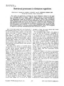
of frequencies, including endothelial NO�related (0.0095–0.02 Hz), neurogenic sympathetic (0.02– 0.046 Hz), myogenic (0.07–0.145 Hz), and passive ranges of frequencies, namely the cardiac or heart (0.8–1.6 Hz) and respiratory (0.2–0.4 Hz) ones. We calculated the activity of the respective regulatory fac� tors such as A/M, where A is the averaged maximum oscillation amplitude in the respective frequency range of the wavelet spectrum, and M is the mean blood (M) or lymph (Ml) flow expressed in perfusion units (perf. un.). All records were obtained from the skin of the palm surface of the nail bone of the third finger (an area enriched with arteriolovenular anastomoses), and the skin of the lower third of the forearm (an area with� out arteriolovenular anastomoses). We performed three measurements in each area of each subject. Skin temperature was measured using an IRTIS� 2000ME infrared thermograph (Russia). Statistical analysis was performed using the Bistat 4.03 software. Comparison of two samples was per� formed using the Mann–Whitney U�test. For correla� tion analysis, we calculated Spearmen rank correla� tion coefficients (r) and Pearson linear (k) correlation coefficients. RESULTS AND DISCUSSION 1. Features of the Spectrum of Oscillation Processes in Lymph Microcirculation of the Human Skin Oscillations of lymph flow in microvessels of the human skin were associated with clear domination of pacemaker phase oscillations originating from the deep subdermal lymphatic vessels containing muscles. In some cases, oscillations of other frequency ranges, such as myogenic or respiratory, were present in the records of wavelet spectra; however, the values of their peak amplitudes never higher than those observed in phase oscillations. This feature was observed indepen� dent of the edema. Examples of typical records of the wavelet spectrum of the skin lymph flow are presented in Fig. 1. In the whole group of subjects, the frequencies of the peak amplitudes of phase oscillations were in the range of 0.016–0.042 Hz: in the skin of the palm sur� face of the nail bone of the third finger, the range was 0.021–0.042 Hz; and in the skin of the forearm, the range was 0.016–0.035 Hz. The frequencies of phase oscillations increased in the case of edema. The mean values of their peak frequencies in the finger skin were 0.031 ± 0.002 and 0.036 ± 0.002 Hz in the case of edema and without it, respectively (р < 0.1); in the forearm skin, they were 0.018 ± 0.003 and 0.026 ± 0.002 Hz in the case of edema and without it, respec� tively (р < 0.05). In the whole group of subjects, the normalized (peak) amplitudes of phase oscillations were in the range of 0.086–0.34 in the skin of the palm surface of the nail bone of the third finger and in the range of
54
KRUPATKIN perf. un. 1.9
(a)
1.7 1.5 1.3 1.1 0.9 0.7 0.5 0.3 0.1 0.01 perf. un.
0.1
1
Hz
1
Hz
(b)
1.0 0.9 0.8 0.7 0.6 0.5 0.4 0.3 0.2 0.1 0.01
0.1
Fig. 1. Wavelet spectrum of oscillations of the blood flow (upper curve) and lymph flow (lower curve) in the skin of the palm sur� face of the nail bone of the third finger of the hand in the subjects with hand edema associated with a complex regional pain syn� drome. Vertical axis, perfusion (perfusion units, p.U); horizontal axis, oscillation frequency (Hz). (a) Moderate activation of phase oscillations (Al/Ml = 0.18, peak frequency 0.039 Hz); (b) prominent activation of phase oscillations (Al/Ml = 0.31, peak frequency 0.037 Hz).
0.061–0.22 in the skin of the forearm. Like the fre� quencies, the amplitudes of phase oscillations were higher in the case of edema. The mean values of the normalized maximum amplitudes in the finger skin were 0.2 ± 0.004 and 0.14 ± 0.001 in the case of edema and without it, respectively (p < 0.05); in the forearm skin, they were 0.16 ± 0.0015 and 0.1 ± 0.002 in the case of edema and without it, respectively (p < 0.05). We compared the range of phase oscillations of the lymph flow and the frequency ranges of the wavelet spectrum of oscillations of the blood flow in the skin and found that the former was similar to the range of oscillations of sympathetic adrenergic origin, which was 0.02–0.046 Hz. However, under the conditions of
a strong deficit of sympathetic innervations, in con� trast to blood flow oscillations, lymph flow oscillations did not disappear in this frequency range of the wavelet spectrum. Moreover, they even predominated in their amplitudes (Fig. 2). These data support the assump� tion that, despite the blood microcirculation, the range of 0.02–0.046 Hz in oscillations of the skin lymph flow has a pacemaker origin rather than a sym� pathetic one. In the subjects with a severe deficit of thin somatic and autonomic nerve fibers due to trauma of the nerve trunks (n = 5), both the amplitude and fre� quency of phase oscillations were lower compared to the mean value in the group without edema. For these subjects, the mean values of the normalized maximum HUMAN PHYSIOLOGY
Vol. 40
No. 1
2014
OSCILLATORY PROCESSES IN LYMPH MICROCIRCULATION IN THE HUMAN SKIN
55
perf. un. 0.9 0.8 0.7 0.6 0.5 0.4 0.3 0.2 0.1 0.01
0.1
1
Hz
Fig. 2. Wavelet spectrum of oscillations of the blood flow (upper curve) and lymph flow (lower curve) in the skin of the palm sur� face of the nail bone of the third finger of the hand in the subject with severe denervation syndrome following trauma of the median nerve and poor recovery of its function but without edema. Vertical axis, perfusion (perfusion units, perf. un.); horizontal axis, oscillation frequency (Hz). We did not find any oscillations of the sympathetic origin (arrow). Clear phase oscillations are present. Both the amplitude and frequency of their activity are decreased (Al/Ml = 0.1, peak frequency 0.028 Hz).
amplitudes in the finger skin were 0.11 ± 0.002 and 0.21 ± 0.003 (p < 0.05) in the subjects with denervation syndrome and intact innervation, respectively, whereas the mean values of peak frequencies were 0.024 ± 0.004 Hz and 0.037 ± 0.003 Hz (p < 0.05) in the subjects with denervation syndrome and continued innervations, respectively. Thus, in spite of the gener� ally accepted opinion that under physiological condi� tions, innervations practically do not influence with the lymph transport, sympathetic nerves affect both the amplitude and frequency of phase oscillations of the lymph flow. Myogenic oscillations in the frequency range of 0.07–0.145 Hz were observed in 43.3% of cases. We did not reveal any interrelationships between their appearance in the wavelet spectrum and the presence or absence of edema in the skin areas studied, includ� ing the finger and forearm. The amplitude of these oscillations was more than half the amplitude of phase oscillations. We suppose that these lymph flow oscilla� tions are related to oscillations of the myogenic tone of the larger lymphatic vessels. Self�sustained clear oscillations of the endothelial origin with frequencies of 0.0095–0.02 Hz were observed rarely in 13.3% of subjects, mostly in those who had edema. These oscillations had low ampli� tudes. Oscillation in the respiratory range of 0.2–0.4 Hz were found in a few records in 13.3% of subjects, more frequently in those who had edema. Due to the high hydrodynamic resistance of the lymphatic nodes, the respiratory rhythms cannot move to the peripheral HUMAN PHYSIOLOGY
Vol. 40
No. 1
2014
lymphatic vessels and microvessels [9]. The most probable explanation of the appearance of a respira� tory rhythm is the transmission to thin�wall lymphatic vessels from the venules and veins closely located to these vessels. We did not observe any cardiac pulse rhythms in the wavelet spectrum of skin lymph flow because, due to anatomical reasons, they cannot be transmitted to the lymph flow. In conclusion, the normalized amplitude and fre� quency of phase oscillations of the lymph flow are the most important indices among the oscillation features that can be used for noninvasive diagnosis of the state of lymph microcirculation in the human skin. The clinical and physiological scheme of interpretation of the changes in the Al/Ml value is presented in Table 1. 2. Interrelationship between Phase Oscillations of Lymph Flow and the Temperature and the Indices of Blood Microcirculation We did not find any correlations between the nor� malized amplitudes of phase oscillations of lymph flow and skin temperature in the whole group of sub� jects or the subgroups with edema or without edema (r < 0.3, p < 0.35). In order to estimate the relationships between phase oscillations of lymph flow and oscillations of blood microcirculation, we calculated the correlations between the normalized amplitude if lymph flow oscil� lations and the amplitudes of the cardiac rhythm (Ac/M), respiratory rhythm (Ar/M), and self�sus�
56
KRUPATKIN
Table 1. Clinical and physiological interpretation of changes in the Al/Ml index Changes in the Al/Ml value Within control values Decrease Increase
Absence of edema
Edema
Ordinary phase activity Decreased activity of phase oscillations Increased activity of phase oscillations
Insufficient activity of phase oscillations Defensive activation of phase oscillations
The control values vary in the range of ±20% of the averaged values. Here and in Table 2, abbreviations are explained in the text.
Table 2. Coefficients of Pearson linear (k) and Spearman rank (r) correlations of the amplitudes of phase lymph flow oscil� lations and oscillations of blood microcirculation in the skin of the third finger of the hand Groups of subjects Whole group (n = 90) Absence of edema (n = 51) Edema (n = 39)
Al/Ml and Ac/M
Al/Ml and Ad/M
Al/Ml and Adom/M Al/Ml and Am/M
k
0.018, p = 0.94
–0.341, p = 0.19
–0.213, p = 0.42
–0.331, p = 0.21
r
0.053, p = 0.84
–0.42, p = 0.11
–0.14, p = 0.59
–0.33, p = 0.21
k
–0.177, p = 0.64
r
–0.23, p = 0.52
–0.7, p = 0.038*
–0.52, p = 0.14
–0.867, p = 0.004*
k
0.355, p = 0.43
0.1, p = 0.81
0.73, p = 0.1
0.43, p = 0.39
r
0.5, p = 0.23
0.37, p = 0.39
–0.6, p = 0.09** –0.407, p = 0.27
–0.9, p = 0.005*
0.5, p = 0.23
0.46, p = 0.26
* p < 0.05; ** p < 0.01. In other cases, p > 0.1. n is the number of measurements.
tained myogenic rhythm (Am/M) normalized to aver� aged perfusion, and the predominate amplitudes of oscillations in the endothelial and neurogenic ranges reflecting the maximum amplitude of oscillations of large and medium arterioles (Adom/M). The data are presented in Table 2. The data in Table 2 demonstrate that, under physi� ological conditions only, in the subgroup in which edema was absent and filtration mechanisms were activated, some correlations of the amplitudes of phase oscillations and blood microcirculation were observed. We found a negative correlation with the activity of self�sustained myogenic oscillations reflect� ing the number of functional capillaries [10], an index directly related to tissue oxidative metabolism. More intense tissue metabolism results in lower accumula� tion of underoxidized products in the interstitial space, lower oncotic pressure there, and matrix hydra� tion. This also means that liquid inflow into the lym� phatic microvessels is lower and the lymph volume and the tension of the walls of the lymphatic vessel are low. The same factors are responsible for the appearance of a negative, though less significant, correlation with the Ad/M value. It has been demonstrated previously that the Ad value is negatively related to the linear rate of capillary flow [10]. We did not find any correlations between lymph flow and blood flow oscillations in the subgroup with edema. This is related to the high impact of impair� ments of the properties of the endothelial barrier, spe� cifically, impairments of the mechanisms of perme� ability and filtration, which are not directly reflected in blood flow oscillations. Moreover, edema is associ�
ated with mechanical compression of thin�wall microvessels, i.e., capillaries and venules, which also impairs correlations between the indices studied. CONCLUSIONS Thus, oscillations of lymph flow in microvessels of the human skin are characterized by the clear domi� nance of pacemaker phase oscillations in the fre� quency range of 0.021–0.042 Hz in the skin of the palm surface of the nail bone of the hand finger and 0.016–0.035 Hz in the forearm skin. Edema was asso� ciated with an increase in the mean values of their peak amplitudes and normalized maximum amplitudes. Low�amplitude oscillations were recorded more rarely in the self�sustained myogenic, endothelial, and respiratory ranges. We did not find any cardiac pulse rhythms in the wavelet spectrum of the lymph flow. The normalized amplitudes of phase oscillations of the lymph flow were not correlated to the skin tempera� ture. Only under physiological conditions, in the sub� group without edema, were the normalized ampli� tudes of phase oscillations of the lymph flow negatively correlated to the self�sustained myogenic oscillations of the blood flow, which reflected the number of func� tional capillaries and activity of oxidative tissue metabolism. In edematous tissue, oscillations of the lymph and blood flow were not correlated to each other. The values of the normalized amplitude and fre� quency of phase oscillations may be used as effective diagnostic tools to study lymph microcirculation. HUMAN PHYSIOLOGY
Vol. 40
No. 1
2014
OSCILLATORY PROCESSES IN LYMPH MICROCIRCULATION IN THE HUMAN SKIN
REFERENCES 1. Zoltzer, H., Morphology and physiology of lymphatic endothelial cells, Microvascular research, Shepro, D., Ed., Elsevier, 2006, vol. I, p. 535. 2. Ardasenov, A.V., Khugaeva, V.K., and Aleksandrov, P.N., Mikrotsirkulyatornoe ruslo kozhi v usloviyakh vospalen� iya i korrektsii metodom limfostimulyatsii (The Micro� circulatory Bed of the Skin under Conditions of Inflammation and Its Correction by the Lymph Stimu� lation Method), Moscow: Nauchnyi Mir, 2004. 3. Földi’s Textbook of Lymphology, Foldi, M. and Foldi, E., Eds., Elsevier, 2006. 4. Scallan, J., Haxley, V.H., and Korthuis, R.J., Capillary Fluid Exchange: Regulation, Functions and Pathology, San Rafael: Morgan and Claypool Life Sciences, 2010. 5. Orlov, R.S., Regulation of lymphatic vessels, in Fiz� iologiya krovoobrashcheniya: Fiziologiya sosudistoi sistemy (Physiology of Blood Circulation: Physiology of the Vascular Sytem), Leningrad: Nauka, 1984.
HUMAN PHYSIOLOGY
Vol. 40
No. 1
2014
57
6. Fischer, M., Franzeck, U.K., Herrig, I., Costanzo, U., Wen, S., Schiesser, M., Hoffmann, U., and Bollinger, A., Flow velocity of single lymphatic capillar� ies in human skin, Am. J. Physiol. Heart Circ. Physiol., 1996, vol. 270, no. 39, p. H358. 7. Franzeck, U.K., Fischer, M., Costanzo, U., Herrig, I., and Bollinger, A., Effect of postural changes on human lymphatic capillary pressure of the skin, J. Physiol., 1996, vol. 494, no. 2, p. 595. 8. Lazernaya dopplerovskaya floumetriya mikrotsirkulyatsii krovi (Laser Doppler Flowmetry of Blood Microcircu� lation), Eds., Krupatkin, A.I. and Sidorov, V.V., Mos� cow: Meditsina, 2005. 9. Lobov, G.I. and Pan’kova, M.N., Role of the lymphatic nodes in lymph transport, Vestnik Limfologii, 2011, no. 3, p. 19. 10. Krupatkin, A.I., Dynamic oscillatory circuit of regula� tion of capillary hemodynamics, Hum. Physiol., 2007, vol. 33, no. 5, p. 595. Translated by M. Stepanichev