Brazilian Journal of Medical and Biological Research (2013) 46: 676-680, http://dx.doi.org/10.1590/1414-431X20132896 ISSN 1414-431X Short Communication
Overexpression of hsa-miR-125b during osteoblastic differentiation does not influence levels of Runx2, osteopontin, and ALPL gene expression M.T. Pinto1,2, L.D.F. Nicolete1, E.S. Rodrigues1,2, P.V.B. Palma1, M.D. Orellana1, S. Kashima1,2 and D.T. Covas1,3 1
Centro Regional de Hemoterapia de Ribeira˜o Preto, Instituto Nacional de Cieˆncia e Tecnologia em Ce´lulas-Tronco e Terapia Celular, Ribeira˜o Preto, SP, Brasil 2 Departamento de Ana´lises Clı´ nicas, Toxicolo´gicas e Bromatolo´gicas, Faculdade de Cieˆncias Farmaceˆuticas de Ribeira˜o Preto, Universidade de Sa˜o Paulo, Ribeira˜o Preto, SP, Brasil 3 Departamento de Clı´ nica Me´dica, Faculdade de Medicina de Ribeira˜o Preto, Universidade de Sa˜o Paulo, Ribeira˜o Preto, SP, Brasil
Abstract Multipotent mesenchymal stromal cells (MSCs) were first isolated from bone marrow and then from various adult tissues including placenta, cord blood, deciduous teeth, and amniotic fluid. MSCs are defined or characterized by their ability to adhere to plastic, to express specific surface antigens, and to differentiate into osteogenic, chondrogenic, adipogenic, and myogenic lineages. Although the molecular mechanisms that control MSC proliferation and differentiation are not well understood, the involvement of microRNAs has been reported. In the present study, we investigated the role of miR-125b during osteoblastic differentiation in humans. We found that miR-125b increased during osteoblastic differentiation, as well as Runx2 and ALPL genes. To study whether the gain or loss of miR-125b function influenced osteoblastic differentiation, we transfected MSCs with pre-miR-125b or anti-miR-125b and cultured the transfected cells in an osteoblastic differentiation medium. After transfection, no change was observed in osteoblastic differentiation, and Runx2, OPN, and ALPL gene expression were not changed. These results suggest that the gain or loss of miR-125b function does not influence levels of Runx2, OPN, and ALPL during osteoblastic differentiation. Key words: Multipotent mesenchymal stromal cells; Osteoblastic differentiation; hsa-miR-125b
Introduction Multipotent mesenchymal stromal cells (MSCs) were first isolated from bone marrow and then from other tissues as colony-forming unit fibroblasts (1,2). MSCs have been isolated from various adult tissues such as placenta, cord blood, deciduous teeth, and amniotic fluid (3-6). Due to the lack of a single definitive marker to define these cells, the International Society for Cellular Therapy has proposed functional criteria for the characterization of MSCs that include adherence to plastic, expression of specific surface antigens, and their ability to differentiate into osteogenic, chondrogenic, adipogenic, and myogenic lineages (7). The molecular mechanisms that control MSC differentiation are not well understood. However, microRNAs
(miRNAs) have been reported to be involved in several important events that occur during differentiation and proliferation. miRNAs are single-stranded RNAs of ,22 nucleotides that derive from an ,70 nucleotide precursor, and have been identified or predicted in a wide variety of organisms, including plants, mice, and humans (8,9). Decreased expression of miR-138 in human MSCs has been reported to be associated with osteogenic (10) and adipogenic differentiation (11). Moreover, overexpression of miR-637 can enhance adipogenic differentiation and suppress osteogenic differentiation by targeting osterix, a transcription factor essential for osteogenesis (12). Another study reported that miR-125b inhibits osteoblastic differentiation in mice by downregulating cell proliferation
Correspondence: S. Kashima, Centro Regional de Hemoterapia de Ribeira˜o Preto, Rua Tenente Cata˜o Roxo, 2501, 14051-140 Ribeira˜o Preto, SP, Brasil. Fax: +55-16-2101-9309. E-mail:
[email protected] Received January 7, 2013. Accepted June 5, 2013. First published online August 26, 2013.
Braz J Med Biol Res 46(8) 2013
www.bjournal.com.br
miR-125b expression during osteoblastic differentiation
(13). With this in mind, the aim of this study was to investigate the role of miR-125b during osteoblastic differentiation in humans.
Material and Methods Isolation and characterization of MSCs A total of 10 mL human bone marrow aspirate was collected from the posterior iliac crest of 7 healthy donors (3 females and 4 males). The study was approved by the Institutional Ethics Committee of the Hospital das Clı´ nicas, Faculdade de Medicina de Ribeira˜o Preto, Universidade de Sa˜o Paulo (process No. 1783/2004), and all individuals signed an informed consent before enrollment. Mononuclear cells were isolated by FicollHypaqueTM PLUS (GE Healthcare Bio-Sciences AB, Sweden). MSCs were separated by plastic adherence during culture in a-minimum essential medium (a-MEM; Invitrogen, USA) supplemented with 15% fetal bovine serum (FBS; Thermo Scientific HyClone, USA), 2 mM L-glutamine, and 100 U penicillin/streptomycin (Sigma, USA). Plastic-adherent MSCs from the third passage were used in the experiments. Cultured MSCs were immunophenotypically characterized using the following monoclonal antibodies: CD45FITC, CD14-PE, CD44-FITC, CD29-PE, CD13-PE, CD90-PE, CD73-PE, CD49e-PE, CD31-FITC, CD34-PE, CD105-PE, HLA-DR-FITC, HLA-ABC-PE, and KDR-PE (Pharmingen, USA). All samples were analyzed on a FacsCalibur flow cytometer (Becton & Dickinson, USA). Osteoblastic differentiation Osteoblastic differentiation was initiated by seeding MSCs in the presence of osteogenic differentiation medium composed of MSC growth medium, i.e., a-MEM supplemented with 7.5% FBS plus 1 M glycerol-2-phosphate (Sigma), 20 mM L-ascorbic acid (Sigma), and 0.1 mM dexamethasone (Sigma). The culture medium was replaced every 3 days during a period of 21 days. During the same period, control cells were kept in standard a-MEM with 7.5% FBS (Thermo Scientific HyClone). All cells were fixed and stained by the von Kossa method (14), which indicates calcium deposition, and were analyzed with an Axioscope 2.0 Zeiss microscope equipped with an AxioCam HR camera (Carl Zeiss, Germany). The cells were harvested and analyzed at 1, 3, 7, 14, and 21 days.
677
was performed (in duplicate) using SYBR Green PCR Master Mix (Applied BioSystems). For quantification of the relative expression of alkaline phosphatase, type liver/ bone/kidney (ALPL), the primers were: forward: 59-CATC GCCTACCAGCTCATG-39; reverse: 59-CTCGTCACTCT CATACTCCACA-39). For osteopontin (OPN) they were: forward: 59-CAGTGATTTGCTTTTGCCTCCT-39; reverse: 59-CAGCATCTGGGTATTTGTTGTAA-39). To normalize sample loading, the differences in threshold cycles (DCt) were derived by subtracting the Ct value for the internal reference (geometric mean of GAPDH and b-actin) from the Ct values of the evaluated genes. The 2-DDCT method was used to calculate relative expression levels (15). qRT-PCR amplification and fluorescence data collection were performed using the ABI 7500 Sequence Detection system (Applied Biosystems). To analyze miR-125b expression, 2.5 ng RNA was reverse transcribed using a high-capacity cDNA archive kit (Applied Biosystems). Stem-loop RT primers specific for miR-125b were used in the qRT-PCR assay. Mature miR-125b was quantified (in duplicate) using the TaqMan MicroRNA assay (Applied Biosystems), according to the manufacturer’s instructions. The geometric means of RNU 24 and RNU 48 were used to normalize the samples. Transfection assay A total of 26105 MSCs were seeded on 6-well culture plates with a-MEM supplemented with 15% FBS. PremiR-125b or anti-miR-125b (Ambion, Canada) were transfected into MSCs at a 40-nM concentration with Lipofectamine 2000 (Invitrogen), according to the manufacturer’s instructions. A negative control was used. Four hours after transfection, all cells were cultured in osteoblastic differentiation medium. For mRNA analysis, cells were harvested 1, 3, and 7 days after transfection. Statistical analysis The results of miRNA and gene expression were compared using the Kruskal-Wallis test followed by the Dunn post-test. Two-way ANOVA followed by the Bonferroni post-test was used to compare results obtained from transfected MSCs. Data were analyzed using GraphPad PRISM, version 5.01 (GraphPad Software, USA), and differences with P values #0.05 were considered to be statistically significant.
Results Quantitative RT-PCR (qRT-PCR) Total RNA was extracted with Trizol1 Reagent (Invitrogen), and then reverse transcribed using a highcapacity cDNA reverse transcription kit (Applied Biosystems, USA), following the manufacturer’s protocol. qRT-PCR for runt-related transcription factor 2 (Runx2; Hs002316925_m1) was done (in duplicate) with TaqMan probes and MasterMix (Applied BioSystems). qRT-PCR
www.bjournal.com.br
MSC characterization Cultured MSCs derived from human bone marrow had a typical MSC immunophenotype. MSCs were 92.4±2.1, 97.9±0.7, 92.4±1.6, 94.7±2.8, 75.8±7.2, 90.5±2.8, 88.3±11.4, and 92.3±1.0% positive for the CD73, CD90, CD29, CD13, CD44, CD49e, CD105, and HLA-ABC markers, respectively. Moreover, MSCs were negative
Braz J Med Biol Res 46(8) 2013
678
for hematopoietic cell markers (CD34, 1.9±0.9%; CD14, 0.9±1.6% and CD45, 0.6±0.4%); endothelial cell markers (CD31, 0.2±0.1%; KDR, 5.3±3.1%), and HLAclass II markers (1.0±0.6%, data not shown). The von Kossa staining method (14) was used to identify biological mineralization in cell culture, and it was observed that MSCs changed their morphology from spindle to cuboid shape after 14 days (Figure 1). miR-125b, Runx2, OPN, and ALPL expression Expression of miR-125b, Runx2, OPN, and ALPL was analyzed by qRT-PCR at different times (0, 1, 3, 7, 14, and 21 days) during osteoblastic differentiation. miR-125b expression increased significantly on day 21 (P#0.05; Figure 2A) compared to day 0. Significant increase in Runx2 was observed on days 3, 7, and 14 (P#0.05, Figure 2B) compared with day zero. Although no statistically significant differences in OPN expression were observed, a continual increase was clearly seen on all 6 days (Figure 2C). In addition, ALPL gene expression was higher on day 21 compared with day 1 (Figure 2D). To study whether the gain or loss of function of miR125b could influence osteoblastic differentiation, we transfected MSCs with pre-miR-125b or anti-miR-125b and cultured them in osteoblastic differentiation medium. First, we analyzed whether transfection of pre-miR-125b or anti-miR-125b could modify the functional level of cellular miR-125b. As expected, transfection of pre-miR125b and anti-miR-125b increased and decreased intracellular levels of miR-125b, respectively (Figure 2E and F). The activity of miR-125b was controlled by transfection of pre-miR-125b and anti-miR-125b for up to 7 days, and after that time the expression was lost. Since pre-miR-125b and anti-miR-125b influenced the cellular level of miR-125b, we thought that osteoblastic markers would be controlled by this system. Therefore, to determine whether changing the functional level of cellular miR-125b influenced osteoblastic differentiation, Runx2, OPN, and ALPL gene expression was analyzed by qRTPCR. MSCs transfected with pre-miR-125b and anti-miR125b and cultured in osteoblastic differentiation medium did not show any differences in Runx2, OPN, and ALPL
M.T. Pinto et al.
expression (Figure 2G-I). Taken together, our results suggest that the gain or loss of function of miR-125b did not influence the levels of Runx2, OPN, and ALPL in osteoblastic differentiation.
Discussion Our goal was to identify miR-125 expression during MSC differentiation in human osteoblasts. We evaluated whether miR-125b expression could interfere with osteoblastic differentiation by analyzing Runx2, OPN, and ALPL gene expression. Differentiation into osteoblasts was confirmed by the development of the specific, black von Kossa color of mineralization nodules (Figure 1D). Although it is an indirect method for confirmation of calcium deposits, we believe that it is suitable for the demonstration of osteoblast differentiation. We found that miR-125b, Runx2 and ALPL gene expression increased during osteoblastic differentiation. Although miRNAs are known to be involved in cell proliferation, development, and differentiation, no change was observed in osteoblastic differentiation or in Runx2, OPN, or ALPL gene expression, when the intracellular level of miR-125b was increased or attenuated. One study reported that miR-125b expression was attenuated during osteoblastic differentiation in mice and that overactivity of miR-125b inhibited osteoblastic differentiation. Also, inhibition of miR-125b promoted osteoblastic differentiation (13). Another study reported that miR-125b inhibited cell proliferation of human breast cancer cells by the inhibition of ErbB2/3 genes (16). Based on the results obtained for expression of the Runx2, OPN, and ALPL genes, we suppose that overexpression of mir-125b probably does not influence human osteoblastic differentiation. Nevertheless, in another study (13) using a mouse model, alkaline phosphatase activity and osteocalcin gene expression were decreased by the overactivity of miR-125b, suggesting an inhibition of osteoblastic differentiation. In this study, we analyzed the expression of Runx2, OPN, and ALPL genes because they are involved in osteoblastic differentiation. Runx2 inhibits adipogenic differentiation and commits MSCs to an osteogenic lineage. After this process, MSCs differentiate into
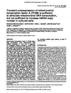
Figure 1. Mesenchymal stromal cells (MSCs) were cultured in osteoblastic differentiation medium and stained by the von Kossa method at 1 day (A), 3 days (B), 7 days (C), and 14 days (D). von Kossa staining showed mineralization nodules (black) and MSCs morphology changed from spindle to cuboid shape after 14 days.
Braz J Med Biol Res 46(8) 2013
www.bjournal.com.br
miR-125b expression during osteoblastic differentiation
679
Figure 2. miR-125b, Runx2, OPN, and ALPL expression during osteoblastic differentiation analyzed by qRT-PCR. A, miR-125b expression was increased on day 21. mRNA levels of osteoblastic markers Runx2 (B), OPN (C), ALPL (D). Significance was determined by the Kruskal-Wallis test and the Dunn post-test (*P#0.05). Pre-miR-125b (E) and anti-miR-125b (F) were transfected into mesenchymal stromal cells (MSCs) and miR-125b expression was measured. Runx2 (G), OPN (H), and ALPL (I) gene expression was analyzed in MSCs transfected with pre-miR-125b and anti-miR-125b. Negative controls were used in all experiments. Cells were cultured in osteoblastic differentiation medium and harvested on the indicated days. Runx2: runt-related transcription factor 2; OPN: osteopontin; ALPL: alkaline phosphatase. Differences in gene expression were analyzed by two-way ANOVA followed by the Bonferroni post-test (*P#0.05).
preosteoblasts. Preosteoblasts express Runx2, distalless homeobox 5, and msh homeobox homologue 2 and differentiate into immature osteoblasts, which express bone morphogenetic protein 2, Osterix, b-catenin, bone matrix proteins, bone sialoprotein, and OPN. At that point, immature osteoblasts develop into mature osteoblasts that express osteocalcin, ALPL, and type I collagen (17).
www.bjournal.com.br
Taken together, we believe that several mechanisms could be involved during osteoblastic differentiation. Although, this study has not shown an association between miR-125b and Runx2, OPN, and ALPL gene expression, miR-125b could control the expression of other genes, since one miRNA may target several different mRNAs (18). In addition, other miRNAs could also be involved in
Braz J Med Biol Res 46(8) 2013
680
osteoblastic differentiation. Understanding the differentiation process is essential to obtain tight control of the process for future clinical applications to avoid undesired side effects. Elucidating the role of hsa-miR-125b during osteoblast differentiation in vivo could contribute to understanding the molecular mechanisms involved in processes such as bone fracture repair and diseases such as osteoporosis.
M.T. Pinto et al.
Acknowledgments The authors thank Saˆmia Rigotto Caruso, Karina Rosa Solano, and Taı´ sa Risque Fernandes for the isolation of MSCs. They are also grateful to the healthy donors who participated in this study. Research supported by Fundac¸a˜o Hemocentro de Ribeira˜o Preto (FUNDHERP) and Centro Regional de Hemoterapia de Ribeira˜o Preto (CRH).
References 1. Luria EA, Panasyuk AF, Friedenstein AY. Fibroblast colony formation from monolayer cultures of blood cells. Transfusion 1971; 11: 345-349, doi: 10.1111/j.1537-2995.1971.tb04426.x. 2. Uccelli A, Moretta L, Pistoia V. Mesenchymal stem cells in health and disease. Nat Rev Immunol 2008; 8: 726-736, doi: 10.1038/nri2395. 3. Li CD, Zhang WY, Li HL, Jiang XX, Zhang Y, Tang P, et al. Isolation and identification of a multilineage potential mesenchymal cell from human placenta. Placenta 2005 (Epub Sept 17 AOP). 4. Bieback K, Kern S, Kluter H, Eichler H. Critical parameters for the isolation of mesenchymal stem cells from umbilical cord blood. Stem Cells 2004; 22: 625-634, doi: 10.1634/ stemcells.22-4-625. 5. Shi S, Gronthos S. Perivascular niche of postnatal mesenchymal stem cells in human bone marrow and dental pulp. J Bone Miner Res 2003; 18: 696-704, doi: 10.1359/ jbmr.2003.18.4.696. 6. In ’t Anker PS, Scherjon SA, Kleijburg-van der Keur C, Noort WA, Claas FHJ, Willemze R, et al. Amniotic fluid as a novel source of mesenchymal stem cells for therapeutic transplantation. Blood 2003; 102: 1548-1549, doi: 10.1182/blood2003-04-1291. 7. Dominici M, Le Blanc K, Mueller I, Slaper-Cortenbach I, Marini F, Krause D, et al. Minimal criteria for defining multipotent mesenchymal stromal cells. The International Society for Cellular Therapy position statement. Cytotherapy 2006; 8: 315-317, doi: 10.1080/14653240600855905. 8. Bartel DP. MicroRNAs: genomics, biogenesis, mechanism, and function. Cell 2004; 116: 281-297, doi: 10.1016/S00928674(04)00045-5. 9. Du T, Zamore PD. microPrimer: the biogenesis and function of microRNA. Development 2005; 132: 4645-4652, doi: 10.1242/dev.02070. 10. Eskildsen T, Taipaleenmaki H, Stenvang J, Abdallah BM,
Braz J Med Biol Res 46(8) 2013
11.
12.
13.
14.
15.
16.
17.
18.
Ditzel N, Nossent AY, et al. MicroRNA-138 regulates osteogenic differentiation of human stromal (mesenchymal) stem cells in vivo. Proc Natl Acad Sci U S A 2011; 108: 6139-6144, doi: 10.1073/pnas.1016758108. Yang Z, Bian C, Zhou H, Huang S, Wang S, Liao L, et al. MicroRNA hsa-miR-138 inhibits adipogenic differentiation of human adipose tissue-derived mesenchymal stem cells through adenovirus EID-1. Stem Cells Dev 2011; 20: 259267, doi: 10.1089/scd.2010.0072. Zhang JF, Fu WM, He ML, Wang H, Wang WM, Yu SC, et al. MiR-637 maintains the balance between adipocytes and osteoblasts by directly targeting Osterix. Mol Biol Cell 2011; 22: 3955-3961, doi: 10.1091/mbc.E11-04-0356. Mizuno Y, Yagi K, Tokuzawa Y, Kanesaki-Yatsuka Y, Suda T, Katagiri T, et al. miR-125b inhibits osteoblastic differentiation by down-regulation of cell proliferation. Biochem Biophys Res Commun 2008; 368: 267-272, doi: 10.1016/ j.bbrc.2008.01.073. Puchtler H, Meloan SN. Demonstration of phosphates in calcium deposits: a modification of von Kossa’s reaction. Histochemistry 1978; 56: 177-185, doi: 10.1007/BF00495978. Pfaffl MW. A new mathematical model for relative quantification in real-time RT-PCR. Nucleic Acids Res 2001; 29: e45, doi: 10.1093/nar/29.9.e45. Scott GK, Goga A, Bhaumik D, Berger CE, Sullivan CS, Benz CC. Coordinate suppression of ERBB2 and ERBB3 by enforced expression of micro-RNA miR-125a or miR-125b. J Biol Chem 2007; 282: 1479-1486, doi: 10.1074/ jbc.M609383200. Zhang Y, Khan D, Delling J, Tobiasch E. Mechanisms underlying the osteo- and adipo-differentiation of human mesenchymal stem cells. Sci World J 2012; 2012: 793-823. Friedman RC, Farh KK, Burge CB, Bartel DP. Most mammalian mRNAs are conserved targets of microRNAs. Genome Res 2009; 19: 92-105, doi: 10.1101/gr.082701.108.
www.bjournal.com.br