CELLULAR & MOLECULAR BIOLOGY LETTERS
Volume 9, (2004) pp 643 – 650 http://www.cmbl.org.pl Received 28 May 2004 Accepted 16 August 2004 Short Communication OXIDATIVE STRESS INDUCES IGF-I RECEPTOR SIGNALING DISTURBANCES IN CULTURED HUMAN DERMAL FIBROBLASTS. A POSSIBLE MECHANISM FOR COLLAGEN BIOSYNTHESIS INHIBITION PAWEŁ SIENKIEWICZ, MACIEJ PAŁKA and JERZY PAŁKA* Department of Medicinal Chemistry Medical Academy of Białystok, ul. Kilinskiego 1, 15-230 Białystok, Poland Abstract: The effects of oxidative stress on collagen and DNA biosynthesis, βgalactosidase and prolidase activities, and the expression of prolidase, β1integrin receptor, FAK, IGF-IR and MAP-kinases (ERK1, ERK2) were evaluated in human dermal fibroblasts. Subconfluent cells were subjected to repetitive stresses with 30µM t-BHP for 1 hour per day over the course of 5 days. It was found that oxidative stress induced the inhibition of collagen biosynthesis in these cells in a time-dependent manner. Exposure of the cells to 5 stresses contributed to a decrease in collagen and DNA biosynthesis to about 30% and 50% of the control values, respectively. Prolidase activity and expression were only suppressed in fibroblasts subjected to 1 and 3 stresses. In these cells prolidase activity was decreased by about 20%. As a result of 5 stresses, no further inhibition of prolidase activity occurred; however, expression of the enzyme was slightly increased, as demonstrated by Western blot analysis. It was found that these phenomena were neither related to the expression of β1-integrin
*Corresponding author; tel: +48 (85) 7485706. fax: +48 (85) 7485416, e-mail:
[email protected]. Abbreviations used: BCIP/NBT – 5-bromo-4-chloro-3-indolyl phosphate/nitro blue tetrazolium liquid substrate reagent; t-BHP – t-Butylhydroperoxide; CBB – Coomassie Briliant Blue R-250; DPBS – Dulbecco’s phosphate buffered saline; DMEM – Dulbecco’s minimal essential medium; EDTA – ethylenediaminetetraacetic acid; ERK1 and ERK2 – extracellular-signal-regulated kinase 1 and kinase 2; FAK – non-receptor focal adhesion kinase pp125FAK; FBS – fetal bovine serum; Grb2 – growth-factor receptor-bound protein 2; IGF-IR – insulin-like growth factor-I receptor, MAPK – mitogen activated protein kinases; PAGE – polyacrylamide gel electrophoresis; SDS – sodium dodecylsulfate; Sos – son of sevenless protein; TBS-T – Tris buffered saline with Tween 20.
644
CELL. MOL. BIOL. LETT.
Vol. 9. No. 4A. 2004
receptor nor to that of FAK. However, the exposure of the cells to 3 and 5 stresses contributed to a distinct decrease in IGF-IR and MAP-kinases (ERK1, ERK2) expression, which is probably responsible for the collagen biosynthesis inhibition. Key Words: Collagen , IGF-IR, β1-integrins, Oxidative Stress, Prolidase INTRODUCTION Oxidative stress is known to evoke collagen biosynthesis disturbances [1]. However, the mechanism of this process remains largely unknown. Collagen biosynthesis in human dermal fibroblasts may depend on the activity of prolidase [2]. Prolidase [EC 3.4.13.9] is a cytosolic enzyme which catalyses hydrolysis of imidodipeptides (mainly derived from collagen degradation), releasing proline, which is used for collagen resynthesis and cell growth [3]. Prolidase activity is known to be induced by β1-integrin receptor activation [4]. Stimulated β1-integrin receptor initiates a signaling cascade, which involves FAK [5], Grb2, Src, Shc, Sos, Ras and Raf proteins and leads to the activation of MAP-kinases (ERK1 and ERK2) [6]. Another factor that strongly stimulates collagen biosynthesis is IGF-I [7]. Stimulation of IGF-IR leads to the activation of MAP-kinases (ERK1 and ERK2) independently of FAK and Src [8]. The effects of IGF-I induce collagen gene expression [9], up-regulation of prolidase activity [10], stimulation of mitotic division and apoptosis prevention [8]. In this study, we examined the effects of oxidative stress on collagen and DNA biosynthesis, β-galactosidase and prolidase activities, and the expression of prolidase, β1-integrin receptor, FAK- and MAP-kinases (ERK1, ERK2) and IGFIR in human dermal fibroblasts. MATERIALS AND METHODS Chemicals were purchased as follows: t-BHP – from Fluka Chemie AG, Germany; DMEM and FBS – from Gibco, USA; glutamine, penicillin and streptomycin – from Quality Biologicals Inc., USA; monoclonal anti-β1-integrin and polyclonal anti-β-actin antibodies – from Santa Cruz Biotechnology Inc., USA; nitrocellulose membrane (0.2 µm), SDS, polyacrylamide, molecular weight standards and CBB – from Bio-Rad Laboratories, USA; L-5[3H] proline (28 Ci/mmol) – from Amersham, UK; anti-Rabbit IgG antibody – from Promega Corp., USA; [3H]thymidine (6.7 Ci/mmol) – from ICN Biomedicals Inc., USA; and Scintillation Coctail "Ultima Gold XR" – from Packard, USA. Polyclonal anti-human prolidase antibody was donated by Dr. James Phang (NCI-Frederick Cancer Research and Development Center, Frederick, MD, USA). All the other chemicals were provided by Sigma Corp., USA. All studies were performed on normal human skin fibroblasts (CRL-1474), purchased from American Type Culture Collection, Manassas, Virginia, USA.
CELLULAR & MOLECULAR BIOLOGY LETTERS
645
Induction of oxidative stress in human dermal fibroblasts Subconfluent cells were subjected to 1, 3 or 5 repetitive oxidative stresses by treatment for 1, 3 or 5 days with 30 µM t-butylhydroperoxide (t-BHP) for 1 hour per day, according to the method described by Dumont et al. [11]. Determination of enzymatic activities The activity of β-galactosidase was determined according to the method described by Zwierz et al. [12]. The activity of prolidase was determined according to the method of Myara et al. [3]. Protein concentration was measured using the method of Lowry et al. [13]. Enzyme activity was reported as nanomoles of released substrate during one minute per milligram of supernatant protein. DNA , collagen and total protein synthesis DNA synthesis was evaluated via measurement of [3H]thymidine incorporation, as described previously [14]. Cells were labeled for 24 h with 5-[3H]proline (5 µCi/ml, 28 Ci/mmol). Its incorporation into proteins was measured as described previously [15]. Incorporation of the tracer into collagen was determined by digesting proteins with purified Clostridium histolyticum collagenase, according to the method of Peterkofsky [16]. Total protein synthesis was calculated from the sum of the radioactivity of the collagenase-resistant proteins and the collagen digest. The results are shown as combined values for a given the cell plus medium fractions. SDS-PAGE and Western blot analysis Slab SDS/PAGE was carried out according to the method of Laemmli [17], using 10% polyacrylamide. Western blot analysis was performed as described previously [14]. RESULTS AND DISCUSSION Elevated activity of β-galactosidase is a biomarker that identifies cells subjected to oxidative stress and senescent cells [11, 18]. As can be seen in Fig. 1, βgalactosidase activity respectively increased by about 62%, 67% and 74% relative to the controls in fibroblasts subjected to 1, 3 and 5 stresses with 30 µM t-BHP. These results suggest a high level of efficiency of t-BHP in inducing oxidative stress in the studied cells. Exposure of the cells to 1 and 3 stresses contributed to a decrease in collagen biosynthesis by about 10% relative to the controls. After 5 stresses, a drastic inhibition of collagen production, by about 50%, was observed (Fig. 2A). This effect seems to be specific for collagen, since at this time, the total protein synthesis only decreased by about 20% (Fig.2B). A similar effect of oxidants on collagen production was previously demonstrated in human cardiac fibroblasts exposed to varying concentrations of hydrogen peroxide or xanthine plus xanthine oxide [1]. In order to explain the mechanism of this process, we considered prolidase as a target enzyme.
646
CELL. MOL. BIOL. LETT.
Vol. 9. No. 4A. 2004
Fig. 1. β-galactosidase activity in subconfluent control human dermal fibroblasts and in cells subjected to 1, 3 or 5 repetitive oxidative stresses by treatment for 1, 3 or 5 days with 30 µM t-butylhydroperoxide (t-BHP) for 1 h per day. The studied cells were cultured in DMEM supplemented with 10% FBS and harvested 24 h after the last scheduled stress. Mean values from three independent experiments done in duplicate are presented.
Fig. 2. 5-[3H]proline incorporation into proteins susceptible to the action of bacterial collagenase (A) and total proteins (B) in subconfluent control human dermal fibroblasts and in cells subjected to repetitive oxidative stresses (see Fig. 1 legend). Mean values from three independent experiments done in duplicate are presented.
Interestingly, the disturbances of collagen biosynthesis in t-BHP-treated cells were not dependent on prolidase activity. Over the course of the experiment, no difference in the enzyme activity was observed. In all these cells, prolidase activity decreased by about 25% (Fig. 3). However, in the cells subjected to 1 and 3 stresses, the decrease in prolidase activity was accompanied by a decrease in expression of that enzyme, while in the cells exposed to 5 stresses, the opposite phenomenon occurred: there was an increase in prolidase expression
CELLULAR & MOLECULAR BIOLOGY LETTERS
647
(Fig. 4A). The mechanism for this phenomenon may involve prolidase phosphorylation after 1 and 3 stresses, or prolidase dephosphorylation after 5 stresses. It has previously been suggested that prolidase is a phosphotyrosine enzyme, the activity of which may be regulated by its phosphorylation/dephosphorylation [19]. Whether this takes place in conditions of oxidative stress in cultured fibroblasts remains to be shown.
Fig. 3. Prolidase activity in subconfluent control human dermal fibroblasts and in cells subjected to repetitive oxidative stresses (See Fig. 1 legend). Mean values from three independent experiments done in duplicate are presented.
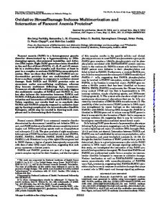
Fig. 4. Western blot analysis for prolidase (A), β1-integrin receptor (B), FAK (C), MAPkinases, ERK1 and ERK2 (D), IGF-IR (E) and β-actin (F) in subconfluent control human dermal fibroblasts (K) and in cells subjected to 1 (1), 3 (3) or 5 (5) repetitive oxidative stresses (See Fig. 1 legend). Samples used for electrophoresis consisted of 20 µg of protein of pooled cell extracts (n = 6). Detection of β-actin was carried out in order to provide a loading control. The arrows indicate the molecular masses of the standards. The intensity of the bands was quantified by densitometric analysis.
648
CELL. MOL. BIOL. LETT.
Vol. 9. No. 4A. 2004
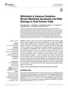
Fig. 5. Time course experiment for DNA biosynthesis (measured using the [3H]thymidine incorporation assay) in subconfluent control human dermal fibroblasts and in cells subjected to repetitive oxidative stresses (See Fig. 1 legend). Mean values from three independent experiments done in duplicate are presented.
Activation of the β1-integrin receptor (which contributes to an increase in prolidase activity [4]) leads to phosphorylation of FAK [5]. However, during the course of the experiment, no differences in the expressions of β1-integrin or FAK were found between the control and t-BHP treated fibroblasts (Fig. 4B and 4C). This suggests that oxidative stress-induced inhibition of collagen biosynthesis is independent of the signal mediated by this receptor. Instead, gradual suppression of MAP-kinase (ERK1 and ERK2) expression was observed (Fig. 4D). The extracellular signal-regulated kinase pathway is a major pathway; through it, growth factor receptors transmit signals to the nucleus. Their phosphorylation in response to treatment with oxidants may lead to activation of the p44/42 MAPK signaling pathway [20]. However, it has recently been shown that oxidative stress can variously modulate ERK activity in a time- and dose-dependent manner [21, 22]. In addition, reactive carbonyl compounds formed extensively in conditions of oxidative stress have been demonstrated to be capable of dephosphorylation of intracellular phospho-ERKs, which results in their inactivation [23]. IGF-I is a strong stimulator of collagen and DNA biosynthesis [7, 8]. It was recently proposed that the activation of IGF-IR may protect cells from oxidative stress [24] and regulate their resistance to the action of oxidants [25]. We found that down-regulation of MAP-kinase (ERK1 and ERK2) expression in fibroblasts subjected to oxidative stress was accompanied by parallel changes in expression of both the α and β subunits of IGF-IR, particularly in cells subjected to 5 stresses (Fig. 4E). Simultaneously, DNA biosynthesis in fibroblasts treated with t-BHP decreased gradually during the course of the experiment, by about 14%, 38% and 68% relative to the control, in cells subjected to 1, 3 and 5 stresses, respectively (Fig. 5). In view of this data, it seems probable that the inhibition of collagen biosynthesis caused by oxidative stress may be mostly a consequence of the disruption of the IGF-I receptor-mediated MAPK (ERK1, ERK2)associated signaling pathway, responsible for collagen gene expression and cell division.
CELLULAR & MOLECULAR BIOLOGY LETTERS
649
REFERENCES 1. Siwik, D.A., Pagano, P.J. and Colucci, W.S. Oxidative stress regulates collagen synthesis and matrix metalloproteinase activity in cardiac fibroblasts. Am. J. Physiol., Cell Physiol. 280 (2001) 53-60. 2. Pałka, J., Miltyk, W., Karna, E. and Wołczyński, S. Modulation of prolidase activity during in vitro aging of human skin fibroblasts the role of extracellular matrix collagen. Tokai J. Exp. Clin. Med. 21 (1996) 207-213. 3. Myara, I., Charpentier, C. and Lemonnier, A. Optimal conditions for prolidase assay by proline colorimetric determination: application to imidodipeptiduria. Clin. Chim. Acta. 125 (1982) 193-205. 4. Pałka, J. and Phang, J. Prolidase activity in fibroblasts is regulated by interaction of extracellular matrix with cell surface integrin receptor. J. Cell. Biochem. 67 (1997) 166-175. 5. Hanks, S.K., Calalb, M.B., Harper, M.C. and Patel, S.K. Focal adhesion protein-tyrosine kinase phosphorylated in response to cell attachment to fibronectin. Proc. Natl. Acad. Sci. USA. 89 (1992) 8487-8491. 6. Seger, R. and Krebs, E.G. The MAPK signaling cascade. FASEB J. 9 (1995) 726-735. 7. Goldstein, R.H., Poliks, C.F., Plich, P.F., Smith, B.D. and Fine, A. Stimulation of collagen formation by insulin-like growth factor-I in cultures of human lung fibroblasts. Endocrinology 124 (1989) 964-970. 8. Butler, A.A., Yakar, S., Gewolb, I.H., Karas, M., Okubo, Y. and LeRoith, D. Insulin-like growth factor-I receptor signal transduction: at the interface between physiology and cell biology. Comp. Biochem. Physiol. B. 121 (1998) 19-26. 9. Tanaka, H., Wakisaka, A., Ogasa, H., Kawai, S. and Liang, C.T. Effect of IGF-I and PDGF administered in vivo on the expression of osteoblastrelated genes in old rats. J. Endocrinol. 174 (2002) 63-70. 10. Miltyk, W., Karna, E., Wołczyński, S. and Pałka, J. Insulin-like growth factor I- dependent regulation of prolidase activity in cultured human skin fibroblasts. Mol. Cell. Biochem. 189 (1998) 177-184. 11. Dumont, P., Burton, M., Chen, Q.M., Gonos, E.S., Frippiat, C., Mazarati, J. B., Eliaers, F., Remacle, J. and Touissant, O. Induction of replicative senescence biomarkers by sublethal oxidative stresses in normal human fibroblast. Free Radic. Biol. Med. 28 (2000) 361-373. 12. Zwierz, K., Gindzieński, A., Głowacka, D. and Porowski, T. The degradation of glycoconjugates in the human gastric mucous membrane. Acta Med. Acad. Sci. Hung. 38 (1981) 145-152. 13. Lowry, O.H., Rosenbregh, N.I., Far, A.L. and Randall, I.R. Protein measurement with the Folin reagent. J. Biol. Chem. 193 (1951) 265-275. 14. Sienkiewicz, P. Bielawski, K., Bielawska, A. and Pałka, J. Amidine analogue of chlorambucil is a stronger inhibitor of protein and DNA synthesis in breast cancer MCF-7 cells than is the parent drug. Eur. J. Pharmacol. 492 (2004) 95-101.
650
CELL. MOL. BIOL. LETT.
Vol. 9. No. 4A. 2004
15. Oyamada, I., Pałka, J., Schalk, E.M., Takeda, K. and Peterkofsky, B. Scorbutic and fasted guinea pig sera contain an insulin-like growth factor I reversible inhibitor of proteoglycan and collagen synthesis in chick embryo chondrocytes and adult human skin fibroblasts. Arch. Biochem. Biophys. 276 (1990) 85-93. 16. Peterkofsky, B., Pałka, J., Wilson, S., Takeda, K. and Shah, V. Elevated activity of low molecular weight insulin-like growth factor- binding proteins in sera of vitamin C- deficient and fasted guinea pigs. Endocrinology 128 (1991) 1769-1779. 17. Laemmli, U.K. Cleavage of structural proteins during the assembly of the head of bacteriophage T4. Nature 227 (1970) 680-685. 18. Dimri, G.P., Lee, X., Basile, G., Acosta, M., Scott, G., Roskeley, C., Medrano, E.E., Linskens, M., Rubelj, I., Pereira-Smith, O., Peacocke, M. and Campisi, J. A biomarker that identifies senescent cells in culture and in aging skin in vivo. Proc. Natl. Acad. Sci. USA. 92 (1995) 9363-9367. 19. Surażyński, A., Pałka, J. and Wołczynski, S. Phosphorylation of prolidase increases the enzyme activity. Mol. Cell. Biochem. 220 (2001) 95-101. 20. Schieven, G.L., Mittler, R.S., Nadler, S.G., Kirihara, J.M., Bolen, J.B., Kanner, S.B. and Ledbetter, J.A., 1994. ZAP-70 tyrosine kinase, CD45, and T cell receptor involvement in UV- and H2O2-induced T cell signal transduction. J. Biol. Chem. 269 (1994) 20718-20726. 21. Li, J. and Holbrook, N.J. Common mechanisms for declines in oxidative stress tolerance and proliferation with aging. Free Radic. Biol. Med. 35 (2003) 292-299. 22. Bai, X., Lu, D., Bai, J., Zheng, H., Ke, Z., Li, X. and Luo, S. Oxidative stress inhibits osteoblastic differentiation of bone cells by ERK and NF-κB. Biochem. Biophys. Res. Commun. 314 (2004) 197-207. 23. Akhand, A.A., Hossain, K., Kato, M., Miyata, T., Du, J., Suzuki, H., Kurokawa, K. and Nakashima, I. Glyoxal and methylglyoxal induce aggregation and inactivation of ERK in human endothelial cells. Free Radic. Biol. Med. 31 (2001) 1228-1235. 24. Hong, F., Kwon, S.J, Jhun, B.S., Kim, S.S., Ha, J., Kim, S.J., Sohn, N.W., Kang, C. and Kang, I., 2001. Insulin-like growth factor-1 protects H9c2 cardiac myoblasts from oxidative stress-induced apoptosis via phosphatidylinositol 3-kinase and extracellular signal-regulated kinase pathways. Life Sci. 68 (2001) 1095-1105. 25. Holzenberger, M., Dupont, J., Ducos, B., Leneuve, P., Geloen, A., Even, P.C., Cervera, P. and Le Bouc, Y. IGF-I receptor regulates lifespan and resistance to oxidative stress in mice. Nature 421 (2003) 182-187.