5.9 + 0.9 mol of acid-labile sulphur, suggesting the presence of two [4Fe-4S] centres, and 0.47 mol of thiamin pyrophosphate. .... Lambda 7 spectrophotometer.
Biochem. J.
529
(1987) 246, 529-536 (Printed in Great Britain)
Purification and characterization of pyruvate :ferredoxin oxidoreductase from the anaerobic protozoon Trichomonas
vaginalis
Kevin WILLIAMS,* Peter N.
LOWEtt and Peter F. LEADLAY*
*Department of Biochemistry, University of Cambridge, Tennis Court Road, Cambridge CB2 IQW, U.K., and tDepartment of Biochemical Microbiology, The Wellcome Research Laboratories, Langley Court, Beckenham, Kent BR3 3BS, U.K.
The pyruvate: ferredoxin oxidoreductase from the anaerobic protozoon Trichomonas vaginalis is an extrinsic protein bound to the hydrogenosomal membrane. It has been solubilized and purified to homogeneity, principally by salting-out chromatography on Sepharose 4B. Low recoveries of active enzyme were caused by inactivation by 02 and the irreversible loss of thiamin pyrophosphate. It is a dimeric enzyme of overall Mr 240000 and subunit Mr 120000. The enzyme contains, per mol of dimer, 7.3 +0.3 mol of iron and 5.9 + 0.9 mol of acid-labile sulphur, suggesting the presence of two [4Fe-4S] centres, and 0.47 mol of thiamin pyrophosphate. The absorption spectrum of the enzyme is characteristic of a non-haem iron protein. The pyruvate: ferredoxin oxidoreductase from T. vaginalis is therefore broadly similar to the 2-oxo acid: ferredoxin (Iavyodoxin) oxidoreductases purified from bacterial sources, except that it is membrane-bound.
INTRODUCTION In the anaerobic protozoa Trichomonas vaginalis and Trichomonas foetus, the enzymes that catalyse the fermentation of pyruvate to acetate are all localized in an unusual type of redox organelle termed a 'hydrogenosome' (Lindmark & Muller, 1973; Muller, 1980; Steinbuchel & Muller, 1986). Substrate-level phosphorylation by this pathway is thought to contribute significantly to the organism's viability (Muller, 1982). Pyruvate: ferredoxin oxidoreductase (pyruvate synthase, EC 1.2.7.1), the first enzyme in the pathway, catalyses the oxidative decarboxylation of pyruvate in a CoA-dependent reaction yielding acetyl-CoA and CO2. The oxidant in vivo is thought to be ferredoxin. The anti-trichomonal drug metronidazole can act as an alternative reductant, and this process is assumed to play an important role in its mechanism of action (Yarlett et al., 1985). Apparently analogous enzymes have been purified from Clostridium acidi-urici (Uyeda & Rabinowitz, 1971) and from Halobacterium halobium (Kerscher & Oesterhelt, 1981a) and shown to contain thiamin and clusters. iron-sulphur pyrophosphate Pyruvate: ferredoxin oxidoreductase and the closely related pyruvate:flavodoxin oxidoreductase have also been detected in a wide range of bacteria and in some other anaerobic protozoa and fungi (Buchanan, 1972; Gehring & Arnon, 1972; Reeves et al., 1977; Lindmark, 1980; Weinbach et al., 1980; Blaschkowski et al., 1982; Neuer & Bothe, 1982; Kerscher & Oesterhelt, 1982; Shah et al., 1983; Yarlett et al., 1983, 1986 ). However, because of the 02-induced lability of many of these enzymes, they are difficult to isolate and have not been extensively studied. The present paper reports the first purification to homogeneity of pyruvate:ferredoxin oxidoreductase from an anaerobic protozoon, and its subsequent characterization. :
To whom correspondence and reprint requests should be sent.
Vol. 246
EXPERIMENTAL Materials Malate dehydrogenase (from pig heart), citrate synthase (from pig heart) and glucose oxidase (from Aspergillus niger), ferredoxin (from Clostridium pasteurianum), 2-oxo acids, coenzymes and proteins used as Mr markers in electrophoresis were obtained from Sigma Chemical Co., Poole, Dorset, U.K. The Bradford (1976) reagent for protein assays was from Bio-Rad Laboratories, Watford, Herts., U.K. The TSK 3000SWG and TSK 2000SWG gel-filtration columns were supplied by Anachem, Luton, Beds., U.K. Ferredoxin was purified from T. vaginalis as described by Gorrell et al. (1984), except that the final gel-filtration step using Sephadex G-75 was replaced by size-exclusion chromatography on a column (600 mm x 7 mm) of TSK 2000SWG.
Organism and growth of cells Trichomonas vaginalis Bushby strain was grown at 37 °C in complex medium (Linstead, 1981) supplemented with 10% (v/v) heat-inactivated horse serum and 360 ,#M-FeSO4 to maintain optimal concentrations for iron-sulphur proteins (Gorrell, 1985). After 48 h, cultures were harvested in late exponential phase (1.5 million cells/ml) by centrifugation at 1200 g for 15 min. The cells were resuspended and washed in 85 mmNaCl/4.6 mM-KCl/ 1.1 mM-MgSO4/222 mM-Na,HPO4/ HCI buffer, pH 7.4, containing 2 g of glucose/litre. The cell pellet could be stored at -20 °C for several months without loss of pyruvate: ferredoxin oxidoreductase activity. Determination of protein, cofactors and acetyl-CoA Protein was generally determined by the Coomassie Blue dye-binding method (Bradford, 1976), calibrated with y-globulin. Samples used for analyses of iron,
K. Williams, P. N. Lowe and P. F. Leadlay
530
sulphide and thiamin pyrophosphate were also assayed for protein by amino acid analysis. Acetyl-CoA was assayed by using malate dehydrogenase and citrate synthase (Decker, 1985). Iron was determined as described by Lovenberg et al. (1963) and sulphide as described by Rabinowitz (1978). Samples of ferredoxin from Clostridium pasteurianum were analysed as controls of these procedures and gave results in accordance with published values (Lovenberg et al., 1963). Thiamin pyrophosphate was released by treatment with 0.3 M-HC104 and determined fluorimetrically after oxidation to thiochrome pyrophosphate (Penttinen, 1979). Total flavin was determined as outlined by Koziol (1971). Enzyme assays Decarboxylation of pyruvate was measured spectrophotometrically by using Methyl Viologen as the electron acceptor, essentially as described by Lindmark & Muller (1973), except that glucose, glucose oxidase and catalase were added to scavenge °2 The assay mixture contained 100 mM-potassium phosphate buffer, pH 7.0, 2.5 mM-sodium pyruvate, 20 mM-dithiothreitol, 0.25 mmCoA, 20 mM-Methyl Viologen, 0.1% Triton X-100, 20 mM-glucose, 10 units of glucose oxidase/ml, 10 units of catalase/ml and enzyme sample in a volume of 1 ml at 30 'C. The mixture was covered with water-saturated n-butanol to exclude 02. The presence of the 02-scavenging system in all assays greatly diminished the lag phase which was present. Enzyme activities were measured from the linear steady-state rates obtained after any lag phase. This rate was independent of the extent of the lag phase and was proportional to the enzyme concentration over a 1000-fold range. The e600 for Methyl Viologen was taken as 13000 M-1 cm-' (Thorneley, 1974). Succinyl-CoA synthetase (ADP-utilizing) was assayed as described by Cha (1969). Hydrogenase was assayed as described by Lindmark & Muller (1973); again an 02-scavenging system was included. One unit of enzyme activity was defined as the amount of enzyme which gave a rate of 1,smol of product formed/min under the conditions specified.
Purification of pyruvate:ferredoxin oxidoreductase from T. vaginalis All operations were carried out at 0-4 'C. All buffers were degassed, sparged with O2-free N2 and supplemented with 2 mM-dithiothreitol and 1 mM-Na2S204 just before use. (1) Preparation of membrane fraction. T. vaginalis cells (20 g wet wt.) were thawed, and 20 ml of 50 mmpotassium phosphate buffer, pH 7, containing 2 mM-EDTA, was added. The mixture was homogenized with 20 passes in a motor-driven Potter-Elvehjem homogenizer at 5000 rev./min. The homogenate was centrifuged at 1500 g for 10 min. Cell membranes were collected by centrifugation of the supernatant at 124 000 g for 1 h. The membranes were washed once with 15 ml of 10 mM-potassium phosphate (pH 7.4)/I mMEDTA/0.1 mM-sodium acetate/30% (v/v) ethanediol buffer, pH 7.4. At this stage membranes could be stored at -20 °C for months without loss of pyruvate :ferredoxin oxidoreductase activity.
(2) Salt wash of membranes. The membrane pellet was resuspended in 15 ml of 10 mM-potassium phosphate/! mM-EDTA/ 1.0 M-sodium acetate buffer, pH 7.4, and stirred for 30 min. After centrifugation at 124000 g for 30 min, the tube contained a golden supernatant and a grey pellet. (3) Salting-out chromatography. Saturated (NH4)2504, adjusted to pH 7.4 with Tris, was added to the supernatant to give an 80% -saturated solution and stirred for 1 h. After centrifugation at 124000 g for 1 h, the pellet was resuspended in 4 ml of 2.4 M-(NH4)2SO4, pH 7.4, and applied at a flow rate of 1O ml/h to a Sepharose 4B column [10 cm x 2 cm, equilibrated with 2.4 M-(NH4)2SO4, pH 7.4]. The column was washed with 60 ml of equilibration buffer. Elution was carried out with a 200 ml descending gradient of (NH4)2SO4 from 2.2 M to 1 M. The active fractions of the eluate were pooled and concentrated in an Amicon ultrafiltration cell fitted with a PM 10 membrane. In the same cell the buffer was changed to 20 mm-Tris/HCl (pH 7.4)/0.4 M-KCl/I mM-EDTA.
(4) Gel filtration. This was carried out with a column (600 mm x 7 mm) of TSK 3000SWG on an LKB liquid chromatograph equilibrated with 20 mM-Tris/HCl (pH 7.4)/0.4 M-KCl/1 mM-EDTA buffer. Three runs were performed, in each of which a 0.5 ml sample was applied and eluted with the same buffer at a flow rate of 0.5 ml/min. Active fractions were pooled and concentrated as before. Purified enzyme was stored in liquid N2. Spectra Absorption spectra of purified pyruvate: ferredoxin oxidoreductase were recorded on a Perkin-Elmer Lambda 7 spectrophotometer. Polyacrylamide-gel electrophoresis Gel electrophoresis in the presence of SDS was performed on Tris/glycine-buffered 10 or 15% -(w/v)polyacrylamide slab gels (Anderson et al., 1973). Gels were stained for protein with Coomassie Brilliant Blue G-250. Amino acid analysis This was performed essentially as described by Perham
(1978). Ultracentrifugation Sedimentation-equilibrium analysis was conducted at 20 °C in a Beckman model E analytical centrifuge fitted with Rayleigh interference optics, the procedures described by Nureddin &-Johnson (1977) being used. Before use, the protein solution (1 mg/ml) was dialysed against 20 mM-Tris/HCI (pH 7.4)/0.4 M-KCl/1 mmEDTA/2 mM-dithiothreitol/ 1 mM-Na2S204 (I 0.42). RESULTS Localization and solubilization of pyruvate:ferredoxin oxidoreductase Subcellular fractionation of trichomonads in iso,osm.Qtic media demonstrated that, pyruvate: ferredoxin oxidoreductase is present solely in their hydrogenosomes 1987
531
Trichomonas pyruvate: ferredoxin oxidoreductase Table 1. Summary of the purification of T. vaginalis pyruvate:ferredoxin oxidoreductase
For full experimental details, see the text.
Cellular extract* (1) Membrane fraction (2) Salt-wash supernatant (3a) (NH4)2S04 pellet (3b) Concentrated saltingout peak (4) Concentrated TSK peak From 20 g wet wt. of T. vaginalis cells. 10- XMr
(mg)
(units)
20 20 16 4 1.5
1310 554 82 25 4.5
992 1100 448 164 33
1.2
1.1
18
Volume (ml)
Step
*
Protein
Total activity
1
2
3
4
205
97 .. .......e.N... .. ...........
67
45
29
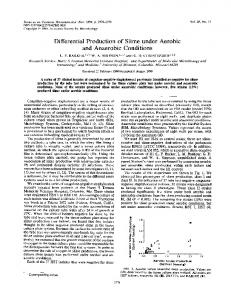
Fig. 1. Purification of pyruvate:ferredoxin oxidoreductase from T. vaginals Electrophoresis was carried out on a 15% -polyacrylamide gel in the presence of SDS. Lane 1, membrane pellet after 0.1 M-sodium acetate salt wash (10 jug of protein); lane 2, 1.0 M-salt-wash supernatant (5/sg); lane 3, concentrated salting-out peak (10,cg); lane 4, concentrated TSK peak (5 ,g).
and that the activity showed a latency revealed by disruption of the hydrogenosomal membrane, e.g. with Triton X-100 (Lindmark & Muller, 1973; Lindmark et al., 1975). We found that vigorous homogenization of Vol. 246
Specific activity
(units/ mg)
0.76 2.0 5.5 6.6 7.3
18.3
Recovery Purification (%) (fold) 100 110 45 17 3.3
1.8
1 2.6 7.2 8.6 9.6 24
T. vaginalis cells, in the absence of osmotic support, allowed measurement of most of the pyruvate :ferredoxin oxidoreductase activity, even in the absence of Triton X-100, and that all of this activity is sedimented at 124000 g for 1 h (Table 1). In contrast, the hydrogenosomal enzyme succinyl-CoA synthetase (ADPutilizing), was not sedimented under these conditions (P. N. Lowe, unpublished work). Thus the pyruvate: ferredoxin oxidoreductase is bound to the hydrogenosomal membrane. A range of methods were tried in order to establish a reliable method for the solubilization of pyruvate: ferredoxin oxidoreductase, using as a criterion of solubilization the appearance of activity in the supernatant after centrifugation at 124000 g for 1 h. Salt (1 M-KCI or 1 M-sodium acetate) or ionic detergents [1% (w/v) deoxycholate or 1% (w/v) cetyltriammonium bromide] were found to be effective solubilizing agents. However, non-ionic detergents [1% (v/v) Triton X-100 or 1% (w/v) n-octyl glucoside) did not solubilize the enzyme (see the Discussion section). Sodium acetate was finally chosen so that the use of detergents could be avoided. Dialysis of the salt-solubilized enzyme against salt-free buffer resulted in aggregation and precipitation of the enzyme protein. However, most of the activity could be regained by redissolving the precipitate in buffer containing 0.4 M-KCI. Therefore, for the purification of the solubilized enzyme, high salt (I > 0.4) was maintained in all buffers. Purification and stability of pyruvate:ferredoxin oxidoreductase The purification of pyruvate: ferredoxin oxidoreductase is summarized in Table 1. The recovery was typically 1-3%, representing an apparent purification of 15-20-fold over the cellular extract. The final specific activity was about 10-20 units/mg of protein. Fig. 1 shows fractions throughout the purification, analysed by SDS/polyacrylamide-gel electrophoresis. No advantage was found in starting from hydrogenosome-enriched fractions (Lindmark & Muller, 1973) rather than whole-cell extracts. The inclusion of ethanediol in the low-salt washing buffer increased the amount of other proteins removed in this step. Typically, about half of the membrane-associated pyruvate: ferredoxin oxidoreductase activity is released by the first salt extraction. The yield was only marginally improved by re-extraction.
532
K. Williams, P. N. Lowe and P. F. Leadlay
Salting-out chromatography on Sepharose 4B afforded excellent purification (Fig. 2) at the expense of a poor recovery of enzyme activity (< 20%). It can be seen by comparison of Fig. 2 with Table 1 that the specific activity of the enzyme was only marginally increased by salting-out chromatography, although the purity was greatly increased. We believe that this is due to an irreversible loss of enzyme activity, as a result of the long run time (20 h) necessary to achieve optimal resolution. The degree of purification shown in Table 1 is thus likely to be an underestimate. The salt extraction also released hydrogenase activity from the membrane, but it was eluted ahead of pyruvate: ferredoxin oxidoreductase and well separated from it. Yields of activity were not improved by including thiamin pyrophosphate/Mg2+,
FeSO4 or antipain [a potent inhibitor of trichomonal proteinases (Coombs & North, 1983)] in the buffers. From 1000-1500 mg of starting protein, 1-1.5 mg of pure pyruvate:ferredoxin oxidoreductase was obtained, which indicates that it represents at least 0.1% of total cell protein. It has been estimated that hydrogenosomes represent 5-10% of T. vaginalis cell protein (Lindmark et al., 1975). Thus the enzyme represents at least 1% of the protein in the organelle. Solubilized pyruvate: ferredoxin oxidoreductase activity was considerably more labile than that of the membrane-bound enzyme. Incubation of solubilized enzyme at 4 °C at pH 7.4 resulted in the irreversible loss of about 20% of activity after 5 h and 80% after 22 h. Maintenance of strict anaerobic conditions using di~.0.
2.2r
(a) _
1.8
F
0.3 V
I
z
1.4
0-
0.2
p
C.c
0>=
-D
1.0 L
x 0)
0.1I
0
0
10
20
30
40
-C
>
60
50
Fraction no. 4-1
cl0
0
V)
a.
Fraction no. ; 18 25 35 36 37 38 39 40 44 46 47 48 50 g -
It
z
8
10-3 XMr
(b)
205 116-
-
0 %WWWOW*www
97-
i.:...;..:..
- .u0,
45..
Dy .:r.o:::
Fig. 2. Purification of pyruvate:ferredoxin oxidoreductae by salting-out chromatography (a) (NH4)2SO4 pellet (60 units, 3 ml) was applied to a Sepharose 4B column (10 cm x 2 cm) equilibrated with 2.4 M-(NH4)2SO4 and eluted as described in the Experimental section. Column fractions (3.5 ml) were assayed for protein ( , A280) and pyruvate: ferredoxin oxidoreductase (0) activity. ----, [(NH4)2SOJ. (b) SDS/polyacrylamide-gel electrophoresis of column fractions. 1987
533
Trichomonas pyruvate: ferredoxin oxidoreductase Table 2. Molecular properties of purified pyruvate:ferredoxin (flavodoxin) oxidoreductases
Cofactors were analysed as described in the text. Abbreviation: ND, not determined. Molecular property
Trichomonas vaginalis
Subunits/Mr
240000 2 x 120000
Fe2+ (mol/mol) S2- (mol/mol) TPP (mol/mol) Flavin (mol/mol) Reference ...
7.3 +0.3(n = 4) 5.9 + 0.9 (n = 3) 0.47 0.005 The present
Mr
Clostridium acidi-urici
Halobacterium halobium
240000t ND
256000 2x86000, 2 x 42000 7.6 5.2 1.3 < 0.005 Kerscher & Oesterhelt (1981)
6 3 0.5-0.8 Trace Uyeda & Rabinowitz (1971)
study *
Klebsiella pneumoniae*
240000: 2 x 120000 8.0+0.6 6.6+0.2 ND ND Shah et al. (1983)
Pyruvate:flavodoxin oxidoreductase.
t The Mr was estimated by Uyeda & Rabinowitz (1971) from the sedimentation coefficient (sO2 w) by assuming a diffusion coefficient (D*20 w) of 3.6 x 10⁻⁷ and a partial specific volume (v) of 0.72 ml/g, and thus might be unreliable. t The M, of?partially purified pyruvate:flavodoxin oxidoreductase from E. coli has an Mr of 200000 (Blaschkowski et al., 1982). apparent subunit
M, was estimated as 120000 on Tris/glycine-buffered gels and 127000 on sodium phosphate-buffered gels. By using the partial specific volume of 0.741 ml/g calculated (Lee & Timasheff, 1979) from its amino acid composition (Table 3 below), sedimentation-equilibrium analysis of the native enzyme gave an estimate of the weight-average Mr as 240000. An independent estimate of Mr of the native enzyme was obtained by gel filtration on a TSK 3000SWG column. A good linear correlation between logMr and Kay for standard proteins was obtained (results not shown). Pyruvate: ferredoxin oxidoreductase was eluted as a broad peak, with an apparent Mr of about 275000. Chemical composition In common with the other 2-oxoacid: ferredoxin oxidoreductases characterized so far, the enzyme from T. vaginalis contains non-haem iron and acid-labile sulphur (Table 2). The absorption spectrum of purified T. vaginalis pyruvate: ferredoxin oxidoreductase exhibited a broad absorption band of low intensity between 350 and 500 nm (Fig. 3) characteristic of iron-sulphur
1.30
1.04
A
0.52
0.26
0
300
350
400
450
500
550
600
Wavelength (nm)
Fig. 3. U.v. and visible absorption spectru The spectrum of pure pyruvate: ferredo un oxidoreductase (1.3 mg of protein/ml), dissolved in 20 mM-Tris/HCl (pH 7.4)/0.4 M-KCl/I mM-EDTA, wass recorded in a cuvette of pathlength 1 cm.
thionite and dithiothreitol decreased the rate of inactivation. Inclusion of phospholipids or glycerol did not prevent inactivation at 4 'C. Enzyme activity was stable for several weeks after storage in liquid N2. Homogeneity and Mr Purified pyruvate: ferredoxin oxidoreductase
was
es-
sentially homogeneous, as judged by polyacrylamide-gel electrophoresis in the presence of SDS (Fig. 1). From comparison with mobilities of standard proteins, its
proteins. Thiamin pyrophosphate in sub-stoichiometric
amounts, but no flavin nucleotide, was detected (Table 2). The low value found for thiamin pyrophosphate suggests losses during purification, since control samples of thiamin pyrophosphate could be analysed with excellent recovery. However, addition of thiamin pyrophosphate to the assay mixture did not increase the activity of the enzyme, suggesting this loss must be irreversible. The amino acid composition of the enzyme is shown in Table 3. Catalytic properties A steady-state kinetic analysis of the purified T. vaginalis enzyme gave a Km for pyruvate of 0.14 mM and a Km for CoA of 2.5 /SM. The enzyme obeyed Bi Bi Ping Pong kinetics (Fig. 4a), except at very high CoA concentrations, where substrate inhibition occurred (Fig. 4b). The inhibition produced by the product, acetyl-CoA, in the presence of saturating CoA, was competitive with respect to pyruvate (Fig. 4c), with K1 = 100 + 20 #M. The
534
K. Williams, P. N. Lowe and P. F. Leadlay
"kinetic parameters of crude membrane preparations (Km for pyruvate, 0.13 mM; Km for CoA, 3.5 gM) were identical, within experimental error, with those for the pure solubilized enzyme. Pure enzyme accepted 2-oxobutyrate, pyruvate or 2-oxoglutarate as substrates in the standard assay, with
Table 3. Amino acid composition of T. vaginalis pyruvate: ferredoxin oxidoreductase
The results (means + S.D.) were obtained from duplicate analyses of 24, 48 and 96 h hydrolysates. The values for threonine and serine were obtained by linear extrapolation to zero hydrolysis time. Isoleucine and valine were taken from 96 h hydrolysates. Cysteine was determined, after performic acid oxidation, as cysteic acid.
Composition (mol/mol of protein)
Amino acid
24 196+8 110+2 187+7 258+6 132+ 12 289+ 13 182+ 14 111+1 33 100+9 157+6 80+3 75+8 63 +6 35* 136+7 101+1
Cys Asx Thr Ser Glx Pro Gly Ala Val Met Ile Leu Tyr Phe His Trp Lys Arg
* Determined spectroscopically (Edelhoch, 1967).
K!mPP- values of 0.1 mm, 0.14 mm and 0.5 mM respectively, and VamP.&. values relative to pyruvate of 0.45, 1.00 and 0.05 respectively. The stoichiometry of the reaction was deduced by assay of the acetyl-CoA formed. The results (Table 4) show that, in the presence of saturating pyruvate and Methyl Viologen, the reaction yields 1 mol of acetyl-CoA for every mol of CoA used. To test its specificity for electron acceptor, pyruvate: ferredoxin oxidoreductase was assayed with a variety of lowpotential electron acceptors (Table 5). The enzyme was unable to use NAD+ or NADP+, but was able to utilize the artificial compounds, Methyl Viologen and Benzyl Viologen, and either bacterial, or protozoal, ferredoxin. DISCUSSION Pyruvate: ferredoxin oxidoreductase has been recognized for a number of years to be an important enzyme in trichomonal energy metabolism, but its purification has been hampered by its instability to 02 (Lindmark & Muller, 1973; Lindmark et al., 1975). An additional problem we encountered stemmed from our observation that the enzyme is bound to the hydrogenosomal membrane; although it can be largely released by washing with buffers of high ionic strength, subsequent decrease in I to below 0.4 resulted in aggregation of the protein, and this limited the choice of purification techniques that could be used. Salting-out chromatography, using a method similar to that used to purify halophilic enzymes from Halobacterium halobium (Mevarech et al., 1976), was a very effective technique, but the time required for this step unavoidably resulted in low recovery of enzyme activity. In this method proteins are adsorbed from solutions of high salt concentration on to agarose gels and resolved by a decreasing salt gradient. The loss of activity (Table 1) during this step was largely accounted for by the lability of the enzyme, and thus does not represent a poor recovery of enzyme protein. A distinctive feature of the T. vaginalis pyruvate:
(a) 2
;
'~~~
0.6
E
0.5
E
0.4
,
' (b)
,
0
--
-,~ ~~ 0.3 022~
1
21
0.20.
1-1
0.1
10 1/[Pyruvate] (mM-')
20
0
0.05 1/[CoA] (pM'1)
0.1
1/[Pyruvate] (mM')
Fig. 4. Kinetic analysis of pyruvate:ferredoxin oxidoreductase (a) Double-reciprocal plot of initial velocity versus pyruvate concentration at various fixed concentrations of CoA: 0.5 gLM (A); 1.0 /SM (0); 2.5CrM (El); 5.0 /sM (A); 10 /SM (0). Citrate synthase and oxaloacetic acid were included to regenerate CoA so as to maintain a constant concentration during the assay. (b) Double-reciprocal plot of initial velocity versus CoA concentration with a saturating concentration of pyruvate (2.5 mM). (c) Double-reciprocal plot of initial velocity versus pyruvate concentration with a saturating concentration of CoA (0.25 mM) with various concentrations of acetyl-CoA: 50/uM (0); 100 /SM (A); 200 ,UM (0); 400 FM (0). 1987
535
Trichomonas pyruvate: ferredoxin oxidoreductase Table 4. Stoichiometry of the forward reaction catalysed by pyruvate:ferredoxin oxidoreductase
Methyl Viologen reduced (/tmol)
CoA added Assay
*
(#mol)
1 39 29.3 2 19.5 3 react with molecules 2 Viologen Assuming Methyl
Activities were calculated using the following absorption coefficients (M-l cm-'): Methyl Viologen, 600 = 13000 (Thorneley, 1974); Benzyl Viologen, c5 = 7550 (Sim & Vignais, 1978); NAD+/NADP+, 6340 = 6220; ferredoxin, 6390 = 17300 (Lindmark & Muller, 1973). The 2-oxo acid substrate was pyruvate in each case.
Methyl Viologen (20 mM) Benzyl Viologen (20 mM) Clostridial ferredoxin (0.5 mg/ml) T. vaginalis ferredoxin (0.2 mg/ml) NAD+ or NADP+ (5 mM)
Specific activity (units/mg of protein) 2.15 2.00 1.50 0.60 0
ferredoxin oxidoreductase is its behaviour as an extrinsic membrane protein, in that it can be solubilized under mild conditions at high ionic strength. Although ionic detergents also solubilized the enzyme, non-ionic detergents did not. We believe that this is because the enzyme aggregates in the presence of Triton X-100. This idea is supported by the observation that salt-solubilized pyruvate: ferredoxin oxidoreductase, when brought out of solution by dialysis against salt-free buffer, could be re-dissolved in either 0.4 M-KCI or 0.1% deoxycholate, but not in 1% Triton X-100. Pyruvate: ferredoxin (flavodoxin) oxidoreductases from Clostridium, Halobacterium, Klebsiella, Escherichia coli, and non-trichomonal protozoa, e.g. Entamoeba histolytica, differ from that from of T. vaginalis in that the enzymes are not apparently membrane-bound. However, the Halobacterium enzyme was only active in the presence of high salt concentrations, and thus, if it has similar properties to the enzyme from T. vaginalis, these might have been overlooked. The value of 240000 for the weight-average Mr is very similar to those values determined by comparable methods for other pyruvate: ferredoxin oxidoreductases and the related enzymes that use flavodoxin instead of ferredoxin (Table 2). The M, of the enzyme from C. acidi-urici reported by Uyeda & Rabinowitz (1971) is only an estimate based on the measurement of its sedimentation coefficient, with no experimental data on partial specific volume, diffusion coefficient or molecular shape, and thus might be unreliable. A value of 120000 for the Mr of the subunit fits well with the suggestion that the Vol. 246
Acetyl-CoA formed
(/Imol)
(Itmol)
42.3 84.6 26.9 53.8 14.6 29.2 I molecule of CoA.
Table 5. Activides of pyruvate:ferredoxin oxidoreductase with different electron acceptors
Electron acceptor
CoA used*
40.5 29.0 18.5
trichomonal enzyme is a homodimer as isolated. A value of 240000 for the Mr was used when calculating cofactor content and amino acid content per mol of enzyme. The homodimeric subunit composition agrees closely with that of the pyruvate: flavodoxin oxidoreductase from Klebsiella pneumoniae (Table 2), but differs from the enzyme from H. halobium, the only other similar enzyme with a known subunit structure. The latter enzyme has an a2cj2 structure; it is noteworthy that the sum of the Mr values of the a- and f8-subunits (128000) is very similar to the individual subunit Mr of the T. vaginalis and K. pneumoniae enzymes. Pyruvate: ferredoxin oxidoreductase from T. vaginalis is similar to equivalent bacterial enzymes (Table 2 and Fig. 3) in being a thiamin pyrophosphate-containing iron-sulphur protein. The instability of anaerobic oxidoreductases to 02 has been previously noted for nitrate reductase (Carlson et al., 1982), nitrogenase (Wang et al., 1985), hydrogenases (e.g. Arp & Burris, 1979) and pyruvate: ferredoxin oxidoreductases [e.g. the enzyme from Dastricha ruminartium (Yarlett et al., 1981), which is completely inactivated by even a 30 s exposure of OJ, and it is suggested that this may be due to irreversible oxidation of the iron-sulphur centres. The sulphide content of the enzyme is likely to be an underestimate (Kerscher & Oesterhelt, 198 la). From the results of the more reliable iron determination it is likely that the enzyme contains two [4Fe-4S] clusters per enzyme molecule. This would be consistent with each subunit bearing one cluster. The thiamin pyrophosphate content of the T. vaginalis enzyme is slightly lower than that estimated for the clostridial enzyme and much lower than that found in the H. halobium enzyme. It is likely that, during the isolation of the T. vaginalis enzyme, an irreversible loss of thiamin pyrophosphate occurred. As with these two bacterial enzymes, addition of thiamin pyrophosphate to the purified enzyme did not result in a recovery of activity. In contrast, the E. coli pyruvate: flavodoxin oxidoreductase showed a total dependence upon exogenous thiamin pyrophosphate (Blaschkowski et al., 1982). The catalytic parameters of the pure solubilized enzyme are identical with those of the membrane-bound form, suggesting that the structure of the enzyme is little altered by removal from the membrane. The Km for CoA of the T. vaginalis enzyme is similar to that of the T. foetus enzyme measured in a large-granule fraction (Lindmark & Muller, 1973), but the Km for pyruvate of the T. vaginalis enzyme (0.14 mM) is much lower than that of the T. foetus enzyme (3.2 mM). The Ping Pong kinetics, substrate inhibition by CoA and competitive inhibition by acetyl-CoA at saturating pyruvate concentrations are consistent with the mechanism of action proposed for the enzyme from H. halobium (Kerscher &
536
Oesterhelt, 198 lb), in which pyruvate decarboxylation precedes binding of CoA. The substrate specificity of the T. vaginalis enzyme resembles that of the H. halobium enzyme (Kerscher & Oesterhelt, 1981a) in that the enzyme catalyses a rapid reaction with 2-oxobutyrate and a slow reaction with 2-oxoglutarate as well as with pyruvate. It differs markedly from the pyruvate: flavodoxin oxidoreductases from E. coli (Blaschkowski et al., 1982) and of K. pneumoniae (Shah et al., 1983), which show a high specificity for pyruvate. With the exception of fungi (Yarlett et al., 1986) and possibly algae, protozoa are the only eukaryotes known to possess pyruvate: ferredoxin oxidoreductase. Respiratory eukaryotes utilize the pyruvate dehydrogenase multienzyme complex, which is distinct in terms of its large size, presence of flavin nucleotide and use of NAD+ as oxidant. The existence in trichomonads of hydrogenase, another common prokaryotic enzyme absent from higher eukaryotes, makes the evolutionary origins of such protozoa and, in particular, the hydrogenosome, an interesting area for speculation. Interestingly, the aspartate aminotransferase from T. vaginalis also shows similarities with its bacterial counterparts (Lowe & Rowe, 1985). The availability of pyruvate: ferredoxin oxidoreductase from a eukaryotic source will allow comparison with its bacterial counterparts and also with the pyruvate dehydrogenase complexes. Inhibition studies will allow us to assess the role of hydrogenosomal energy metabolism for the viability of T. vaginalis. K. W. holds a Science and Engineering Research Council (CASE) postgraduate studentship. We are indebted to Dr. S. E. Harding (University of Nottingham) for the analyticalultracentrifugation experiments, to Mrs. M. Walters and Mr. T. Steward for cultivation of T. vaginalis, and to Mr. J. B. Lester for amino acid analyses.
REFERENCES Anderson, C. W., Baum, P. R. & Gesteland, R. F. (1973) J. Virol. 12, 241-252 Arp, D. J. & Burris, R. H. (1979) Biochim. Biophys. Acta 570, 221-230 Blaschkowski, H. P., Neuer, G., Ludwig-Festl, M. & Knappe, J. (1982) Eur. J. Biochem. 123, 563-569 Bradford, M. M. (1976) Anal. Biochem. 72, 248-254 Buchanan, B. B. (1972) Enzymes 3rd Ed. 6, 193-216 Carlson, C. A., Ferguson, L. P. & Ingraham, J. L. (1982) J. Bacteriol. 151, 162-171 Cha, S. (1969) Methods Enzymol. 13, 62-69 Coombs, G. H. & North, M. J. (1983) Parasitology 86, 1-6 Decker, K. (1985) in Methods of Enzymatic Analysis (Bergmeyer, H. U., ed.), 3rd edn., vol. 7, pp. 186-193, Academic Press, London and New York
K. Williams, P. N. Lowe and P. F. Leadlay Edelhoch, H. (1967) Biochemistry 6, 1948-1954 Gehring, U. & Arnon, D. I. (1972) J. Biol. Chem. 247, 6963-6969 Gorrell, T. E. (1985) J. Bacteriol. 161, 1228-1230 Gorrell, T. E., Yarlett, N. & Muller, M. (1984) Carlsberg Res. Commun. 49, 259-268 Kerscher, L. & Oesterhelt, D. (1981a) Eur. J. Biochem. 116, 587-594 Kerscher, L. & Oesterhelt, D. (1981b) Eur. J. Biochem. 116, 595-600 Kerscher, L. & Oesterhelt, D. (1982) Trends Biochem. Sci. 7, 371-374 Koziol, J. (1971) Methods Enzymol. 188, 253-285 Lee, J. C. & Timasheff, S. N. (1979) Methods Enzymol. 61, 49-57 Lindmark, D. G. (1980) Mol. Biochem. Parasitol. 1, 1-12 Lindmark, D. G. & Muller, M. (1973) J. Biol. Chem. 248, 7724-7728 Lindmark, D. G., Muller, M. & Shio, H. (1975) J. Parasitol. 63, 552-554 Linstead, D. (1981) Parasitology 83, 125-137 Lovenberg, W., Buchanan, B. B. & Rabinowitz, J. C. (1963) 238, 3899-3913 Lowe, P. N. & Rowe, A. F. (1985) Biochem. J. 232, 689-695 Mevarech, M., Leicht, W. & Werber, M. M. (1976) Biochemistry 15, 2383-2386 Muller, M. (1980) Symp. Soc. Gen. Microbiol. 30, 127-142 Muller, M. (1982) Surgery 93, 165-171 Neuer, G. & Bothe, H. (1982) Biochim. Biophys. Acta 716, 358-365 Nureddin, A. & Johnson, P. (1977) Biochemistry 16, 1730-1737 Penttinen, H. K. (1979) Methods Enzymol. 62, 58-59 Perham, R. N. (1978) in Techniques in Protein and Enzyme Biochemistry (Kornberg, H. L., Metcalfe, J. C., Northcote, D. H., Pogson, C. I. & Tipton, K. I., eds.), vol. BIl/l, pp. 1-39, Elsevier, Amsterdam Rabinowitz, J. C. (1978) Methods Enzymol. 52, 275-277 Reeves, R. E., Warren, L. G., Susskind, B. & Lo, H.-S. (1977) J. Biol. Chem. 252, 726-731 Shah, V. K., Stacey, G. & Brill, W. J. (1983) J. Biol. Chem. 258, 12064-12068 Sim, E. & Vignais, P. M. (1978) Biochimie 60, 307-314 Steinbuchel, A. & Muller, M. (1986) Mol. Biochem. Parasitol. 20, 57-65 Thorneley, R. N. F. (1974) Biochim. Biophys. Acta 333, 487-496 Uyeda, K. & Rabinowitz, J. C. (1971) J. Biol. Chem. 246, 3111-3119 Wang, Z. C., Bums, A. & Watt, G. D. (1985) Biochemistry 24, 214-221 Weinbach, E. C., Claggett, C. E., Keister, D. B. & Diamond, L. S. (1980) J. Parasitol. 66, 347-350 Yarlett, N., Hann, A. C., Lloyd, D. & Williams, A. (1981) Biochem. J. 200, 365-372 Yarlett, N., Hann, A. C., Lloyd, D. & Williams. A. G. (1983) Comp. Biochem. Physiol. 74B, 357-364 Yarlett, N., Gorrell, T. E., Marczak, R. & Muller, M. (1985) Mol. Biochem. Parasitol. 14, 29-40 Yarlett, N., Orpin, C. G., Munn, E. A., Yarlett, N. C. & Greenwood, C. A. (1986) Biochem. J. 236, 729-739
Received 24 February 1987/20 May 1987; accepted 28 May 1987
1987