REVIEW Cellular Logistics 2:2, 117–125; April/May/June 2012;
G
2012 Landes Bioscience
PAK in Alzheimer disease, Huntington disease and X-linked mental retardation Qiu-Lan Ma,1,2 Fusheng Yang,1,2 Sally A. Frautschy1,2 and Greg M. Cole1,2,* 1
Department of Neurology; University of California Los Angeles; Los Angeles, CA USA; 2Geriatric Research and Clinical Center; Greater Los Angeles Veterans Affairs Healthcare System; West Los Angeles Medical Center; Los Angeles, CA USA
Keywords: Alzheimer disease, curcumin, PAK, ROCK, signaling pathways, synapses Abbreviations: AD, Alzheimer disease; Aβ, amyloid-β; NFT, neurofibrillary tangles; PAK, p21-activated kinase; APP, amyloid precursor protein; ADDLs, β-amyloid-derived oligomers; MR, mental retardation; CRMP-2, collapsin response mediator protein-2; LTP, long term potentiation; LIMK, LIM-Kinase; NMDARs, N-methyl-D-aspartate receptors; BBB, blood brain barrier; CA1, Cornu Ammonis 1; DHA, docosahexaenoic acid; CTCL, cutaneous T-cell lymphoma; HDAC, histone deacetylase; HD, Huntington disease; XLMR, X-linked mental retardation; MRX, nonsyndromic mental retardation; FXS, fragile X syndrome; FMR1, the fragile X mental retardation 1
Developmental cognitive deficits including X-linked mental retardation (XLMR) can be caused by mutations in P21activated kinase 3 (PAK3) that disrupt actin dynamics in dendritic spines. Neurodegenerative diseases such as Alzheimer disease (AD), where both PAK1 and PAK3 are dysregulated, may share final common pathways with XLMR. Independent of familial mutation, cognitive deficits emerging with aging, notably AD, begin after decades of normal function. This prolonged prodromal period involves the buildup of amyloid-b (Ab) extracellular plaques and intraneuronal neurofibrillary tangles (NFT). Subsequently region dependent deficits in synapses, dendritic spines and cognition coincide with dysregulation in PAK1 and PAK. Specifically proximal to decline, cytoplasmic levels of actin-regulating Rho GTPase and PAK1 kinase are decreased in moderate to severe AD, while aberrant activation and translocation of PAK1 appears around the onset of cognitive deficits. Downstream to PAK1, LIM kinase inactivates cofilin, contributing to cofilin pathology, while the activation of Rho-dependent kinase ROCK increases Ab production. Ab activation of fyn disrupts neuronal PAK1 and ROCK-mediated signaling, resulting in synaptic deficits. Reductions in PAK1 by the anti-amyloid compound curcumin suppress synaptotoxicity. Similarly other neurological disorders, including Huntington disease (HD) show dysregulation of PAKs. PAK1 modulates mutant huntingtin toxicity by enhancing huntingtin aggregation, and inhibition of PAK activity protects HD as well as fragile X syndrome (FXS) symptoms. Since PAK plays critical roles in learning and memory and is disrupted in many cognitive disorders, targeting PAK signaling in AD, HD and XLMR may be a novel common therapeutic target for AD, HD and XLMR.
*Correspondence to: Greg M. Cole; Email:
[email protected] Submitted: 03/07/12; Revised: 07/12/12; 07/17/12; Accepted: 07/24/12 http://dx.doi.org/10.4161/cl.21602
www.landesbioscience.com
Alzheimer Disease Alzheimer disease (AD) is the most prevalent neurodegenerative disease of aging but has many common mechanisms with other neurodegenerative diseases. AD is characterized clinically by progressive cognitive decline and pathologically by prodromal accumulation of neuritic plaques containing amyloid-β (Aβ) protein and neurofibrillary tangles containing tau protein aggregates comprising paired helical filaments. Proximal to cognitive decline, there is a selective loss of synapses, especially excitatory synapses and vulnerable neurons in networks required for learning and memory. Synaptic loss is accelerated at early stages of AD clinical symptoms and is more closely related to cognitive deficits than neuronal loss or amyloid buildup.1,2 Although soluble aggregated Aβ forms called β-amyloid-derived oligomers (ADDLs) or Aβ oligomers, including dimers, trimers and dodecamers (12-mer or Aβ *56) are implicated in synaptic dysfunction and loss in AD patients and AD animal models3-6 and Aβ immunoneutralization rescues synaptic defects in AD animal models,7,8 there are major gaps in our understanding the mechanisms controlling synaptic loss in AD and other neurodegenerative diseases, which remain under active investigation. Here we explore the potential overlap of dysregulation in PAK kinases that cause mental retardation, with synaptic deficits in other neurodegenerative diseases, which link GTPases to cytoskeletal reorganization and to nuclear signaling. The RAC/CDC42-activated kinase PAK1 is a key regulator for actin cytoskeleton and dendritic spine morphogenesis. We first reported a loss of PAK1 and PAK3 in cytoplasm of AD brain specimens, as well as in AD animal and cellular models, suggesting PAKs might play crucial roles in dendritic spine/synapses loss and cognitive defects in AD.9 Synaptic plasticity is dependent on the regulation of the actin cytoskeleton in dendritic spines.10-12 The regulation of F-actin cytoskeleton involves various actin-binding proteins and the molecular regulators of actin dynamics by membrane receptors and their downstream signaling cascades. In particular, Rho family GTPases, Rho, RAC and CDC42, play a
Cellular Logistics
117
central role in regulating actin reorganization.13,14 The balance between RhoA and RAC/CDC42’s reciprocal effects are linked to distinct upstream and downstream regulators to regulate morphogenesis of dendritic spines and synaptic plasticity. RhoA activates the kinase ROCK which also promotes processing of amyloid precursor protein (APP) to its derivative toxic species, Aβ42.15 Normally, both PAK1 and PAK 3 show diffuse distribution in cell bodies and dendrites and they are activated by upstream RAC and CDC42, but reduced in the cytoplasm of AD brain specimens and in AD animal and cellular models. This suggests that disruption in the signaling of both PAKs might play crucial roles in deficits of dendritic spines, synapses and cognition in AD.9 While PAK1 regulates actin cytoskeleton through LIMK1 control of cofilin, notably in dendritic spines, PAK3 shows activity-dependent recruitment into dendritic spines where it regulates dynamics16 but also uses the adaptor protein NCK2/ Grb4 downstream from Ephrin B to regulate synaptic transmission.17 Most significantly, both have redundant and overlapping functions so that knockout of either PAK1 or PAK3 alone does not cause a robust morphological phenotype in mice or loss of pcofilin and F-actin, while, in contrast, dual PAK1 and 3 knockout causes a dramatic marked loss of dendritic and neuronal arbor, pcoflin and F-actin, brain shrinkage and defects in dendritic spines, LTD, LTP and cognition but without global neuron loss.18 Thus, both PAK1 and PAK3 deficits in AD are relevant to synaptic and cognitive deficit. However, we also found that PAK1 showed aberrant activation in AD, and this activated PAK1 was translocated from cytoplasm to membrane, probably to granular structures in a complex with its activators, GTPases RAC/ CDC42.8 Therefore, while here we mainly review the pathologic activation of PAK1 and its downstream LIM kinase signaling pathways and potential therapeutic interventions for targeting these PAK1-LIM kinase pathway defects in AD, we acknowledge the significant role of PAK3. Alteration of PAKs in AD Cognitive decline has been directly linked to the synaptic dysfunction, especially to synapses, postsynaptic and dendritic spine loss in AD and mental retardation (MR) syndromes.1,19 A primary role of dendritic spine defects in MR has been demonstrated by the discovery of multiple mutant X-linked MR genes.19 These MR genes reveal a clustering of proteins in the postsynaptic pathways regulating spine actin assembly and disassembly and spine morphogenesis. PAK3 is one of these MR genes and missense mutation in PAK3 causes severe X-linked nonspecific MR.20,21 Animal models of MR syndromes created using the AID (auto-inhibitory domain) of PAK1, which blocks PAK1–3,22 or knockout of its downstream LIM-kinase both show defects in dendritic spines and cognition.23 These observations suggest the essential role of the PAK1–3/LIM-kinase pathway in regulating synaptic plasticity. Similar to MR, in AD, postsynaptic and dendritic spine defects are an early event in memory circuits and therefore spatiotemporally situated to play a critical role in cognitive deficits. For example, although the overall estimate of neuronal loss in AD
118
hippocampus ranges from 5–40%, albeit with higher losses in CA1, overall loss of postsynaptic proteins on Westerns from whole hippocampus, such as the actin-regulating developmentallyregulated brain protein (drebrin), have been found to reach 70– 95%.24-26 Selective drebrin loss results from an attack on excitatory synapses since drebrin regulates actin assembly and drebrindependent actin filaments in dendritic filopodia govern synaptic targeting of PSD-95 and dendritic spine morphogenesis.27 Two major neuronal isoforms of PAK exist in the brain, PAK1 and PAK3. Normally, both show diffuse distribution in cell bodies and dendrites. However, in AD brain, significant losses of PAK1 (35% ± 6) and PAK3 (55–69%) were observed in hippocampus, while PAK3 was also significantly decreased in AD temporal cortex (63– 77%). However, the loss of PAK1’s kinase activity clearly exceeds that of its protein level. The auto-phosphorylated PAK1 at Ser 141, an index of activity, was reduced by 73% in AD temporal cortex.9 In addition, PAK1 also showed aberrant activation in AD, and this activated PAK1 was translocated from cytoplasm to membrane, probably to granular structures in a complex with its activators, GTPases RAC/CDC42.8 An increase in the total protein level of PAK1–3 at early stages of AD but a reduction of both total and cytoplasmic phospho-PAK1 in late-stage severe AD was observed by Nguyen and collaborators.28 Moreover, a similar reduction and sub-cellular translocation of PAK1 was observed in a transgenic AD mouse model.8 Similarly, cytoplasmic phosphoPAK1 (pPAK) was significantly reduced in a triple transgenic mouse model of AD that develops both plaque and tangle pathology and this loss of pPAK, and cognitive deficits were improved by dietary docosahexaenoic acid (DHA).29 Since PAK1–3 defects are sufficient to cause cognitive deficits, these data suggest that dys-regulation (hyperactivation) of PAK1 followed by a loss of soluble pPAK may also play an important role in dendritic spine/synapse loss and cognitive defects in AD. To characterize the likely mechanism behind the cytoplasmic PAK deficits in AD, cultured hippocampal neurons were treated with β-amyloid (Aβ) oligomers, where we then observed a rapid abnormal PAK activation/ translocation followed by subsequent loss of cytoplasmic pPAK. This aberrant activation was accompanied by a rapid loss of F-actin and dendritic spines unlike the response to normal activation of PAK1 by RAC/ CDC42. The wild-type PAK1, but not its kinase–dead mutant (K299A), prevented the pathological changes in spines, providing evidence that functional PAK1 recruitment and signaling is blocked by Aβ exposure.8 In addition, it has been observed that fibrillar Aβ42 treatment of hippocampal neurons can also activate PAK1.30 These results suggest that β-amyloid species could be the primary elements responsible for PAK dysfunction in AD. These results also indicated that PAK1 has a significant functional role in neuronal plasticity, many important functions can be compensated by PAK3 and aberrant and chronic PAK1 activation should be considered “bad” in AD and Huntington disease (HD) that will be discussed later. Since PAK1 aberrant activated at early stages of AD, inhibition of PAK1 could reverse, partially reverse or delay clinical signs in AD. However, since PAK1 and PAK3 were eventually lost at latestage severe AD, this dynamic alteration of PAK at different AD
Cellular Logistics
Volume 2 Issue 2
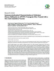
stages may also limit PAK inhibitors as a consistent drug for AD therapy. RAC/PAK1/LIMK/cofilin signaling pathway in AD. The regulation of actin cytoskeletal dynamics is through the phosphorylation of cofilin at Ser 3 by LIM-Kinase (LIMK), in a pathway where PAK1 activates LIMK.31 PAK/LIMK signaling inactivates cofilin, which depolymerizes actin filament (F-actin). Our data suggest this may enable other proteins, for example, drebrin, to bind and regulate actin in postsynaptic spines. Pathologic intracellular inclusion bodies (Hirano bodies) containing cofilin and smaller actin rods decorated by other actin-binding proteins are prominent features in the hippocampus and cortex of AD brains.32,33 We have observed that confocal co-labeling of pPAK and cofilin in AD hippocampus show cells with different stages of pPAK and cofilin pathologies; for instance, some cells exhibit increasingly intense cofilin labeling associated with progressively decreased diffuse pPAK accompanied by granular structure staining (Fig. 1A). The severe pPAK and cofilin pathologies in AD are associated with the reduction in the dendritic spine actin-regulating protein drebrin (70–95%).25-27 This is consistent with the hypothesis that translocation and loss of the cytosolic pPAK can lead to local pathology related to cofilin aggregation, drebrin loss and synaptic defects observed in AD brain.9 Cofilin labeling in and around Aβ plaques is also observed in AD model APPswe transgenic mouse hippocampus. Triple labeling of pPAK (green), cofilin (red) and Aβ plaques (blue with 10G4 antibody) in Tg+ mouse hippocampus indicated intense central-plaque pPAK staining sometimes associated with local cofilin puncta (Fig. 1B), similar to that observed in AD hippocampus. In addition, a large 62% loss in drebrin was also observed in these mice.9 Therefore, pPAK and cofilin pathology and severe drebrin loss are found in both AD and aged AD
Figure 1. Confocal co-labeling of pPAK and cofilin in AD hippocampus and in amyloid plaques from AD APPswe transgenic mice hippocampus. (A) Confocal co-labeling of pPAK and cofilin in AD hippocampus. Some neurons exhibit intense cofilin labeling (red) and granular pPAK staining (green). Blue represents DAPI. (B) Confocal co-labeling of pPAK and cofilin in amyloid plaques in AD APPswe transgenic mice hippocampus. Triple labeling of pPAK (green), cofilin (red) and amyloid plaques (blue with 10G4 antibody) showed that both pPAK and coffilin were present in amyloid plaques. Magnification: 100X.
www.landesbioscience.com
APPswe mice, suggesting that the dysregulation of PAK/LIMK/ cofilin signaling pathway might play a significant role in the regulation of synaptic defects and memory deficits in AD. Both Aβ oligomers and fibrillar Aβ42 treatment of hippocampal neurons can activate PAK1, which in turn activates LIMK1 in vitro.9,30 In addition, Heredia et al. (2006) observed that fibrillar Aβ could activate LIMK1 and induce ADF/cofilin phosphorylation in cultured neurons, suggesting LIM kinase is required for the neurotoxicity of Aβ.34 They also demonstrated that in AD brain, the number of pLIMK-positive neurons was significantly increased in those regions affected with AD pathology.34 Collectively, these data suggest that dysregulation of the RAC/ PAK/LIMK/cofilin signaling pathway occurs in AD brain. Rho/ROCK/LIMK/cofilin signaling pathway in AD. ROCKs, Rho-activated Ser/Thr kinases, are implicated in Rho-mediated actin reorganization. For the maintenance of synaptic balance, ROCK mediates signals to retract the growth cones and dendritic spines via its downstream target LIMK. Recent studies have found that ROCKs can induce the processing of APP to the toxic Aβ42 species and inhibitors of ROCKs, such as statins and certain NSAIDs, can significantly suppress this amyloidogenic APP processing.15,32,35 Our earlier study also found that chronic orally administered ibuprofen, the most commonly used non-aspirin NSAID, fed to a mutant APPswe transgenic AD mouse model resulted in significant reductions of Aβ deposits,36,37 This might also relate to ROCK signaling although the doses required for ROCK inhibition in vitro are higher than the low micromolar levels we have measured in brain. One of the ROCK effectors, CRMP-2 (collapsin response mediator protein-2) displays a prominent hyperphosphorylation in AD, but CRMP-2 can also be phosphorylated by known tau kinases, GSK3β and Cdk538 (Fig. 2). These observations suggest aberrant ROCK and downstream CRMP2 signaling could also play a role in AD pathogenesis. Petratos et al. (2008) observed that Aβ increases while a ROCK inhibitor prevents the RhoA-GTP response and CRMP-2 phosphorylation observed in cultured neuroblastoma cells. RhoA and phospho-CRMP-2 levels are increased in neurons surrounding amyloid plaques in the cerebral cortex of the APPswe mice.32,39 These observations support the hypothesis that Aβ increases the Rho GTPase activity via ROCK2 activation that enhances CRMP-2 phosphorylation to inhibit neurite outgrowth and synapse formation. Since the RhoA/ROCK pathway also activates ADF/cofilin-mediated actin depolymerization via LIMK, dysregulation of the Rho-ROCK/LIMK/cofilin signaling pathway may also play a role in the pathogenesis of synaptic defects in AD. In addition, although conventionally angiotensin receptors serve to regulate vasodilation and blood pressure, AT2 receptors linked to RhoA inhibition are found on neurons and have been implicated in X-linked mental retardation and the regulation of actin dynamics in dendritic spines.40 The possibility of using selective AT2 receptor agonists to modulate synaptic plasticity and potentially treat AD has been recently reviewed.41 Connecting NMDA receptors, FYN and PAK signaling in AD. The Rho family interacts with N-methyl-D-aspartate receptors (NMDARs). NMDARs are a subtype of ionotropic
Cellular Logistics
119
Figure 2. Proposed Rho family pathways involved in actin disorganization in AD pathogenesis. Both b-amyloid (Ab) oligomers and fibrillar amyloid can activate ROCK and PAK1, which in turn activates LIMK1 and induces cofilin phosphorylation to mediate actin depolymerization. ROCKs can also induce the processing of APP to the toxic Ab42 species and inhibitors of ROCKs, such as NSAIDs, can significantly suppress this amyloidogenic APP processing. Curcumin may indirectly inhibit PAK1 activity via suppressing Ab oligomer and fibril toxicity. This suggests that dysregulation of Rho-ROCK/LIMK/cofilin and RAC/PAK/LIMK/cofilin signaling pathways might play a significant role in the regulation of synaptic defects and memory deficits in AD pathogenesis. In addition, one of the ROCK effectors, CRMP-2 (collapsin response mediator protein2) displays a prominent hyperphosphorylation in AD, and CRMP-2 can be phosphorylated by known tau kinases, GSK3b and Cdk5. Furthermore, NMDA receptors mediate Ab oligomer-induced effects on dendritic spine and synaptic marker loss though SRC family Tyr-kinase FYN link to PAK. FYN activation has been implicated in soluble Ab oligomer induced LTP defects in vitro, and synaptotoxicity and cognitive deficits in APP transgenic mice.
glutamate receptor that play a critical role in synaptic mechanisms of learning and memory.42 NMDARs are directly anchored to the PSD and thus to the actin cytoskeleton. They flux Ca2+ and are central to dendrite spine formation induced by neuronal activity. Recent studies have shown that Ca2+ influx can stimulate the CaMKK/CaMKI cascade, which activates GIT1 and a RAC GEF, and subsequently downstream PAK activation involved in the induction of synaptogenesis.43 Thus cross-talk between NMDARs and the Rho family via calcium signaling to activate RAC/PAK occurs during synaptic plasticity underlying new synapse formation. NMDARs subunit NR2A and NR2B mRNA levels are decreased in hippocampus and entorhinal cortex from AD brains.44 It was also found that decreased protein subunits of NMDARs, for example NR2B as well as the scaffold PSD-95 and activated a-CaMKII occur in postsynaptic density preparations of APP[V717I] AD transgenic mice. This was associated with
120
impaired NMDA-dependent long-term potentiation (LTP), a major cellular mechanism required for learning and memory and with decreased NMDA- and AMPA-receptor currents in hippocampal CA1 region.45 These observations on the NMDA receptor link to RAC/PAK appear directly relevant to in vitro studies that show NMDA receptors mediate Aβ oligomer-induced effects on dendritic spine and synaptic marker loss in cultured neurons.46,47 The SRC family Tyr-kinase FYN phosphorylates NMDA receptor subunits NR2A and NR2B. Robust phosphorylation of NR2B at Tyr1472 by FYN has been implicated in the long-term potentiation (LTP).48 We found that after 5 h Aβ oligomers decreased pNR2B Tyr1472 levels in both membrane and cytosol fractions without altering the membrane/cytosol ratio. SRC family Tyr-kinase inhibitor PP2 didn’t block these oligomer effects, but significantly blocked both RAC and pPAK translocation in Aβ oligomer-treated primary neurons. Thus, Aβ oligomer-induced RAC/PAK changes appear downstream from SRC/FYN.8 In fact, PP1, a PP2 derivative, blocks both RAS-induced PAK1 activation and growth of RAS-induced transformants (cancers) in vivo without any adverse effect.49 Although the responsible direct target of PP1 among SRC family kinases needs to be clarified, FYN appears to be among its likely targets, because the IC 50 of PP1 for blocking PAK1 is around 10 nM, which is very close to the IC50 for FYN.50 Oncogenic RAS expression was found to upregulate FYN mRNA dramatically (. 100-fold) and its kinase activity through AKT while either siRNA for FYN or the SRC/ FYN inhibitor PP2 strongly inhibits the PAK1-dependent metastasis/invasion of human breast cancers.51 FYN activation has been implicated in soluble oligomer (ADDLs) induced LTP defects in vitro and synaptoxicity and cognitive deficits in APP transgenic mice. Collectively, these data suggest that the rapid FYN-dependent abnormal activation and translocation of RAC/PAK1 is likely to contribute to synaptic dysfunction and excitatory synaptic deficits involved in a pathway dysregulating NMDARs and FYN in AD. PAK1 inhibitors or blockers for AD therapy. Several PAK inhibitors such as TAT-PAK18, IPA-3 and PF-3758309 have been developed for the therapy of mainly non-brain solid tumors. These inhibitors block the growth of PAK1-dependent solid tumor cells selectively without affecting the growth of normal cells.52-55 However, the most potent inhibitors fail to pass the BBB. Thus, it is rather unlikly that this class of drug would be useful for AD therapy. Further, since PAK signaling plays a critical role in synaptic plasticity, learning and memory, there could be limitations for inhibiting PAK1 as a direct drug target for AD or other brain diseases. Despite this caveat, based on the dynamic alteration of PAK1 in different AD stages, PAK1 inhibitors might be still useful for AD intervention to block abnormal PAK1 activation and translocation, albeit with a narrow therapeutic window. One can also (and perhaps more safely) target the upstream Aβ oligomers. We found that a pleiotropic natural compound, curcumin, inhibited Aβ-induced PAK1 activity suppressing persistent phospho-PAK translocation to granules in CA1 neurons evaluated in aged APPswe Tg2576 mice. Curcumin also
Cellular Logistics
Volume 2 Issue 2
suppressed punctate anti-Aβ staining and pPAK translocation induced by Aβ42 oligomers in cultured hippocampal neurons.8 Since curcumin has been reported as an effective anti-amyloid and anti-Aβ oligomer agent in vivo and in vitro,56,57 curcumin’s activity on PAK1 is likely through the reduction of upstream Aβ aggregates. Curcumin is an anti-cancer drug with logP~2.5 (XLogP3-AA = 3.2), consistent with BBB permeability; however, curcumin alone has a very poor bioavailability and has to be encapsulated with liposomes or formulated for clinical application, for example for good oral absorption.58 In 2011, a Gonzalez-Billault group found that fibrillar Aβ42 can activate LIMK, through RAC/CDC42 and PAK1, leading to the inactivation of cofilin.30 Furthermore, a cofilin phosphatase called Slingshot (SSH), which antagonizes LIMK, blocks the neuro-cytotoxicity of fibrillar Aβ42.30 In short, fibrillar Aβ42 block cofilin’s F-actin severing activity through the SRC-Tiam1RAC/CDC42-PAK1-LIMK signaling pathway, and SSH could reverse this neurodegenerative pathway. Thus, in principle an SSH activator, in addition to a water-soluble (aminohexyl) derivative of SRC family kinase inhibitors such as PP1 and PP2, or PAK1/LIM kinase blockers, could be useful candidates for oral therapy, particularly for early stage AD. Although neither the potent PAK1 blocker IPA-3 nor the PAK1 inhibitor PF-3758309 is available on the market for neurodegenerative diseases, several natural PAK1 blockers are inexpensively available. One of them is berberine chloride, which has been shown to ameliorate spatial memory impairment in a rat Aβ infusion model of AD.59 Huntington Disease Huntington disease (HD) is an autosomal dominant progressive neurodegenerative disorder that prominently affects the basal ganglia, leading to clinically significant motor function, cognitive and behavioral deficits. HD is caused by an expanded CAG repeat encoding a polyglutamine (polyQ) tract in exon 1 of the HD gene Htt coding for huntingtin (htt). Normal HD alleles have 37 or fewer glutamines in this polymorphic tract, more than 37 of these residues cause HD.60 A polyQ repeat expansion of more than 37 units as observed in HD results in a very large protein (. 348 kDa) of 3,145 or more amino acids aggregated in HD. The length of the CAG tract is directly correlated with disease onset, with longer expansions leading to earlier onset of HD. The onset age in HD patients with CAG repeats below 60 units varies considerably. Although the hallmark of HD is motor disability that features chorea, HD and AD patients share many of the same clinical manifestations. These include behavioral and psychiatric disturbances (including depression and apathy) in the early stages of the diseases, as well as cognitive defects in later stages that result in forgetfulness, impaired judgment, disorientation and confusion. Cognitive deficits in patients with HD are usually less severe than in AD. Currently, the exact mechanisms of cellular toxicity caused by mutant htt are not completely understood. A number of studies have shown that wild-type htt reduced the cellular toxicity of
www.landesbioscience.com
mutant huntingtin in vitro and in vivo and protects neurons with a mechanism that involves inhibition of procaspase-9 processing or caspase 3.61-64 Wild type htt also prevents PAK2 cleavage by caspase-3 and caspase-8, which activates PAK2 by releasing a constitutively active C-terminal kinase domain that mediates cell death.65 Thus, it has been proposed that loss-of-function of htt might contribute to neuronal toxicity resulting from the polyQ expansion.66 In contrast, genetic and transgenic data argue that the primary toxicity caused by the mutation of the HD gene is via a gain-of-function caused by intracellular aggregates of mutant htt protein. It remains unclear whether the toxic effects of this protein are due to loss of function or soluble monomers, or oligomers or insoluble species.67 In this respect, the same questions arise with AD and tau aggregates. And as with AD, PAK1 appears to modulate toxic pathways in HD. Recently, PAK1 was identified as an htt interactor that modifies mutant htt (muhtt) toxicity.68 PAK1 promoted soluble mutant huntingtin self-interaction that enhances toxicity in HD cellular models,68 suggesting PAK1 may play an important role in HD pathogenesis. PAK1 co-localized with muhtt aggregates in cell models and in human HD brains. PAK1 overexpression not only enhanced the aggregation of muhtt, but also promotes soluble wild-type htt (wthtt)-wthtt, wthtt-muhtt and muhtt-muhtt interactions. Moreover, PAK1 overexpression enhanced htt toxicity in cell models and neurons in parallel with its ability to promote aggregation, while PAK1 knockdown suppressed both aggregation and toxicity. Interestingly, overexpression of either kinase-dead or wild-type PAK enhanced both aggregation and toxicity of muhtt protein. The domains of PAK1 that bind htt also facilitate oligomerization/aggregation, and no enhanced toxicity was observed with PAK1 domains that do not bind htt. More importantly, PAK1 also enhances dimerization of wt htt, but this does not lead to any large or toxic aggregates. This suggests that PAK1 plays a key role in enhancing htt-htt interactions in a way that synergizes with the effects of the “sticky” expanded polyQ tract to enhance aggregated muhtt toxicity.68 In addition, the PAK-interacting exchange factor (a PIX/ Cool2) was also identified as a novel htt interacting protein.69 Similar to PAK1, a PIX binds to both the N-terminal region of wthtt and muthtt, and colocalizes with muthtt in cells where it accumulates in the muthtt aggregates. Deletion analysis suggested that the dbl homology (DH) and pleckstrin homology (PH) domains of a PIX are required for its interaction with htt. Overexpression of a PIX enhanced muthtt aggregation by inducing SDS-soluble muthtt-muthtt interactions. Conversely, knocking down a PIX attenuated muhtt aggregation.69 These findings suggest that a PIX plays an important role in muthtt aggregation, and targeting PAK1 or a PIX could be a useful strategy for HD therapy (Fig. 3). X-Linked Mental Retardation Mental retardation (MR) is characterized by significantly impaired cognitive function affecting 2~3% of the population in Western countries. Unlike AD and HD, MR often occurs before
Cellular Logistics
121
Figure 3. Diagram of PAK1 or PAK-interacting exchange factor (PIX) in HD pathogenesis. PAK1 or PIX interact with wild-type and mutant htt protein, promote htt aggregation and enhance htt neuronal toxicity, which induces clinical behavioral and psychiatric disturbances and cognitive defects in HD. PAK1 inhibitors or compounds acting upstream like trehalose and curcumin could suppress both htt aggregate accumulation and neuronal toxicity.
adulthood and 25–35% of MR might have a genetic background with mutations on the X chromosome X-linked mental retardation (XLMR). XLMR are commonly subdivided into two forms of non-syndromic and syndromic on the basis of clinical presentation. The distinction between these two forms of XLMR is becoming less clear since clinical phenotypes are described from several mutations of the genes implicated in both nonsyndromic and syndromic XLMR pedigrees.70 Dysfunction of PAK might be involved in the pathogenesis of both forms of XLMR, because mutations in the PAK3 gene were found in nonsyndromic Xlinked mental retardation (MRX)20,21 and inhibition of PAK activity was found to rescue symptoms of fragile X syndrome (FXS) in FXS mouse models. Rho GTPases signaling and PAK3 mutation in nonsyndromic X-linked mental retardation. Several X-linked forms of nonsyndromic mental retardation (MRX) have been mapped on the X chromosome. MRX syndromes are clinically homogeneous but genetically diverse. The lack of distinctive clinical features for MRX syndrome causes each example to be represented by essentially a single pedigree. At present more than 60 MRX pedigrees have been mapped to a variety of loci on the X chromosome. It has been estimated that these 60 pedigrees define at least 8–10 genetic loci.71,72 Among them, two MRX genes play critical roles in pre- or postsynaptic functions involved in Rho GTPases signaling. The first gene, GDI1, which encodes a GDPdissociation inhibitor for Rab3a, is mutated in two MRX pedigrees that map to Xq28.73 Rab3a is a small G protein that regulates vesicular transport through the secretory pathway, thus GDI1 mutations may alter the exocytic events associated with synaptic transmission. Another gene found to be mutated in MRX pedigrees mapping to Xq12 encodes oligophrenin, a protein that includes a GTPase activation domain (GAP) for Rho GTPases. GAP proteins stimulate the intrinsic GTPase activity of small G proteins, so that inactivation of GAP proteins potentially causes constitutive activity of the corresponding G protein. Oligophrenin shows GAP
122
activity for Rho, RAC and CDC42, which are G proteins implicated in the control of actin cytoskeletal organization and cell shape and motility.74 Since the Rho GTPases have been implicated directly in the control of axon outgrowth, the shape and size of dendrites and dendritic spines, the implication of a Rho GAP in human mental retardation suggests that the Rho GTPases is directly involved in the synaptic and dendritic mechanisms of neuronal plasticity in MRX.21 In addition, PAK3 was isolated from Xq22 in MRX families.75 The PAK3 gene is predominantly expressed in fetal and adult brain, and it has been implicated as a critical downstream effector of Rho GTPases via the actin cytoskeleton and MAP kinase cascades. Currently, there are two disease-causing mutations of PAK3 found in MRX pedigrees, one is a nonsense mutation75 and another is missense mutation,76 suggesting a direct link between PAK3 and related pathways and the pathogenesis of MRX. PAK in fragile X syndrome. Recent studies have found that physiological activation of synaptic RAC/PAK signaling is defective in a mouse model of fragile X syndrome (FXS)77 and inhibition of PAK activity in this model directly ameliorated several cellular and behavioral deficits, including FXS-related abnormalities present at the levels of synaptic morphology, synaptic plasticity and behavioral abnormalities such as locomotor activity, stereotypy, anxiety and trace fear conditioning.78 This observation suggested that defects of PAK signaling might directly contribute to human FXS pathogenesis. FXS is the most common inherited form of mental retardation with an estimated incidence of 1 in 4,000 males and 1 in 6,000~8,000 females.79,80 FXS is caused by the expansion of the CGG repeat in the 5-untranslated region of the fragile X mental retardation 1 (FMR1) gene located on the X chromosome.81,82 The length of the CGG is the major genetic factor to determine FXS or the carrier status of individuals. Usually, individuals with . 200 CGG repeats are classified as having FXS-associated cognitive deficits and abnormal cortical dendritic spines.83 This expansion of the CGG repeats in the X-linked FMR1 gene results in the silencing of transcription of the gene to cause the loss of the FMR1 protein (FMRP) and clinically to present the fragile X syndrome phenotype. FMRP is a selective RNA-binding protein that is mainly expressed in the brain and gonads where it is mostly confined to the cytoplasm.84,85 Several studies have shown that FMRP plays a critical role in regulating mRNA translation, transport and stability.86,87 In neurons, FMRP regulates the local translation of a subset of mRNAs at synapses in response to activation of Gp1 metabotropic glutamate receptors and possibly other receptors essential processes for learning and intellectual development. However, in the absence of FMRP, dysregulated mRNA translation leads to altered synaptic function and loss of protein synthesis-dependent synaptic plasticity.86,88,89 Patients with FXS display long, thin and immature dendritic spines, which are similar to the dendritic spine morphology of FMR1 knockout (KO) mice.90-92 FMR1 KO mice also showed similar behavioral defects to those found in human FXS such as anxiety, hyperreactivity to auditory stimuli, impaired motor coordination and impairment in spatial learning.92-94 Thus, the interaction of
Cellular Logistics
Volume 2 Issue 2
PAK with FMRP78 and the defects of synaptic RAC/PAK signaling in FMR1 KO mice suggest a direct link between altered PAK function and defective synaptic plasticity in human FXS. FMRP has also been implicated in regulation of APP expression via mGluR5 and mGluR5 antagonists currently used to treat FXS, lower Aβ and audiogenic seizures in vivo.95-97 Since mouse models of FXS (FMR1 KO) recapitulate the cellular and behavioral phenotypes observed in human FXS, the genetic rescue of the phenotypes of FXS by inhibiting PAK activity suggest that targeting PAK signaling pathway could be a potential therapeutic strategy for development of new drugs for FXS. In conclusion, evidence from genetic, biochemical and animal data suggest that normal learning and memory require functional PAK and related pathways that are disrupted with the major dementia of aging (AD) as well as with HD and fragile X and other syndromes with developmental cognitive deficits. Preliminary preclinical data suggest that PAK inhibition may be an interesting approach for the treatment of AD, HD and fragile References 1.
2.
3.
4.
5.
6.
7.
8.
9.
Selkoe DJ. Alzheimer’s disease is a synaptic failure. Science 2002; 298:789-91; PMID:12399581; http:// dx.doi.org/10.1126/science.1074069 Coleman P, Federoff H, Kurlan R. A focus on the synapse for neuroprotection in Alzheimer disease and other dementias. Neurology 2004; 63:1155-62; PMID: 15477531; http://dx.doi.org/10.1212/01.WNL. 0000140626.48118.0A Cleary JP, Walsh DM, Hofmeister JJ, Shankar GM, Kuskowski MA, Selkoe DJ, et al. Natural oligomers of the amyloid-β protein specifically disrupt cognitive function. Nat Neurosci 2005; 8:79-84; PMID: 15608634; http://dx.doi.org/10.1038/nn1372 Lesné S, Koh MT, Kotilinek L, Kayed R, Glabe CG, Yang A, et al. A specific amyloid-β protein assembly in the brain impairs memory. Nature 2006; 440:352-7; PMID:16541076; http://dx.doi.org/10.1038/nature04533 Lue LF, Kuo YM, Roher AE, Brachova L, Shen Y, Sue L, et al. Soluble amyloid β peptide concentration as a predictor of synaptic change in Alzheimer’s disease. Am J Pathol 1999; 155:853-62; PMID:10487842; http:// dx.doi.org/10.1016/S0002-9440(10)65184-X McLean CA, Cherny RA, Fraser FW, Fuller SJ, Smith MJ, Beyreuther K, et al. Soluble pool of Abeta amyloid as a determinant of severity of neurodegeneration in Alzheimer’s disease. Ann Neurol 1999; 46:860-6; PMID:10589538; http://dx.doi.org/10.1002/15318249(199912)46:6,860::AID-ANA8.3.0.CO;2-M Klyubin I, Walsh DM, Lemere CA, Cullen WK, Shankar GM, Betts V, et al. Amyloid β protein immunotherapy neutralizes Abeta oligomers that disrupt synaptic plasticity in vivo. Nat Med 2005; 11:55661; PMID:15834427; http://dx.doi.org/10.1038/ nm1234 Ma QL, Yang F, Calon F, Ubeda OJ, Hansen JE, Weisbart RH, et al. p21-activated kinase-aberrant activation and translocation in Alzheimer disease pathogenesis. J Biol Chem 2008; 283:14132-43; PMID:18347024; http://dx.doi.org/10.1074/jbc. M708034200 Zhao L, Ma QL, Calon F, Harris-White ME, Yang F, Lim GP, et al. Role of p21-activated kinase pathway defects in the cognitive deficits of Alzheimer disease. Nat Neurosci 2006; 9:234-42; PMID:16415866; http://dx.doi.org/10.1038/nn1630
www.landesbioscience.com
X syndrome based on abnormal PAK activation in these diseases. PAK inhibitors are hypothesized to exert beneficial effects on improving cognitive impairment via modulation of dendritic spine morphology and/or synaptic function. This suggests that abnormal PAK activation contributes to symptoms in several neurological diseases and raises the possibility of common treatment strategies that correct PAK dysregulation. Acknowledgments
We thank Dr. Hiroshi Maruta for comments on our manuscript. This work was supported by AT003008 (G.M.C.), NIA AG16570 (G.M.C.), RC1AG035878 (G.M.C. and S.A.F), AG13471 (G.M.C.), VA Merit BX000542 (G.M.C.), VA Merit BX001257 (S.A.F.), U0128583 (S.A.F.), AG021975 (S.A.F.), Alzheimer Association NIRG-07-59659 (Q.L.M.), UCLA ADRC Easton Drug Discovery Program. Easton Translational Center for Alzheimer, UCLA and the UCLA Older Americans Independence Center, NIH/NIA Grant P30AG028748. The authors have no financial conflicts of interest.
10. Carlisle HJ, Kennedy MB. Spine architecture and synaptic plasticity. Trends Neurosci 2005; 28:182-7; PMID:15808352; http://dx.doi.org/10.1016/j.tins. 2005.01.008 11. Schubert V, Dotti CG. Transmitting on actin: synaptic control of dendritic architecture. J Cell Sci 2007; 120: 205-12; PMID:17215449; http://dx.doi.org/10.1242/ jcs.03337 12. Sekino Y, Kojima N, Shirao T. Role of actin cytoskeleton in dendritic spine morphogenesis. Neurochem Int 2007; 51:92-104; PMID:17590478; http://dx.doi.org/10.1016/j.neuint.2007.04.029 13. Narumiya S, Ishizaki T, Watanabe N. Rho effectors and reorganization of actin cytoskeleton. FEBS Lett 1997; 410:68-72; PMID:9247125; http://dx.doi.org/ 10.1016/S0014-5793(97)00317-7 14. Hall A. Rho GTPases and the actin cytoskeleton. Science 1998; 279:509-14; PMID:9438836; http://dx. doi.org/10.1126/science.279.5350.509 15. Tang BL, Liou YC. Novel modulators of amyloid-β precursor protein processing. J Neurochem 2007; 100: 314-23; PMID:17241154; http://dx.doi.org/10.1111/ j.1471-4159.2006.04215.x 16. Dubos A, Combeau G, Bernardinelli Y, Barnier JV, Hartley O, Gaertner H, et al. Alteration of synaptic network dynamics by the intellectual disability protein PAK3. J Neurosci 2012; 32:519-27; PMID:22238087; http://dx.doi.org/10.1523/JNEUROSCI.3252-11. 2012 17. Thévenot E, Moreau AW, Rousseau V, Combeau G, Domenichini F, Jacquet C, et al. p21-Activated kinase 3 (PAK3) protein regulates synaptic transmission through its interaction with the Nck2/Grb4 protein adaptor. J Biol Chem 2011; 286:40044-59; PMID: 21949127; http://dx.doi.org/10.1074/jbc.M111. 262246 18. Huang W, Zhou Z, Asrar S, Henkelman M, Xie W, Jia Z. p21-Activated kinases 1 and 3 control brain size through coordinating neuronal complexity and synaptic properties. Mol Cell Biol 2011; 31:388-403; PMID: 21115725; http://dx.doi.org/10.1128/MCB.00969-10 19. Ramakers GJ. Rho proteins, mental retardation and the cellular basis of cognition. Trends Neurosci 2002; 25: 191-9; PMID:11998687; http://dx.doi.org/10.1016/ S0166-2236(00)02118-4
Cellular Logistics
20. Bienvenu T, des Portes V, McDonell N, Carrié A, Zemni R, Couvert P, et al. Missense mutation in PAK3, R67C, causes X-linked nonspecific mental retardation. Am J Med Genet 2000; 93:294-8; PMID:10946356; http://dx.doi.org/10.1002/10968628(20000814)93:4,294::AID-AJMG8.3.0.CO;2F 21. Allen KM, Gleeson JG, Bagrodia S, Partington MW, MacMillan JC, Cerione RA, et al. PAK3 mutation in nonsyndromic X-linked mental retardation. Nat Genet 1998; 20:25-30; PMID:9731525; http://dx.doi.org/10. 1038/1675 22. Hayashi ML, Choi SY, Rao BS, Jung HY, Lee HK, Zhang D, et al. Altered cortical synaptic morphology and impaired memory consolidation in forebrainspecific dominant-negative PAK transgenic mice. Neuron 2004; 42:773-87; PMID:15182717; http:// dx.doi.org/10.1016/j.neuron.2004.05.003 23. Meng Y, Zhang Y, Tregoubov V, Janus C, Cruz L, Jackson M, et al. Abnormal spine morphology and enhanced LTP in LIMK-1 knockout mice. Neuron 2002; 35:121-33; PMID:12123613; http://dx.doi.org/ 10.1016/S0896-6273(02)00758-4 24. Fiala JC, Spacek J, Harris KM. Dendritic spine pathology: cause or consequence of neurological disorders? Brain Res Brain Res Rev 2002; 39:29-54; PMID:12086707; http://dx.doi.org/10.1016/S01650173(02)00158-3 25. Shim KS, Lubec G. Drebrin, a dendritic spine protein, is manifold decreased in brains of patients with Alzheimer’s disease and Down syndrome. Neurosci Lett 2002; 324:209-12; PMID:12009525; http://dx. doi.org/10.1016/S0304-3940(02)00210-0 26. Hatanpää K, Isaacs KR, Shirao T, Brady DR, Rapoport SI. Loss of proteins regulating synaptic plasticity in normal aging of the human brain and in Alzheimer disease. J Neuropathol Exp Neurol 1999; 58:637-43; PMID:10374754; http://dx.doi.org/10.1097/ 00005072-199906000-00008 27. Takahashi H, Sekino Y, Tanaka S, Mizui T, Kishi S, Shirao T. Drebrin-dependent actin clustering in dendritic filopodia governs synaptic targeting of postsynaptic density-95 and dendritic spine morphogenesis. J Neurosci 2003; 23:6586-95; PMID:12878700
123
28. Nguyen TV, Galvan V, Huang W, Banwait S, Tang H, Zhang J, et al. Signal transduction in Alzheimer disease: p21-activated kinase signaling requires C-terminal cleavage of APP at Asp664. J Neurochem 2008; 104: 1065-80; PMID:17986220; http://dx.doi.org/10. 1111/j.1471-4159.2007.05031.x 29. Arsenault D, Julien C, Tremblay C, Calon F. DHA improves cognition and prevents dysfunction of entorhinal cortex neurons in 3xTg-AD mice. PLoS One 2011; 6:e17397; PMID:21383850; http://dx.doi. org/10.1371/journal.pone.0017397 30. Mendoza-Naranjo A, Contreras-Vallejos E, Henriquez DR, Otth C, Bamburg JR, Maccioni RB, et al. Fibrillar amyloid-β1-42 modifies actin organization affecting the cofilin phosphorylation state: a role for Rac1/cdc42 effector proteins and the slingshot phosphatase. J Alzheimers Dis 2012; 29:63-77; PMID:22204905 31. Edwards DC, Sanders LC, Bokoch GM, Gill GN. Activation of LIM-kinase by Pak1 couples Rac/Cdc42 GTPase signalling to actin cytoskeletal dynamics. Nat Cell Biol 1999; 1:253-9; PMID:10559936; http://dx. doi.org/10.1038/12963 32. Salminen A, Suuronen T, Kaarniranta K. ROCK, PAK, and Toll of synapses in Alzheimer’s disease. Biochem Biophys Res Commun 2008; 371:587-90; PMID: 18466762; http://dx.doi.org/10.1016/j.bbrc.2008.04. 148 33. Bamburg JR, Wiggan OP. ADF/cofilin and actin dynamics in disease. Trends Cell Biol 2002; 12:598605; PMID:12495849; http://dx.doi.org/10.1016/ S0962-8924(02)02404-2 34. Heredia L, Helguera P, de Olmos S, Kedikian G, Solá Vigo F, LaFerla F, et al. Phosphorylation of actindepolymerizing factor/cofilin by LIM-kinase mediates amyloid β-induced degeneration: a potential mechanism of neuronal dystrophy in Alzheimer’s disease. J Neurosci 2006; 26:6533-42; PMID:16775141; http:// dx.doi.org/10.1523/JNEUROSCI.5567-05.2006 35. Zhou Y, Su Y, Li B, Liu F, Ryder JW, Wu X, et al. Nonsteroidal anti-inflammatory drugs can lower amyloidogenic Abeta42 by inhibiting Rho. Science 2003; 302:1215-7; PMID:14615541; http://dx.doi.org/10. 1126/science.1090154 36. Lim GP, Yang F, Chu T, Gahtan E, Ubeda O, Beech W, et al. Ibuprofen effects on Alzheimer pathology and open field activity in APPsw transgenic mice. Neurobiol Aging 2001; 22:983-91; PMID:11755007; http://dx. doi.org/10.1016/S0197-4580(01)00299-8 37. Lim GP, Yang F, Chu T, Chen P, Beech W, Teter B, et al. Ibuprofen suppresses plaque pathology and inflammation in a mouse model for Alzheimer’s disease. J Neurosci 2000; 20:5709-14; PMID:10908610 38. Cole AR, Noble W, van Aalten L, Plattner F, Meimaridou R, Hogan D, et al. Collapsin response mediator protein-2 hyperphosphorylation is an early event in Alzheimer’s disease progression. J Neurochem 2007; 103:1132-44; PMID:17683481; http://dx.doi. org/10.1111/j.1471-4159.2007.04829.x 39. Petratos S, Li QX, George AJ, Hou X, Kerr ML, Unabia SE, et al. The β-amyloid protein of Alzheimer’s disease increases neuronal CRMP-2 phosphorylation by a Rho-GTP mechanism. Brain 2008; 131:90-108; PMID:18000012; http://dx.doi.org/10.1093/brain/ awm260 40. Gallo-Payet N, Guimond MO, Bilodeau L, Wallinder C, Alterman M, Hallberg A. Angiotensin II, a Neuropeptide at the Frontier between Endocrinology and Neuroscience: Is There a Link between the Angiotensin II Type 2 Receptor and Alzheimer’s Disease? Front Endocrinol (Lausanne) 2011; 2:17; PMID:22649365; http://dx.doi.org/10.3389/fendo. 2011.00017
124
41. Maul B, von Bohlen und Halbach O, Becker A, Sterner-Kock A, Voigt JP, Siems WE, et al. Impaired spatial memory and altered dendritic spine morphology in angiotensin II type 2 receptor-deficient mice. J Mol Med (Berl) 2008; 86:563-71; PMID:18335189; http:// dx.doi.org/10.1007/s00109-008-0316-4 42. Bliss TV, Collingridge GL. A synaptic model of memory: long-term potentiation in the hippocampus. Nature 1993; 361:31-9; PMID:8421494; http://dx. doi.org/10.1038/361031a0 43. Saneyoshi T, Wayman G, Fortin D, Davare M, Hoshi N, Nozaki N, et al. Activity-dependent synaptogenesis: regulation by a CaM-kinase kinase/CaM-kinase I/ betaPIX signaling complex. Neuron 2008; 57:94-107; PMID:18184567; http://dx.doi.org/10.1016/j.neuron. 2007.11.016 44. Bi H, Sze CI. N-methyl-D-aspartate receptor subunit NR2A and NR2B messenger RNA levels are altered in the hippocampus and entorhinal cortex in Alzheimer’s disease. J Neurol Sci 2002; 200:11-8; PMID: 12127670; http://dx.doi.org/10.1016/S0022-510X (02)00087-4 45. Dewachter I, Filipkowski RK, Priller C, Ris L, Neyton J, Croes S, et al. Deregulation of NMDA-receptor function and down-stream signaling in APP[V717I] transgenic mice. Neurobiol Aging 2009; 30:241-56; PMID:17673336; http://dx.doi.org/10.1016/j.neurobiolaging.2007.06.011 46. Shankar GM, Bloodgood BL, Townsend M, Walsh DM, Selkoe DJ, Sabatini BL. Natural oligomers of the Alzheimer amyloid-β protein induce reversible synapse loss by modulating an NMDA-type glutamate receptordependent signaling pathway. J Neurosci 2007; 27: 2866-75; PMID:17360908; http://dx.doi.org/10. 1523/JNEUROSCI.4970-06.2007 47. De Felice FG, Wu D, Lambert MP, Fernandez SJ, Velasco PT, Lacor PN, et al. Alzheimer’s disease-type neuronal tau hyperphosphorylation induced by A β oligomers. Neurobiol Aging 2008; 29:1334-47; PMID: 17403556; http://dx.doi.org/10.1016/j.neurobiolaging. 2007.02.029 48. Nakazawa T, Komai S, Tezuka T, Hisatsune C, Umemori H, Semba K, et al. Characterization of Fyn-mediated tyrosine phosphorylation sites on GluR epsilon 2 (NR2B) subunit of the N-methyl-D-aspartate receptor. J Biol Chem 2001; 276:693-9; PMID: 11024032; http://dx.doi.org/10.1074/jbc. M008085200 49. He H, Hirokawa Y, Levitzki A, Maruta H. An anti-Ras cancer potential of PP1, an inhibitor specific for Src family kinases: in vitro and in vivo studies. Cancer J 2000; 6:243-8; PMID:11038144 50. Schindler T, Sicheri F, Pico A, Gazit A, Levitzki A, Kuriyan J. Crystal structure of Hck in complex with a Src family-selective tyrosine kinase inhibitor. Mol Cell 1999; 3:639-48; PMID:10360180; http://dx.doi.org/ 10.1016/S1097-2765(00)80357-3 51. Yadav V, Denning MF. Fyn is induced by Ras/PI3K/ Akt signaling and is required for enhanced invasion/ migration. Mol Carcinog 2011; 50:346-52; PMID: 21480388; http://dx.doi.org/10.1002/mc.20716 52. He H, Hirokawa Y, Manser E, Lim L, Levitzki A, Maruta H. Signal therapy for RAS-induced cancers in combination of AG 879 and PP1, specific inhibitors for ErbB2 and Src family kinases, that block PAK activation. Cancer J 2001; 7:191-202; PMID: 11419027
Cellular Logistics
53. Jones RL, Judson IR. The development and application of imatinib. Expert Opin Drug Saf 2005; 4:183-91; PMID:15794712; http://dx.doi.org/10.1517/ 14740338.4.2.183 54. Nheu TV, He H, Hirokawa Y, Tamaki K, Florin L, Schmitz ML, et al. The K252a derivatives, inhibitors for the PAK/MLK kinase family selectively block the growth of RAS transformants. Cancer J 2002; 8:32836; PMID:12184411; http://dx.doi.org/10.1097/ 00130404-200207000-00009 55. Murray BW, Guo C, Piraino J, Westwick JK, Zhang C, Lamerdin J, et al. Small-molecule p21-activated kinase inhibitor PF-3758309 is a potent inhibitor of oncogenic signaling and tumor growth. Proc Natl Acad Sci U S A 2010; 107:9446-51; PMID:20439741; http:// dx.doi.org/10.1073/pnas.0911863107 56. Yang F, Lim GP, Begum AN, Ubeda OJ, Simmons MR, Ambegaokar SS, et al. Curcumin inhibits formation of amyloid β oligomers and fibrils, binds plaques, and reduces amyloid in vivo. J Biol Chem 2005; 280:5892-901; PMID:15590663; http://dx.doi. org/10.1074/jbc.M404751200 57. Lim GP, Chu T, Yang F, Beech W, Frautschy SA, Cole GM. The curry spice curcumin reduces oxidative damage and amyloid pathology in an Alzheimer transgenic mouse. J Neurosci 2001; 21:8370-7; PMID:11606625 58. Gota VS, Maru GB, Soni TG, Gandhi TR, Kochar N, Agarwal MG. Safety and pharmacokinetics of a solid lipid curcumin particle formulation in osteosarcoma patients and healthy volunteers. J Agric Food Chem 2010; 58:2095-9; PMID:20092313; http://dx.doi.org/ 10.1021/jf9024807 59. Zhu F, Qian C. Berberine chloride can ameliorate the spatial memory impairment and increase the expression of interleukin-1β and inducible nitric oxide synthase in the rat model of Alzheimer’s disease. BMC Neurosci 2006; 7:78; PMID:17137520; http://dx.doi.org/10. 1186/1471-2202-7-78 60. Rubinsztein DC, Leggo J, Coles R, Almqvist E, Biancalana V, Cassiman JJ, et al. Phenotypic characterization of individuals with 30-40 CAG repeats in the Huntington disease (HD) gene reveals HD cases with 36 repeats and apparently normal elderly individuals with 36-39 repeats. Am J Hum Genet 1996; 59:16-22; PMID:8659522 61. Ho LW, Brown R, Maxwell M, Wyttenbach A, Rubinsztein DC. Wild type Huntingtin reduces the cellular toxicity of mutant Huntingtin in mammalian cell models of Huntington’s disease. J Med Genet 2001; 38:450-2; PMID:11432963; http://dx.doi.org/ 10.1136/jmg.38.7.450 62. Leavitt BR, Guttman JA, Hodgson JG, Kimel GH, Singaraja R, Vogl AW, et al. Wild-type huntingtin reduces the cellular toxicity of mutant huntingtin in vivo. Am J Hum Genet 2001; 68:313-24; PMID: 11133364; http://dx.doi.org/10.1086/318207 63. Rigamonti D, Sipione S, Goffredo D, Zuccato C, Fossale E, Cattaneo E. Huntingtin’s neuroprotective activity occurs via inhibition of procaspase-9 processing. J Biol Chem 2001; 276:14545-8; PMID:11278258; http://dx.doi.org/10.1074/jbc.C100044200 64. Rigamonti D, Bauer JH, De-Fraja C, Conti L, Sipione S, Sciorati C, et al. Wild-type huntingtin protects from apoptosis upstream of caspase-3. J Neurosci 2000; 20: 3705-13; PMID:10804212
Volume 2 Issue 2
65. Luo S, Rubinsztein DC. Huntingtin promotes cell survival by preventing Pak2 cleavage. J Cell Sci 2009; 122:875-85; PMID:19240112; http://dx.doi.org/10. 1242/jcs.050013 66. Cotteret S, Jaffer ZM, Beeser A, Chernoff J. p21Activated kinase 5 (Pak5) localizes to mitochondria and inhibits apoptosis by phosphorylating BAD. Mol Cell Biol 2003; 23:5526-39; PMID:12897128; http://dx. doi.org/10.1128/MCB.23.16.5526-5539.2003 67. Nagai Y, Inui T, Popiel HA, Fujikake N, Hasegawa K, Urade Y, et al. A toxic monomeric conformer of the polyglutamine protein. Nat Struct Mol Biol 2007; 14: 332-40; PMID:17369839; http://dx.doi.org/10.1038/ nsmb1215 68. Luo S, Mizuta H, Rubinsztein DC. p21-activated kinase 1 promotes soluble mutant huntingtin selfinteraction and enhances toxicity. Hum Mol Genet 2008; 17:895-905; PMID:18065495; http://dx.doi. org/10.1093/hmg/ddm362 69. Eriguchi M, Mizuta H, Luo S, Kuroda Y, Hara H, Rubinsztein DC. alpha Pix enhances mutant huntingtin aggregation. J Neurol Sci 2010; 290:80-5; PMID: 19969308; http://dx.doi.org/10.1016/j.jns.2009.11. 003 70. Lisik MZ, Sieron AL. X-linked mental retardation. Med Sci Monit 2008; 14:RA221-9; PMID:18971887 71. Antonarakis SE, Van Aelst L. Mind the GAP, Rho, Rab and GDI. Nat Genet 1998; 19:106-8; PMID: 9620758; http://dx.doi.org/10.1038/450 72. Gedeon AK, Donnelly AJ, Mulley JC, Kerr B, Turner G. How many X-linked genes for non-specific mental retardation (MRX) are there? Am J Med Genet 1996; 64:158-62; PMID:8826466; http://dx.doi.org/10. 1002/(SICI)1096-8628(19960712)64:1,158::AIDAJMG26.3.0.CO;2-L 73. D’Adamo P, Menegon A, Lo Nigro C, Grasso M, Gulisano M, Tamanini F, et al. Mutations in GDI1 are responsible for X-linked non-specific mental retardation. Nat Genet 1998; 19:134-9; PMID:9620768; http://dx.doi.org/10.1038/487 74. Nobes CD, Hall A. Rho, rac, and cdc42 GTPases regulate the assembly of multimolecular focal complexes associated with actin stress fibers, lamellipodia, and filopodia. Cell 1995; 81:53-62; PMID:7536630; http://dx.doi.org/10.1016/0092-8674(95)90370-4 75. Gu XX, Decorte R, Marynen P, Fryns JP, Cassiman JJ, Raeymaekers P. Localisation of a new gene for nonspecific mental retardation to Xq22-q26 (MRX35). J Med Genet 1996; 33:52-5; PMID:8825049; http://dx. doi.org/10.1136/jmg.33.1.52 76. Ryan SG, Chance PF, Zou CH, Spinner NB, Golden JA, Smietana S. Epilepsy and mental retardation limited to females: an X-linked dominant disorder with male sparing. Nat Genet 1997; 17:92-5; PMID:9288105; http://dx.doi.org/10.1038/ng0997-92 77. Chen LY, Rex CS, Babayan AH, Kramár EA, Lynch G, Gall CM, et al. Physiological activation of synaptic Rac.PAK (p-21 activated kinase) signaling is defective in a mouse model of fragile X syndrome. J Neurosci 2010; 30:10977-84; PMID:20720104; http://dx.doi. org/10.1523/JNEUROSCI.1077-10.2010
www.landesbioscience.com
78. Hayashi ML, Rao BS, Seo JS, Choi HS, Dolan BM, Choi SY, et al. Inhibition of p21-activated kinase rescues symptoms of fragile X syndrome in mice. Proc Natl Acad Sci U S A 2007; 104:11489-94; PMID:17592139; http://dx.doi.org/10.1073/pnas.0705003104 79. Coffee B, Keith K, Albizua I, Malone T, Mowrey J, Sherman SL, et al. Incidence of fragile X syndrome by newborn screening for methylated FMR1 DNA. Am J Hum Genet 2009; 85:503-14; PMID:19804849; http://dx.doi.org/10.1016/j.ajhg.2009.09.007 80. Peprah E. Fragile X syndrome: the FMR1 CGG repeat distribution among world populations. Ann Hum Genet 2012; 76:178-91; PMID:22188182; http://dx. doi.org/10.1111/j.1469-1809.2011.00694.x 81. Fu YH, Kuhl DP, Pizzuti A, Pieretti M, Sutcliffe JS, Richards S, et al. Variation of the CGG repeat at the fragile X site results in genetic instability: resolution of the Sherman paradox. Cell 1991; 67:1047-58; PMID: 1760838; http://dx.doi.org/10.1016/0092-8674(91) 90283-5 82. Verkerk AJ, Pieretti M, Sutcliffe JS, Fu YH, Kuhl DP, Pizzuti A, et al. Identification of a gene (FMR-1) containing a CGG repeat coincident with a breakpoint cluster region exhibiting length variation in fragile X syndrome. Cell 1991; 65:905-14; PMID:1710175; http://dx.doi.org/10.1016/0092-8674(91)90397-H 83. Kronquist KE, Sherman SL, Spector EB. Clinical significance of tri-nucleotide repeats in Fragile X testing: a clarification of American College of Medical Genetics guidelines. Genet Med 2008; 10:845-7; PMID:18941415; http://dx.doi.org/10.1097/GIM. 0b013e31818c2606 84. Khandjian EW, Fortin A, Thibodeau A, Tremblay S, Côté F, Devys D, et al. A heterogeneous set of FMR1 proteins is widely distributed in mouse tissues and is modulated in cell culture. Hum Mol Genet 1995; 4: 783-9; PMID:7633436; http://dx.doi.org/10.1093/ hmg/4.5.783 85. Verheij C, Bakker CE, de Graaff E, Keulemans J, Willemsen R, Verkerk AJ, et al. Characterization and localization of the FMR-1 gene product associated with fragile X syndrome. Nature 1993; 363:722-4; PMID: 8515814; http://dx.doi.org/10.1038/363722a0 86. Bassell GJ, Warren ST. Fragile X syndrome: loss of local mRNA regulation alters synaptic development and function. Neuron 2008; 60:201-14; PMID:18957214; http://dx.doi.org/10.1016/j.neuron.2008.10.004 87. De Rubeis S, Bagni C. Fragile X mental retardation protein control of neuronal mRNA metabolism: Insights into mRNA stability. Mol Cell Neurosci 2010; 43:43-50; PMID:19837168; http://dx.doi.org/ 10.1016/j.mcn.2009.09.013 88. Bassell GJ, Gross C. Reducing glutamate signaling pays off in fragile X. Nat Med 2008; 14:249-50; PMID: 18323845; http://dx.doi.org/10.1038/nm0308-249
Cellular Logistics
89. Antar LN, Dictenberg JB, Plociniak M, Afroz R, Bassell GJ. Localization of FMRP-associated mRNA granules and requirement of microtubules for activity-dependent trafficking in hippocampal neurons. Genes Brain Behav 2005; 4:350-9; PMID:16098134; http://dx.doi.org/10. 1111/j.1601-183X.2005.00128.x 90. Comery TA, Harris JB, Willems PJ, Oostra BA, Irwin SA, Weiler IJ, et al. Abnormal dendritic spines in fragile X knockout mice: maturation and pruning deficits. Proc Natl Acad Sci U S A 1997; 94:5401-4; PMID: 9144249; http://dx.doi.org/10.1073/pnas.94.10.5401 91. Grossman AW, Aldridge GM, Lee KJ, Zeman MK, Jun CS, Azam HS, et al. Developmental characteristics of dendritic spines in the dentate gyrus of Fmr1 knockout mice. Brain Res 2010; 1355:221-7; PMID:20682298; http://dx.doi.org/10.1016/j.brainres.2010.07.090 92. Baker KB, Wray SP, Ritter R, Mason S, Lanthorn TH, Savelieva KV. Male and female Fmr1 knockout mice on C57 albino background exhibit spatial learning and memory impairments. Genes Brain Behav 2010; 9:56274; PMID:20398059 93. Mineur YS, Huynh LX, Crusio WE. Social behavior deficits in the Fmr1 mutant mouse. Behav Brain Res 2006; 168:172-5; PMID:16343653; http://dx.doi.org/ 10.1016/j.bbr.2005.11.004 94. Peier AM, McIlwain KL, Kenneson A, Warren ST, Paylor R, Nelson DL. (Over)correction of FMR1 deficiency with YAC transgenics: behavioral and physical features. Hum Mol Genet 2000; 9:1145-59; PMID:10767339; http://dx.doi.org/10.1093/hmg/9.8. 1145 95. Lee EK, Kim HH, Kuwano Y, Abdelmohsen K, Srikantan S, Subaran SS, et al. hnRNP C promotes APP translation by competing with FMRP for APP mRNA recruitment to P bodies. Nat Struct Mol Biol 2010; 17:732-9; PMID:20473314; http://dx.doi.org/ 10.1038/nsmb.1815 96. Westmark CJ, Westmark PR, Malter JS. Alzheimer’s disease and Down syndrome rodent models exhibit audiogenic seizures. J Alzheimers Dis 2010; 20:100913; PMID:20413855 97. Malter JS, Ray BC, Westmark PR, Westmark CJ. Fragile X Syndrome and Alzheimer’s Disease: Another story about APP and β-amyloid. Curr Alzheimer Res 2010; 7:200-6; PMID:20088809; http://dx.doi.org/10. 2174/156720510791050957
125