Intestinal Metaplasla. Further Mutation. Dysplasia. Individua Su biblty l. Gender .... Dill S, Payne-James JJ, Misiewicz JJ, Grimble GK, McSwiggan D, Pathak K, Wood AJ, .... Dammann H, Fischer M, Greiner L, Haase W, Hogeboom-Verdegal A,.
THE YALE JOURNAL OF BIOLOGY AND MEDICINE 65 (1992), 625-638
Pathophysiology and Clinical Relevance of Helicobacter pylori FRED HALTER, M.D., STEFAN HURLIMANN, M.D., AND WERNER INAUEN, M.D.
Gastrointestinal Unit, University Hospital, Inselspital, Bern, Switzerland Received May 11, 1992 Considerable knowledge has recently accumulated on the mechanisms by which Helicobacter pylon (H. pylon) induces chronic gastritis. Although H. pylon is not an invasive bacterium, soluble surface constituents can provoke pepsinogen release from gastric chief cells or trigger local inflammation in the underlying tissue. Urease appears to be one of the prime chemoattractants for recruitment and activation of inflammatory cells. Release of cytokines, such as tumor necrosis factor alpha, interleukin 1 and 6, and oxygen radicals, leads to a further tissue inflammation accompanied by a potent systemic IgA and IgG type of immune response. Chronic inflammation and antigens on glandular epithelial cells lead to a progressive destruction with loss of the epithelial barrier function. Within the gastric mucosa, patches of intestinal metaplasia develop, which may be a risk factor for subsequent development of gastric carcinoma. Hyperacidity in duodenal ulcer patients induces gastric metaplasia in the duodenal bulb, which represents a target for H. pylon colonization and ulcer formation. H. pylon can be detected in the majority of patients with peptic ulcers and, compared to age-matched healthy people, it is also found more often in patients with dyspepsia and gastric carcinoma. Although H. pylon can be detected in healthy people, the marked reduction of the ulcer recurrence rate by eradication of H. pylon (80 percent versus 20 percent relapse within one year) suggests that H. pylon is a major risk factor for duodenal ulcer formation. The potential role of H. pylon in non-ulcer dyspepsia and carcinogenesis is under investigation. Current regimens aimed at eradicating H. pylon use a combination of several drugs that are potentially toxic. Since the risk of complications may exceed the potential benefit in most patients, eradication treatment should be limited to clinical trials and to patients with aggressive ulcer disease. New drug regimens, e.g., the combination of proton pump inhibitors with one antibiotic, may provide less toxic alternatives. Beyond ulcer treatment, effective and well-tolerated eradication regimens may have a place in prophylaxis of gastric carcinoma.
INTRODUCTION Our knowledge about the pathogenesis of the major gastric disorders such as peptic ulcer disease and gastric malignancy is still very fragmentary. The extensive research which has evolved since the first reports of Warren [1] and Marshall [2], in the early 1980s, on gastric colonization by Campylobacterpyloridis, now referred to as Helicobacterpylori (H. pylon), has partly unraveled a few hitherto poorly understood phenomena accompanying gastric pathology, but has also raised many new questions. It is now well established that H. pylon colonizations represents one of the most widespread human infections. It occurs worldwide, with considerable variation, 625 Abbreviations: EGF: epidermal growth factor FMLP: N-formylated oligopeptides GM: gastric metaplasia IL: interleukin IM: intestinal metaplasia LPS: lipopolysaccharides 02-: superoxide anions TNF: tumor necrosis factor Address reprint requests to: F. Halter, M.D., Gastrointestinal Unit, University Hospital, Inselspital, CH-3010 Bern, Switzerland Copyright © 1992 by The Yale Journal of Biology and Medicine, Inc. All rights of reproduction in any form reserved.
626
HALTER ET AL.
depending on hygienic standards. The latter also influences the time of onset, which is considerably earlier in underdeveloped countries. In the industrialized world, approximately 50 percent of people above the age of 60 are infected [3]. It is now widely accepted that H. pylon infection is the most important cause of gastritis [1,4] and that it is strongly associated with peptic ulcer disease [5] and gastric cancer [6]. The majority of subjects infected do, however, never develop a peptic ulcer or gastric malignancy, indicating that H. pylon infection does not represent more than a co-factor in the pathogenesis of both diseases. Little is known of the source and the spread of H. pylon. It appears to be a primate pathogen, since it has been detected mainly in humans and rhesus monkeys [7]. Based on endoscopic and serological data of family members, direct transmission by contact has been proposed [8,9]. Unique properties of H. pylon such as its ability to alter gastric mucus or to induce structural changes of epithelial cells, as well as several clinical observations demonstrating the induction of gastritis in human volunteers and a therapeutic effect of eradication of the bacteria, indicate that H. pylon plays a major role in the pathogenesis of gastroduodenal diseases [10]. This review is focused on the current knowledge of the pathophysiological mechanisms and the clinical relevance of H. pylon. PATHOPHYSIOLOGICAL MECHANISMS H. pylori Discovery: More Questions than Answers H. pyloni is a gram-negative, curved, S-shaped, or spiral bacillus with one to five sheathed unipolar flagellae. Its length is approximately 2.5-3.5 ,um, its diameter 0.5-1.0 ,um. Discovery of H. pylon has given a clear answer as to why patients suffering from peptic ulcer disease in general have an increased ammonia concentration in the gastric juice. While this condition was initially attributed to urease production by gastric epithelial cells [11], the fact is now well established that it is caused by the almost ubiquitous colonization of the stomachs of these patients by H. pyloni, a bacterium with high urease activity which splits urea to ammonia [12]. The high urease activity facilitates detection of this organism. There are now a variety of direct and indirect diagnostic tests available for the detection of this bacterium, but H. pylon culture still represents the gold standard. Detection based on staining of histological sections or determination of urease activity in mucosal biopsies yields highly specific results, but the sensitivity varies between 69 and 100 percent [13,14]. The high urease activity of H. pylon allowed the development of breath tests based on hydrolysis of urea labeled with carbon isotopes 13C and 14C [15]. During hydrolysis, CO2 is produced and can be recovered from the expired air as measurable 13CO2 or 14CO2. Breath tests have a sensitivity of about 90 percent and a specificity that varies between 90-94 percent [16,17]. A series of serological tests has also been developed, and several ELISAs based on sonicated and acid-extracted antigens from H. pyloni are commercially available. Their sensitivity and specificity vary considerably, but, with newer techniques utilizing purified antigens, both parameters are close to 100 percent [18]. These sereological tests are particularly useful for screening. They do, however, not reflect the actual inflammatory activity in the gastric mucosa and tend to fall only after three or more months following successful eradication of H. pylon
[19,20].
It is now proven beyond doubt that H. pylori is the principal cause of the so-called
PATHOPHYSIOLOGY OF HELICOBACTER PYLORI
627
type B gastritis [4] and that it is an important co-factor in associated diseases, including gastric malignancy. The discovery of H. pylon has changed the previous concept that duodeno-gastric reflux is the main cause of the gastritis found in ulcer patients. Indeed, duodeno-gastric reflux can today at best be considered as a co-factor of H. pylon-induced damage of the gastric mucosa, especially in reflux gastritis following gastric surgery. H. pylon is mainly found in gastric mucus and at intercellular junctions. It only rarely invades epithelial cells [21], and it was initially considered by many as an innocent bystander in previously damaged mucosa. From many incidental observations, including those made on two human subjects who intentionally ingested H. pylon [22,23], it is, however, now well established that following H. pylon infection the gastric mucosa is infiltrated with inflammatory cells, including neutrophils, eosinophils, basophils, macrophages, monocytes, plasma cells, and mast cells. The inflammatory response is accompanied by a substantial systemic IgA and IgG antibody response, which is the proof that H. pylon is not simply a commensal organism [4]. Despite this immunologically hostile microenvironment, H. pylon appears to persist lifelong [24]. The presence of specific antibodies does not even protect against recurrence of infection after a temporary clearance [24]. This fact suggests that the organism is either inaccessible to the host immune response or that the responses are rendered ineffective. It is now well established that type B gastritis is largely reversible, following eradication of H. pylon at any stage of the infection [9,25]. It has also been shown that H. pylon can causally elevate plasma gastrin [26]. The original concept that the ammonia produced by H. pylon urease neutralizes gastric acid and therefore interferes with the acid-gastrin feedback is probably an oversimplification [27,28]. Of special interest are recent observations suggesting that patients with H. pylon-associated gastritis have a selective suppression of the D cells with subsequent loss of the normal inhibition of D cells on G cells [29-31]. H. pylon is also responsible for elevation of serum pepsinogen I levels in duodenal ulcer disease [32] because, similar to gastrin levels, those of pepsinogen can be reversed to control levels following H. pylon eradication. In contrast, neither maximum acid output nor the sensitivity of the parietal cell to gastrin appears to be influenced by H. pylon [33].
Mechanisms by Which H. pylori Damages Gastric Mucosa
Of particular interest are the mechanisms by which H. pylon damages gastric mucosa. Locomotion is presumably required to allow H. pylon to move from the gastric lumen into the viscous environment of gastric mucus [34]. Curved bacteria have been detected within the phagocytic vacuoles of neutrophils located between gastric epithelial cells, but invasion of H. pylon below the basal membrane has not been observed [35]. These findings suggest that H. pylon recruits inflammatory cells by releasing cell components or by shedding membrane components which, after absorption into the mucosa, attract and activate leukocytes, monocytes, and macrophages (Fig. 1). Among the surface proteins, purified H. pylon urease has been identified as an activator of leukocytes [35]. Like other gram-negative bacteria, H. pylori contains lipopolysaccharides (LPS) and N-formylated oligopeptides (FMLP), which are both potent chemotactic agents [36]. They stimulate monocytes and macrophages which then release cytokines, such as tumor necrosis factor (TNF alpha) and interleukins (IL-1 and -6), and oxygen radicals, such as superoxide anions (02) [36-38]. All these cell products and probably additional unknown factors lead
628
HALTER ET AL.
H. pylor and mucosal Injury
H. pylorl
Gastric chief cells
Chemoattatns - Surface prteins
- Urem - Upopolysaccharddes (LPS) r - N-forTnylated oligopepes (FMLP)
Monoc tes/Macrophages 4 Cyloknes
necoss 1.factor Intedeukdn ~~~~~~~~~~~~~-
t ,
Pepsinogen
Tumor
Oxygen radlcals
0O
- Inferfeuldn 6
Recruitment and activation of inflammatory cells e
1
Tissue Inflammation 1 ; Mucosal injury
FIG. 1. Hypothetical se-
quence of cellular and biochemical changes leading from H. pylon infection to mucosal injury.
to recruitment and activation of inflammatory cells, tissue inflammation, and tissue injury. These in vitro findings help to explain the histopathological observation of H. pyloni-associated gastritis. Bacterial LPS appear to be strategic in pepsinogen stimulation. As recently shown by Young and co-workers, H. pylon LPS induced a fiftyfold increase of pepsinogen secretion, whereas E. coli LPS produced a much lower response [39]. An increase of pepsin secretion was also observed when isolated glands from rabbit gastric mucosa were exposed to a sonicated H. pylori preparation [40]. These in vitro findings support the hypothesis that elevated serum pepsinogen I levels in patients with duodenal ulcer disease are due to H. pyloni infection. The stimulation of pepsin secretion appears to be an additional mechanism by which H. pylon produces mucosal injury
(Fig. 1). A pathogenetic role has been proposed for several other virulence factors including catalase, phospholipase, adhesins, and cytotoxin [41]. When exposed to cultured epithelial cells, culture filtrates from H. pylori strains found in patients with duodenal ulcer produced intracytoplasmatic vacuoles, whereas culture filtrates from H. pylon strains found in non-ulcer patients produced less cytotoxic effects [42]. Final proof whether such factors modulate the virulence of H. pyloni strains might come from manipulation of the H. pyloni genome [43]. Identification of virulence factors is limited by the lack of satisfactory animal models of H. pyloni infection. H. pylori: Degree and Extension of Gastnitis
H. pyloni-induced chronic inflammation of the gastric mucosa has important cellular consequences. In acute type B gastritis, polymorph leukocytes tend to congregate specifically around the gland neck. The damage to the stem cells located at this site may provoke an increased cell turnover, as observed in chronic gastritis. Although mucosal injury appears to induce secretion of mitogenic factors, such as epidermal growth factor (EGF), preliminary in vitro experiments indicate that H. pyloni inhibits EGF-stimulated epithelial cell proliferation [44-50]. The interaction
PATHOPHYSIOLOGY OF HELICOBACTER PYLORI
629
between H. pylon and mucosal growth factors is largely unknown and needs further investigation [51]. Various degrees of antral and body gastritis may develop over time, and it is well established that, in patients with type B gastritis, mucosal atrophy increases with age. It appears that secondary immunological damage contributes to tissue destruction. Antibodies produced against H. pylori are common but are not effective in the defense against the bacillus. Moreover, it appears that the antibodies against H. pylon cross-react with the antigens on glandular epithelial cells, leading to destruction of the epithelial structure and loss of the epithelial barrier function [52]. Glandular atrophy mainly affects gastric mucosa of the body and, over the years, the corpus-antrum border moves proximally [53]. Extension of gastritis leads to a decreased acid secretory capacity of the stomach, which may explain the "burnout phenomenon" of peptic ulcer disease [54,5]. It appears, however, that the continuous advancement of gastritis from the antrum to the body and fundus is more a feature of gastric than of duodenal ulcer disease [55]. Patches of intestinal metaplasia (IM) are common in chronic atrophic gastritis. Since IM is found in many patients with gastric cancer, especially the intestinal type of adenocarcinoma, it has been concluded that IM represents a pre-malignant condition [56]. The notion that H. pylon may, at least in part, be responsible for IM comes from observations made on dyspeptic patients in whom IM was more frequent in H. pylon-infected subjects. H. pylon does not, however, adhere to metaplastic areas, and it is thus possible that intestinal metaplasia represents a defense response; i.e., replacement of gastric epithelium by an epithelium which is less susceptible to chronic injury. A less pronounced inflammation was indeed found in the lamina propria below patches of intestinal metaplasia [57-59]. It is likely that the metaplastic epithelium does not present antigenic material to the same degree as the inflamed H. pylon-positive gastric epithelium, and this possibility may be a reason why "end-stage" gastritis often becomes H. pylon-negative. The question remains open whether different strains of H. pylon vary in their damaging effect on gastric mucosa, e.g., why two-thirds of H. pylon strains isolated from patients with peptic ulcers produce a cytotoxin, while this condition was found in only 30 percent of patients without ulcers [41]. Gastric metaplasia (GM), i.e., gastric-type epithelium within the duodenal bulb mucosa, is common in patients with duodenal ulcer disease. It is mainly found in the first part of the duodenum but may extend to the second part in patients with extensive involvement [59]. Gastric metaplasia is closely related to a low pH in the fasting gastric juice [59]. Since duodenal ulcer disease is generally accompanied by gastric hyperacidity, it is likely that gastric metaplasia develops as a result of increased acid delivery from the stomach. Organisms passing into the duodenum from the infected gastric mucosa may infect the metaplastic areas and provoke an inflammatory cell response. The resultant cytotoxic injury reduces mucosal resistance below a critical level so that acid-pepsin exposure leads to erosive and ulcerative damage. H. pylon has a predilection for gastric-type epithelium. Whether found in the stomach, the duodenum, or in Barrett's esophagus [60], only gastric epithelial cells appear to possess receptor sites capable of binding the bacterial attachment molecules or ligands. Thus, there is a direct sequence of events from H. pylori-induced cellular injury through diminished mucosal defense and enhanced
630
HALTER ET AL.
mucosal susceptibility to injurious agents (e.g., acid, pepsin, inflammatory agents) to mucosal erosion or ulcers.
drugs like non-steroidal
CLINICAL RELEVANCE OF H. PYLORI
Peptic Ulcer Disease The possibility that H. pyloni is the sole cause of duodenal ulcer is far from being established. Infection with H. pylori in the presence of normal gastric acid secretion is, however, considered to be the most important factor in the etiology of duodenal ulcer disease. Close to 100 percent of all patients suffering from duodenal ulcer, and some 80 percent of those with gastric ulcer, harbor this infectious agent in their stomachs. Nevertheless, despite H. pyloni infection, most ulcers heal if treated with potent inhibitors of gastric acid secretion such as H2-receptor antagonists or proton pump inhibitors, and chronic use of the same drugs is effective for prevention of ulcer relapse. The main clinical implication of H. pyloni infection comes from the observation that eradication of H. pylori currently represents the only treatment modality to keep duodenal ulcers healed. This fact would make prophylactic use of acidinhibiting drugs or surgery unnecessary in most cases. Eradication of H. pyloni, defined as absence of H. pylori infection one month after the end of treatment, leads to a long-term remission, whereas the relapse rate of duodenal ulcers is 70-80 percent if H. pylor is not eradicated [61]. From studies performed before H. pylori was identified, it is well known that the recurrence rate was smaller in patients treated with colloidal bismuth as compared to those treated with H2-receptor antagonists [61-63]. When bismuth was subsequently found to suppress H. pyloni, it was hypothesized that the therapeutic gain resulting from bismuth was related to the eradication of H. pyloni. Bismuth alone, however, suppresses H. pylori only temporarily [64], and more aggressive therapeutic regimens, generally a three-drug combination of bismuth with metronidazole and an additional antibiotic such as tetracycline, amoxycillin, or erythromycin, given for two to four weeks, yields the best eradication rates of 80-90 percent (Table 1). Complete eradication of H. pylori will keep approximately four-fifths of all ulcers healed for several years, whereas only one-fourth to one-fifth of the patients without antimicrobial therapy or failed eradication attempt remain relapse-free [65-72]. Arguments against a general propagation of the rather cumbersome and expensive multi-drug therapy are difficulties with patient compliance, emergence of resistant strains, and potentially dangerous side effects such as diarrhea or Clostridium difficile-associated colitis. Therefore, we currently limit treatment of H. pyloni to patients with complicated or recurrent peptic ulcer disease. In an attempt to make treatment of H. pylori more acceptable to the patient, Logan et al. developed a one-week treatment regimen consisting of tripotassium dicitrato bismuthate, amoxycillin, and metronidazole [73] (Table 1). This regimen yielded an eradication rate of 72 percent (median follow-up, 9.3 months), and the best eradication rate was found in patients with metronidazole-sensitive H. pylori strains (93 percent). Recently it has become apparent that the proton pump inhibitor omeprazole possesses antimicrobial activity in vitro. Given in mono-therapy, omeprazole could suppress but not eradicate H. pylon infection (Table 2) [74-77,79]. Combined with amoxycillin, eradication rates rose to 82 percent [78,79], which makes this
631
PATHOPHYSIOLOGY OF HELICOBACTER PYLORI
TABLE 1 Standard Triple Therapy and Eradication of Helicobacter pylori
Author [Ref.]
Marshall et al. [67] 1988
Borody et al. [69] 1988
Rauws and Tytgat [71] 1990
Logan et al. [73] 1991
No. of Patients
Drug Combination (dose/duration)
Eradication (%)
22 29 22
Cimetidine/placebo Cimetidine/tinidazole CBS/placebo
0 4 27
27
CBS, 1 tablet qds/8 weeks Tinidazole, 500 mg bid/day 1-10
74
If sensitive to tinidazole
85
Tripotassium dicitrato bismuthate, qds/4 weeks Tetracycline, 500 mg qds/4 weeks Metronidazole, 200 mg qds/2 weeks
94
26
CBS, 1 tablet qds/4 weeks
10
24
CBS, 1 tablet qds/4 weeks Amoxicillin, 350 mg tds/4 weeks Metronidazole, 500 mg tds/day 18-28
88
100
106
Tripotassium dicitrato bismuthate, 120 mg qds/1 week Amoxicillin, 500 mg qds/1 week Metronidazole, 400 mg x 5/day 5-7
72
Metronidazole-sensitive
93
CBS, colloidal bismuth subcitrate
treatment regimen an acceptable alternative. These results are based on small patient groups and need further confirmation. Non-Ulcer Dyspepsia It has long been debated whether chronic gastritis is responsible for dyspeptic symptoms [80]. Studies performed before the recognition of H. pylori have generally yielded a negative result [81]. No satisfactory answer to the question whether H. pylon infection is more common in patients with chronic dyspepsia than in asymptomatic subjects is currently available. This difficulty is related to various factors, such as differences in classification of the degree of gastritis, the increase of the prevalence of H. pylon infection with age, and the wide variations of clinical symptomatology. Several studies show a slight preponderance of infected individuals in the chronic dyspepsia group compared with controls, but the difference is small [82]. Some investigators found that some symptoms like "burping" [83], postprandial bloating [84,85], or epigastric burning and fullness [86] were found more often in H. pylon-positive dyspeptic patients. In contrast, no significant differences in symptoms could be found between H. pylon-positive and H. pylon-negative patients in several other studies [87-89]. It is thus likely that H. pylon infection does not cause a constant and specific dyspeptic symptomatology. Indeed, very large population
632
HALTER ET AL.
TABLE 2 Omeprazole and Eradication of Helicobacterpylori
Author [Ref.]
Unge et al. [74] 1989
De Koster et al. [75] 1991
No. of Patients
0
7
Amoxicillin, 750 mg bd/2 weeks
14
8
Omeprazole, 40 mg om/2 weeks Amoxicillin, 750 mg bd/2 weeks
63
CBS, 120 mg qds/1 week Omeprazole, 40 mg om/1 week
0
9
20
20
16 14
Bayesd6rffes et al. [79]
Eradication (%)
Omeprazole, 40 mg om/2 weeks
12
Bell et al. [77] 1991
(dose/duration)
8
18
Lamouliatte et al. [76] 1991
Drug Combination
CBS, 120 mg qds/1 week Omeprazole, 40 mg om/1 week Erythromycin, 500 mg qds/1 week
50
CBS, 120 mg qds/ 1 week Omeprazole, 40 mg om/ 1 week Amoxicillin, 500 mg qds/1 week Minocyclin, 100 mg bd/1 week
50
Omeprazole, 20 mg daily/4 weeks Amoxicillin, 2 g daily/2 weeks Tinidazole, 1 g daily for 10 days
90
Ranitidine, 300 mg daily/6 weeks Amoxicillin, 2 g daily/2 weeks Tinidazole, 1 g daily/2 weeks
80
Omeprazole, 20 mg at night/4 weeks Amoxicillin, syrup, 250 mg tds/2 weeks
31
Omeprazole, 20 mg at night/4 weeks CBS, 240 mg bd/4 weeks
14
27
Omeprazole, 40 mg bd for 10 days Amoxicillin, 1 g bd for 10 days followed by omeprazole, 20 mg daily/6 weeks
82
26
Omeprazole, 40 mg bd for 10 days followed by omeprazole, 20 mg daily/6 weeks
0
1992
CBS, colloidal bismuth subcitrate
samples would be necessary to confirm or refute the possibility of a specific H. pylori-associated syndrome. Many clinicians consider routine biopsy sampling aimed at identifying H. pylori infection in patients with common upper gastrointestinal symptoms as premature, since the risk of complications from the current multi-drug regimen may well exceed the potential benefits. Gastric Malignancy Recently, evidence has accumulated that H. pylori increases the risk of gastric carcinoma [90-92,6]; it appears that population groups infected at an early age are
PATHOPHYSIOLOGY OF HELICOBACTER PYLORI
H.
633
Pylon
? Other mucoa
Irrtants
tar
so
Chronic
MUCSQA
AOOOO
Gastit (Lf(elong)
cell tumover Mucosal
LUE 4 Vimin C t Niti t pH (wih at
Instability
)
DNA/ Mutagen -/ Interaction
'I Mutation
Intestinal Metaplasla Further Mutation
Dysplasia
l I
Individua Su
biblty
Gender I eASO group
Oncogenen DNA re r Thole
L_
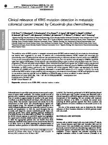
FIG. 2. Hypothetical sequence of cellular and
GaiicCncr|biochemical changes leading from H. pylon infection to gastric cancer (from [96], with permission).
particularly at risk [93-95]. In American men of Japanese ancestry in Hawaii, affected with gastric carcinoma, the odds ratio of having serum antibodies against H. pylon was 6.0 as compared to matched controls without gastric tumor [92]. Nevertheless, most human subjects infected with H. pylori will not develop gastric cancer, and there are even data indicating that the presence of a peptic ulcer protects patients infected with H. pylori from developing gastric carcinoma [91]. It is therefore evident that H. pylon is neither sufficient nor necessary for gastric carcinogenesis, and environmental factors and carcinogens are likely to be important co-factors [96] (Fig. 2). There is circumstantial evidence that long-standing H. pylon infection predisposes to formation of intestinal metaplasia. In a recent study including 533 subjects, intestinal metaplasia was found in 135 patients (25 percent) and H. pylon in 289 patients (54 percent) [55]. Intestinal metaplasia was encountered more often in H. pylon-positive patients as compared with H. pylon-negative subjects (34 percent versus 15 percent, p < 0.001). The prevalence of intestinal metaplasia and H. pylon was age-related, being more common in patients over 50 years as compared with patients under 50 years. The mean age of patients with intestinal metaplasia who were also H. pylon-positive was 64 years, whereas the mean age of patients with intestinal metaplasia who were H. pylon-negative was 72 years (p < 0.005). Screening for growth factor expression in gastric tissue may have a potential to detect patients at risk. Immunohistochemical studies revealed an increase in expression of growth factors close to the proliferative zone in inflamed antral mucosa, especially in H. pylon-positive patients [97]. Epidermal growth factor (EGF) may have an oncogenic potential in the stomach [98,99]. EGF receptors have been verified in a poorly differentiated gastric cancer cell line [100] and in surgically
634
HALTER ET AL.
resected gastric carcinomas [101]. In this context, it would be of interest to compare growth factor expression in different stages of intestinal metaplasia and to evaluate whether this comparison could serve as a screening method in high-risk patients. A still open question is why only a small fraction of subjects infected with H. pylon develop gastric cancer. It has been suggested that differences in the virulence of H. pylon strains might determine their carcinogenic potential [102]. A similar open question is why, in some individuals, the lymphoid infiltrates which accumulate in the gastric mucosa in response to H. pylon infection undergo malignant transformation and develop to malignant lymphoma [103].
OUTLOOK Progress on H. pylon is hampered by several shortcomings. Of particular importance are the lack of a cheap small animal model, the lack of methods for identifying H. pylon strains with different virulence, and the unavailability of a simple non-toxic therapeutic regimen for eradication of H. pylon. Current assay methods for H. pylon need to be improved. Progress is in sight with regard to new assays based on in situ hybridization [104] or on the polymerase chain reaction [105-107]. Once these molecular biology techniques are established as the gold standard, both the sensitivity and specificity of current assay methods may have to be revised. These new developments might also help to evaluate the existence of particularly virulent strains. From a clinical standpoint, the prospect of simpler and less toxic therapeutic measures aimed at eradication of H. pylon is of special importance. The promising results obtained with a treatment regimen combining proton pump inhibitors with a non-toxic antibiotic urgently need further confirmation and extension. A serological test able to detect seroconversion to normal shortly after eradication of H. pylon would greatly help to evaluate new treatment regimens. REFERENCES 1. Warren JR: Unidentified curved bacilli on gastric epithelium in active chronic gastritis. Lancet i:1273, 1983 2. Marshall B: Unidentified curved bacilli on gastric epithelium in active chronic gastritis. Lancet i:1273-1275, 1983 3. Blaser MJ: Epidemiology of Helicobacter pylori infection. In Helicobacter pylori, Gastritis and Peptic Ulcer. Edited by P Malferteiner, H Ditschuneit. Berlin, Germany, Springer-Verlag, 1990, pp 38-40 4. Dooley CP: Helicobacter pylori: Review of research findings. Aliment Pharmacol Therap 5 (Supplement 1):129-143, 1991 5. Graham DY: Campylobacter pylori and peptic ulcer disease. Gastroenterology 96:615-625, 1989 6. Correa P: Is gastric carcinoma an infectious disease? N Engl J Med 325:1170-1171, 1991 7. Baskerville A, Newell DG: Naturally occurring gastritis and C. pylori infection in the rhesus monkey: A potential model for gastritis in men. Gut 29:465-472, 1988 8. Mitchell IM, Bohane TD, Kerkowicz J: Antibody to campylobacter pylori in families of index children with gastrointestinal illness due to C. pylori. Lancet ii:681-682, 1987 9. Oderda G, Vaira D, Holton J, Ainley C, Smith A, Altare F, Boero M, Ansaldi N: Campylobacter pylori in families of children with peptic ulcer (Abstract). Gut 30:A1472, 1989 10. Rauws EAJ, Langenberg W, Houthoff HJ, Zanen HC, Tytgat GNJ: Campylobacter pyloridisassociated chronic active antral gastritis: A prospective study of its prevalence and the effects of antibacterial and antiulcer treatment. Gastroenterology 94:33-40, 1988 11. Conway EJ, Fitzgerald 0, McGeeney K, Geoghegan F: The location and origin of gastric urease. Gastroenterology 37:449-456, 1959
PATHOPHYSIOLOGY OF HELICOBACTER PYLORI
635
12. Triebling AT, Korsten MA, Dlugosz JW, Paronetto F, Lieber CS: Severity of Helicobacter-induced gastric injury correlates with gastric juice ammonia. Dig Dis Sci 36:1089-1096, 1991 13. Marshall BJ, Guerrant RJ, Plankey MW, Dye KR, Barrett L, Frierson HF, Hoffman SR, McCallum RW: Comparison of 14C-urea breath test, microbiology and histology for the diagnosis of Campylobacter pylori (Abstract). Gastroenterology 94:A370, 1988 14. Vaira D, Holton J, Cairns S, Polydoro A, Falzon M, Dowsett J, Salmon PR: Urease test for Campylobacter pylori: Care in interpretation. J Clin Path 41:812-813, 1988 15. Graham DY, Klein PD, Evans DG, Alpert IC, Opekun AR, Boutton TW: Campylobacter pyloridis detected noninvasively by the 13C-urea breath test. Lancet i:1174-1177, 1987 16. Dill S, Payne-James JJ, Misiewicz JJ, Grimble GK, McSwiggan D, Pathak K, Wood AJ, Scrimgeour CM, Rennie MJ: Evaluation of 13C-urea breath test in the detection of Helicobacter pylori and in monitoring the effect of tripotassium dicitratobismuthate in non-ulcer dyspepsia. Gut 31:1237-1241, 1990 17. Rauws EAJ, Tytgat GNJ, Royen EA, Langenberg W: C-14 urea breath test for the non-invasive detection of campylobacter pylori colonization (Abstract). Gastroenterology 94:A370, 1988 18. Megraud F: Comparison of different tests for Campylobacter pylori. Scand J Gastroenetrol 23 (Supplement 142):64-68, 1988 19. Evans DJ, Evans DG, Graham DY, Klein PD: Use of high molecular weight cell-associated protein antigens with urease activity in a highly specific ELISA, which distinguishes between Campylobacter pylori-infected and non-infected individuals. In Workshop Gastroduodenal Pathology and Campylobacter pylori. Edited by F Megraud, H Lamouliatte. Bordeaux, France, 1988, p 51 20. Newell DG, Bell GD, Weil J, Jones P, Grant P, Harrison G: The effect of treatment on circulating anti-Helicobacter pylori antibodies.-A two year follow-up study. In Helicobacter pylori, Gastritis and Peptic Ulcer. Edited by P Malferteiner, H Dischuneit. Berlin, Germany, Springer-Verlag, 1990, pp 172-175 21. Andersen LP, Holck S: Possible evidence of invasiveness of Helicobacter (Campylobacter) pylori. Europ J Clin Microbiol Infect Dis 9:135-138, 1990 22. Marshall BJ, Armstrong JA, McGechie DBN, Glancy RJ: Attempt to fulfil Koch's postulates for pyloric campylobacter. Med J Aust 142:436-439, 1985 23. Morris A, Nicholson G: Ingestion of Campylobacter pyloridis causes gastritis and raised fasting gastric pH. Am J Gastroenterol 82:192-199, 1987 24. Langenberg W, Rauws EAJ, Houthoff HJ, Oudbier JH, van Boehmen CG, Tytgat GNJ, Rietra PJGM: Follow-up of individuals with untreated campylobacter pylori-associated gastritis and of non-infected persons with non-ulcer dyspepsia. Infec Dis 157:1245-1248, 1988 25. Langenberg W, Rauws EAJ, Widjojokusumo A, Tytgat GNJ, Zanen HC: Identification of campylobacter pyloridis isolate by restriction endonuclease DNA analysis. Clin Microbiol 24:414-417, 1986 26. Humphreys H, Bourke S, Dooley C, McKenna D, Power B, Keane CT, Sweeney EC, O'Morain C: Effect of therapy on campylobacter pylori in peptic disease: A randomized prospective trial. Gut 29:279-283, 1988 27. Chittajallu RS, Ardill JES, McColl KEL: The degree of hypergastrinaemia induced by Helicobacter pylori is the same in duodenal ulcer patients and asymptomatic volunteers. Eur J Gastroenterol Hepatol 4:49-53, 1992 28. Chittajallu RS, Dorrian CA, Neithercut WD, Daghill S, McColl KEL: Is Helicobacter pylori associated hypergastrinaemia due to the bacterium's urease activity or the antral gastritis? Gut 32:1286-1290, 1991 29. Harume K, Sumii K, Okamoto S, Yosihara M, Tari A, Teixera CR, Takehara Y, Sumii M, Hou W, Kishimoto S, Kajiyama G: Helicobacer pylori infection causes low antral somatostatin content: Pathogenesis of inappropriate hypergastrinemia (Abstract). Gastroenterology 102:A80, 1992 30. Murty UK, Linscheer R, Co C: The hypergastrinemia in helicobacter pylori gastritis is due to a decrease in antral D-cell density and D:G cell ratio (Abstract). Gastroenterology 102:A 130, 1992 31. Moss SF, Legon S, Calam J: Helicobacter pylori infection decreases gastric somatostatin mRNA in duodenal ulcer patients. Gut 33 (Supplement 2):S27, 1992 32. Chittajallu RS, Dorrian CA, Ardill JES, McColl KEL: Effect of Helicobacter pylori on serum pepsinogen I and plasma gastrin in duodenal ulcer patients. Scand J Gastroenterol 27:20-24, 1992 33. Chittajallu RS, Harwood J, Dorrian CA, McColl KEL: Is Helicobacter pylori related hypergastrinaemia due to the bacterium inhibiting parietal cell function? (Abstract). Gut 31:1206, 1991 34. Eaton KA, Morgan DR, Krakowka S: Campylobacter pylori virulence factors in gnotobiotic piglets. Infect Immun 57:1119-1125, 1989
636
HALTER ET AL.
35. Mai UEH, Perez-Perez GI, Allen JB, Wahl SM, Blaser MJ, Smith PD: Surface proteins from Helicobacter pylori exhibit chemotactic activity for human leukocytes and are present in gastric mucosa. J Exp Med 175:517-525, 1992 36. Mooney C, Keenan J, Munster D, Wilson I, Allardyce R, Bagshaw 0, Chapman B, Chadwick V: Neutrophil activation by Helicobacter pylori. Gut 32:853-857, 1991 37. Mai UEH, Perez-Perez GI, Wahl LM, Wahl SM, Blaser MJ, Smith PD: Soluble surface proteins from Helicobacter pylori activate monocytes/macrophages by lipopolysaccharide-independent mechanism. J Clin Invest 87:894-900, 1991 38. Crabtree JE, Shallcross TM, Heatley RV, Wyatt JI: Mucosal tumour necrosis factor alpha and interleukin-6 in patients with Helicobacter pylori associated gastritis. Gut 32:1473-1477, 1991 39. Young GO, Stemmet N, Lastovica A, van den Merwe EL, Louws JA, Modlin IM, Marks IN: Helicobacter pylori lipopolysaccharide stimulates gastric mucosal pepsinogen secretion. Aliment Pharmacol Therap 6:169-179, 1992 40. Cave TR, Cave DR: Helicobacter pylori stimulates pepsin secretion from isolated rabbit gastric glands. Scand J Gastroenterol 26 (Supplement 181):9-14, 1991 41. Leunk RD, Johnson PT, David BC, Kraft WG, Morgan DR: Cytotoxic activity in broth-culture filtrates of Campylobacter pylori. J Med Microbiol 26:93-99, 1988 42. Figura N, Guglielmetti P, Rossolini A, et al: Cytotoxin production by Campylobacter pylori strains isolated from patients with peptic ulcers and from patients with chronic gastritis only. J Clin Microbiol 27:225-226, 1989 43. Lee A: Infectious causes of gastroduodenal inflammation in humans. Submitted for publication 44. Wright NA, Pike C, Elia G: Induction of a novel epidermal growth factor-secreting cell lineage by mucosal ulceration in human gastrointestinal stem cell. Nature 343:82-85, 1990 45. Wright NA, Pike CM, Elia G: Ulceration induces a novel epidermal growth factor-secreting cell lineage in human gastrointestinal mucosa. Digestion 46 (Supplement 2):125-133, 1990 46. Dreschner EE, Lipin M: Proliferation and differentiation of gastrointestinal cells in health and disease. In Gastrointestinal Tract Cancer. Edited by M Lipkin, RA Good. New York, Plenum Medical Book, 1978, pp 3-24 47. Hopwood D: A histometric analysis of gastric biopsies from patients treated with Gastritex: A new drug active against acute or chronic gastritis (Abstract). J Pathol 154:86a, 1988 48. Winawer SJ, Lipkin M: Cell proliferation kinetics in the gastrointestinal tract of man. IV. Cell renewal in the intestinalized gastric mucosa. JNCI 42:9-17, 1969 49. Ames BN, Gold LS: Too many rodent carcinogens: Mitogenesis increases mutagenesis. Science 249:970-971, 1990 50. Nakajima N, Kuwayama H, Tanaka N, Nakajima M, Eastwood GL: Campylobacter pylori filtrate inhibits epidermal growth factor-stimulated gastric epithelial proliferation in vitro (Abstract). Gastroenterology 98:94, 1990 51. Kawano S, Tsujii M, Fusamoto H, Sato N, Kamada T: Chronic effect of intragastric ammonia on gastric mucosal structures in rats. Dig Dis Sci 36:33-38, 1991 52. Negrini R, Lisato L, Zanella I, Cavazzini L, Gullini S, Villanacci V, Poiesi C, Albertini A, Ghielmi S: Helicobacter pylori infection induces antibodies cross-reacting with human gastric mucosa. Gastroenterology 101:437-445, 1991 53. Fujishima K, Misumi A, Akagi M: Histopathologic study on development and extension of atrophic change in the gastric mucosa. Gastroenterol Jap 19:9-17, 1984 54. Fry J: Peptic ulcer disease: A profile. Br Med J 2:809-812, 1964 55. Siurala M, Sipponen P, Kekki M: Chronic gastritis: Dynamic and clinical aspects. Scand J Gastroneterol 20 (Supplement 109);69-76, 1985 56. Craanen ME, Dekker W, Blok P, Ferwerda J, Tytgat GNJ: Intestinal metaplasia and H pylori: An endoscopic bioptic study of the gastric antrum. Gut 33:16-20, 1992 57. Wyatt JI, Dixon MF: Campylobacter-associated chronic gastritis. In Pathology Annual (Part 1). Edited by PP Rosen. New York, Year Book Publishers, 1990, p 75 58. Wyatt JI, Rathbone BJ, Dixon MF, Heatley RV: Campylobacter pyloridis and acid induced gastric metaplasia in the pathogenesis of duodenitis. J Clin Path 40:841-848, 1987 59. Wyatt JI, Rathbone BJ, Sobala GM: Gastric epithelium in the duodenum: Its association with Helicobacter pylori and inflammation. J Clin Path 43:981-986, 1990 60. Price AP: Histological aspects of campylobacter pylori colonization and infection of gastric and duodenal mucosa. Scand J Gastroenterol 23 (Supplement 142):21-24, 1988 61. Axon ATR: Helicobacter pylori therapy: Effect of peptic ulcer disease. J Gastroenterol Hepatol 6:131-137, 1991
PATHOPHYSIOLOGY OF HELICOBACTER PYLORI
637
62. Koelz HR, Bauerfeind P: Mucosal protecting agents: First choice in uncomplicated ulcer disease. Scand J Gastroenterol 23 (Supplement 153):71-80, 1988 63. Lee Fl, Samloff IM, Hardmann M: Comparison of tri-potassium dicitrato bismuthate tablets with ranitidine in healing and relapse of duodenal ulcers. Lancet i:1299-1302, 1985 64. Logan RPH, Polson RJ, Baron JH, Misiewicz JJ: Follow-up after anti-Helicobacter pylori treatment. Lancet 337:562-563, 1991 65. Coghlan JG, Humphries H, Dooley C, Keane C, Gilligan D, McKenna D, Sweeney E, O'Morain C: Campylobacter pylori and recurrence of duodenal ulcers-a 12-month follow-up study. Lancet ii:1109-1111, 1987 66. Lambert JR, Borromeo M, Korman MG, Hansky J, Eaves ER: Effect of colloidal bismuth (De-Nol) on healing and relapse of duodenal ulcers-role of Campylobacter pyloridis (Abstract). Gastroenterology 92:1489, 1987 67. Marshall BJ, Warren JR, Blincow ED, Phillips M, Goodwin CS, Murray R, Blackbourn SJ, Waters TE, Sanderson CR: Prospective double-blind trial of duodenal ulcer relapse after eradication of Campylobacter pylori. Lancet ii:1437-1442, 1988 68. Smith AC, Price AB, Borriello P, Levi AJ: A comparison of ranitidine and tripotassium dicitratobismuth (TDB) in relapse rates of duodenal ulcer. The role of Campylobacter pylori (CP) (Abstract). Gastroenterology 94:431, 1988 69. Borody T, Cole P, Noonan S, Morgan A, Ossip G, Maysey J, Brandl S: Long-term campylobacter pylori recurrence post-eradication (Abstract). Gastroenterology 94:43, 1988 70. Borody TJ, Cole P, Noonan S, Morgan A, Lenne J, Hyland L, Brandl S, Borody EG, George LL: Recurrence of duodenal ulcer and Campylobacter pylori infection after eradication. Med J Aust 151:431-435, 1989 71. Rauws EAJ, Tytgat GNJ: Cure of duodenal ulcer associated with eradication of Helicobacter pylori. Lancet i:1233-1235, 1990 72. Blum AL, Armstrong D, Dammann H, Fischer M, Greiner L, Haase W, Hogeboom-Verdegal A, Liszkay M, Stolte M, Sulser H, Simon B, and the Talcicamp Study Group: The effect of Helicobacter pylori on the healing and relapse of duodenal ulcer (Abstract). Gastroenterology 98:22, 1990 73. Logan RPH, Gummett PA, Misiewicz JJ, Karim QN, Walker MM, Baron JH: One week eradication regimen for Helicobacter pylori. Lancet 338:1249-1252, 1991 74. Unge P, Gad H, Gnarpe H, Olsson J: Does omeprazole improve antimicrobial therapy directed towards gastric Campylobacter pylori in patients with antral gastritis? Scand J Gastroenterol 24 (Supplement 167):49-54, 1989 75. De Koster E, Burette A, Nyst JF, Glupczynski Y, Deprez C, Jonas C, Otero J, De Reuck M, Deltenre M: HP treatment: Bismuth, omeprazole, antibiotics (Abstract). Gastroenterology 100:52, 1991 76. Lamouliatte H, Bernard PH, Boulard A, Megraud F, De Mascarel A, Quinton A: Controlled study of omeprazole-amoxicillin-tinidazole vs ranitidine-amoxicillin-tinidazole in Helicobacter pylori associated duodenal ulcers (Abstract). Gastroenterology 100:104, 1991 77. Bell GD, Powell K, Weil J, Burridge SM, Morden A, Harrison G, Gant PW, Jones PH, Trowell JE: Experience with omeprazole in combination with either amoxycillin or colloidal bismuth subcitrate in patients with metronidazole-resistant Helicobacter pylori. Eur J Gastroenterol Hepatol 3:923926, 1991 78. Labenz J, Gyenes E, Ruhl GH, Borsch G: Amoxycillin-omeprazole treatment for eradication of Helicobacter pylori. Eur J Gastroenterol Hepatol 3 (Supplement 1):10, 1991 79. Bayesdorffes E, Mannes GA, Sommer A, Hochter W, Weingart J, Hatz R, Lehn N, Ruckdeschel G, Dirschedl P, Stolte M: High omeprazole treatment combined with amoxicillin eradicates Helicobacter pylori. Eur J Gastroenterol Hepatol 4:697-702, 1992 80. Villako K, Ihamaki T, Tamm A, Tammur R: Upper abdominal complaints and gastritis. Ann Clin Res 16:192-194, 1984 81. Cheli R, Perasso A, Giacosa A: Dyspepsia and chronic gastritis. Hepato-Gastroenterology 30:21-23, 1983 82. Tytgat GNJ, Noach LA, Rauws EAJ: Is gastroduodenitis a cause of chronic dyspepsia? Scand J Gastroenterol 26 (Supplement 182):33-39, 1991 83. Marshall BJ, Warren JR: Unidentified curved bacilli in the stomach of patients with gastritis and peptic ulceration. Lancet i: 1311-1315, 1984 84. Rokkas T, Pursey C, Uzoechina E, Dorrington L, Simmons NA, Filipe MI: Campylobacter pylori and non-ulcer dyspepsia. Am J Gastroenterol 82:1149-1152, 1987 85. Rokkas T, Pursey C, Simmons NA, Filipe MI, Sladen GE: Non-ulcer dyspepsia and colloidal bismuth subcitrate therapy: The role of Campylobacter pyloridis (Abstract). Gastroenterology 92:1599, 1987
638
HALTER ET AL.
86. Tucci A, Tosetti C, Stanghellini V: Helicobacter pylori infection and delayed gastric emptying of solids identify two subsets of patients with chronic idiopathic dyspepsia. Rev Esp Enferm Apar Dig 78 (Supplement 1):65-70, 1990 87. Loffeld RJLF, Potters HVPJ, Stobberingh E, Flendrig JA, Van Spreeuwel JP, Arends JW: Campylobacter associated gastritis in patients with non-ulcer dyspepsia: A double blind placebo controlled trial with colloidal bismuth subcitrate. Gut 30:1206-1212, 1989 88. Borsch G, Wegener M, Schmidt G, Sandmann M, Adamek R, Reitemeyer E: Prospective analysis of clinical and histologic factors associated with Campylobacter pylori colonization (Abstract). Gastroenterology 94:44, 1988 89. Jeena CP, Simjee AE, Pettengell KE, Spitaels JM, Naran AD, Miller NM, Manion GL: Comparisons of symptoms in Campylobacter pylori positive and negative patients presenting with dyspepsia for upper gastrointestinal endoscopy (Abstract). S Afr Med J 73:659, 1988 90. Forman D, Newell DG, Fullerton F, Yarnell JWG, Stacey AR, Wald N, Sitas F: Association between infection with Helicobacter pylori and risk of gastric cancer: Evidence from a prospective investigation. Br Med J 302:1302-1305, 1991 91. Parsonnet J, Friedman GD, Vandersteen DP, Chang Y, Vogelman JH, Orentreich N, Sibley RK: Helicobacter pylori infection and the risk of gastric carcinoma. N Engl J Med 325:1127-1131, 1991 92. Nomura A, Stemmerman GN, Chyou P-H, Kato I, Perez-Perez GI, Blaser MJ: Helicobacter pylori infection and gastric carcinoma among Japanese Americans in Hawaii. N Engl J Med 325:1132-1136, 1991 93. Fox JG, Correa P, Taylor NS, Zavala D, Fontham E, Janney F, Rodriguez E, Hunter F, Diavolitsis S: Campylobacter pylori-associated gastritis and immune response in a population at increased risk of gastric carcinoma. Am J Gastroenterol 84:775-781, 1989 94. The Gastrointestinal Physiology Working Group: Helicobacter pylori and gastritis in Peruvian patients: Relationship to socioeconomic level, age and sex. Am J Gastroenterol 85:819-823, 1990 95. Perez-Perez GI, Taylor DN, Bodhidatta L, Wongsrichanalai J, Baze WB, Dunn BE, Echeverria PD, Blaser MJ: Seroprevalence of Helicobacter pylori infections in Thailand. J Infect Dis 161:1237-1241, 1990 96. O'Connor HJ: Helicobacter pylori and gastric cancer: A review and hypothesis. Eur J Gastroenterol Hepatol 4:103-109, 1992 97. Jankowski J, Hopwood D, Wormsley KG: Flow cytometric analysis of growth regulatory peptides and their receptors in Barrett's oesophagus and oesophageal adenocarcinoma. Scand J Gastroenterol, in press 98. Tahara E, Yokozaki H: Growth factors as biological markers of malignancy. Gan To Kagaku Ryoho 15:1102-1108, 1988 99. Yasui W, Hata J, Yokozaki H, Nakatani H, Ochiai A, Ito H, Tahara E: Interaction between epidermal growth factor and its receptor in progression of human gastric carcinoma. Int J Cancer 41:211-217, 1988 100. Fukuyama R, Minoshima S, Ochiai A, Tahara E, Shimizu N: Flow cytometric analysis of the expression of 9A3 antigen, E-cadherin and EGF receptor in TMK-1 stomach cancer cells. Int J Cancer 48:81-84, 1991 101. Hirose K, Arai M, Nakagawara G: Expression of human epidermal growth factor and DNA ploidy pattern in gastric carcinoma. Nippon Geka Gakkai Zasshi 92:122-126, 1991 102. Burnie J, Lee W, McNulty C, Dent J: Virulence of Campylobacter strains and degree of gastritis. Lancet i:302, 1988 103. Wotherspoon AC, Oritz-Hidalgo C, Falzon MR, Isaacson PG: Helicobacter pylori-associated gastritis and primary B-cell gastric lymphoma. Lancet 338:1175-1176, 1991 104. Wetherall BL, McDonalds PJ, Johnson AM: Detection of Campylobacter pylori DNA by hybridization with non-radioactive probes in comparison with a 32P labelled probe. J Med Microbiol 26:257-263, 1988 105. Ho DS, Lewis FA, Wyatt JI, Dixon MF: Helicobacter pylori detection by PCR of the gene encoding 16S ribosomal RNA in fresh paraffin embedded material (Abstract). J Pathol 161:351, 1990 106. Valentine JL, Arthur RR, Mobley HL, Dick JD: Detection of Helicobacter pylori by using the polymerase chain reaction. J Clin Microbiol 29:689-695, 1991 107. Clayton C, Kleanthous K, Tabaqchali S: Detection and identification of Helicobacter pylori by the polymerase chain reaction. J Clin Pathol 44:515-516, 1991