base of observations to build our theo- ries on. ..... LITERATURE. Aikens, R.S., Agard, D.A., and Sedat, J.W.: .... Vardi, E. and Grover, N.B.: Aggregation of.
GRONINGEN REDUCTION OF IMAGE DATA: A MICROBIOLOGICAL IMAGE PROCESSING SYSTEM WITH APPLICATIONS IN IMMUNOFLUORESCENCE AND MORPHOMETRY MICHAEL H.F. WILKINSON, GIJSBERT J. JANSEN, and DIRK VAN DER WAAIJ Laboratory for Medical Microbiology, University of Groningen, Groningen, The Netherlands SUMMARY The interaction between the intestinal flora and the immune system is complex, and as yet little understood. The Groningen Reduction of Image Data (GRID) image processing system is a relatively new tool in the investigation of this interaction. The image processing approach allows measurement of morphological and immunological characteristics of faecal bacteria, which have not been cultured, and should therefore represent the flora in the intestinal lumen well. In this review, the main application programs of GRID in the field of bacterial morphology and (immuno-)fluorescence detection are presented. Its low cost hardware set-up, based on ordinary personal computers is described. Examples of the research done and data acquired with the system are given. Future plans include multi-colour fluorescence measurement. The system allows rapid quantification of morphology and immunofluorescence, and can combine both types of data in "fluoromorphometry": quantifying patterns of fluorescence as a function of shape. These patterns could lead to new insights into the interaction between intestinal flora and immune system, though the interpretation is as yet not simple. INTRODUCTION It has been pointed out by numerous authors (e.g. Taylor and Heimer, 1975; Hiraoka et al., 1987; Aikens et al., 1989; Tanke, 1989), that one of the major problems when viewing microscopic slides of any kind is the lack of quantitative methods of describing what is seen. Without such quantitative information, it becomes very difficult to compare results obtained by different observers, even when viewing the same slide, on the same microscope. Things invariably become worse when different
microscopes and different slides are used. One particular area where quantitative measurements are useful is in immunofluorescence. Usually the titre of a serum is judged visually from immunofluorescence slides. The observer must estimate which of a series of wells, containing a dilution sequence of serum, has a positive fluorescence. The highest dilution still judged to be "positive" is designated the titre. Each observer will tend to use his or her own criteria when estimating the level of
117
fluorescence, and these criteria will not even remain constant with one observer. The adaptation of the eye to darkness, eye-fatigue, changes in ambient light conditions, and many other factors will tend to change a human observer's idea of what is positive or negative fluorescence. It is difficult to estimate the number or percentage of bacteria, which are positive, and impossible to distinguish autofluorescence from immunofluorescence. It has been shown by Apperloo-Renkema et al. (1990) that with a single observer there may be as much as a factor of four difference on average between duplicate measurements (i.e. 2 titre steps on a 2 log dilution sequence). It was also shown that a computerised system was at least twice as accurate. When estimating the morphological characteristics in a similar subjective manner, the classification problems are equally bad. Distinguishing "coccoid rods", from "slightly rod-like cocci" is just one example of a distinction that is impossible to make reliably and reproducibly with the naked eye (Bacquero et al., 1988). Visual assessments do of course have a solid place within microbiology, and the fact that they can be useful is not contested. Their main drawback is the unknown, but in any case large, amount of noise, or random error, inherent in such a subjective approach. The main advantage of automating the assessment of microscopic slides is the acquisition of quantitative data, which should be more accurate, for which error margins can be properly estimated, and which can be reproduced elsewhere. We can conclude that, though it is by no means useless to make visual assessments of bacteriological (and other) slides, deriving quantitative data from such slides
118
can provide a much more solid scientific base of observations to build our theories on. Many others have pointed this out (for a review see Tanke, 1989), and a lot has been done in the field of image processing of slides of eukaryote cells and tissues (e.g. Gross and Loew, 1989; Swanson, 1989; Lamaziere et al. 1993), but only recently has much work been done on the (smaller) prokaryotes, and much of that either using electron micrographs (e.g. Vardi and Grover, 1992), or bacterial colonies or cultures (e.g. Waterhouse et al., 1993) or limit the image analysis to a simple counting (Singh et al., 1989; Evans-Hurrell et al., 1993). Many of these image processing efforts are not very much automated, as in the case of Vardi and Grover (1992), who measured bacterial lengths by pointing at start and end point on the screen (for thousands of bacteria), or Evans-Hurrell et al. (1993) who counted the bacteria manually, from the video screen. This situation lead to the design of the GRID image processing system. It was designed first of all to measure bacterial shapes from ordinary (light) microscopic slides quantitatively (Meijer et al., 1990). This found applications in the assessment of changes in the gut flora, both in healthy volunteers and during antibiotic treatment (Meijer et al., 1991a, 1991b). Extensions to the system, for use in immunofluorescence work, have been made by ApperlooRenkema et al. (1990), and this work is being continued by the authors. In this paper, the set-up of the GRID system, its hardware, its software, its design goals and its applications, will be reviewed.
THE GRID SYSTEM Hardware When the GRID system was first envisaged in about 1983, most image processing was done on large computers (mini-computers and upwards, e.g. PDP 11-70 for the GIPSY system [Allen and Terlouw, 1981]). At that time, personal computers were generally thought of as completely inadequate for the computing speed and data storage requirements of image processing. Partly because of lack of funds, the GRID system was first conceived on an Apple II compatible computer (Unicom), with a video capture board, which could only distinguish two shades of grey (black and white), and an expansion board with a Motorola 68000 processor. The system ran under the CP/M68K operating system. On this minimal hardware set-up, all the basic functions of morphology were first implemented and tested. Much of this code is still used in our bacterial morphology measurements today. As faster platforms became more and more affordable this first system was abandoned and we moved to IBM PC-AT compatible machines. It is a fairly recent development that the most powerful IBM PCAT compatible computers are regularly being used for image processing (e.g. Groen et al., 1988, Froehling, 1990, Hewison et al., 1993, Lamaziere et al., 1993). At this moment 5 different computer systems are used to run GRID software in our laboratory, and a sixth has recently been installed at the Department of Dermatology, University Hospital, Groningen. Though the system could be ported to many different computer environments, our hardware consists of a IBM PC-AT compatible computers based on Intel 80286, 80386 or 80486 main processors, preferably supported by a mathematical co-processor, run-
ning Microsoft MS-DOS versions 3.30 and upwards (2.0 should work as well). The memory of the smallest (and oldest system) is 640 kilobytes (kB) but 2 megabytes (MB) is recommended as a minimum. Hard disk in our computers range from a mere 20 MB to 120 MB. Considering the data bulk gathered on the faster systems each day (60 MB of raw data!), more is better, when it comes to disk sizes. A tape-streamer is incorporated in two systems, as backup and permanent mass storage device. The key element in each computer system is an expansion board, which allows the computer to accept input from standard video cameras. Two different types of these so-called frame-grabbers are used in our system. The earlier type is the PIP-1024(A or B) frame grabber (MATROX Ltd., Dorval, Quebec, Canada). It can accept either European or American black and white video signals, and can hold up to 4 images with a resolution of 512 by 512 pixels and 256 grey levels. This board does not perform many extra image processing functions itself. The main burden of the image processing falls on the main processor of the computer. The second type is the MVP-AT board (also by MATROX). This can do everything the PIP-1024 can, but has powerful image processing functions implemented in the hardware of the board. This greatly enhances the performance of the image processing functions in the system. As an example, subtracting two images from each other takes 4 to 7 seconds on the PIP-1024 systems, but only 1/30th of a second on a MVP-AT. The MVPAT also supports full colour (RGB) video signals, at a resolution of 512 by 512 pixels and 16.7 million colours per pixel. Quite obviously, the latter board is the preferred one. The cameras connected to the systems are almost exclu-
119
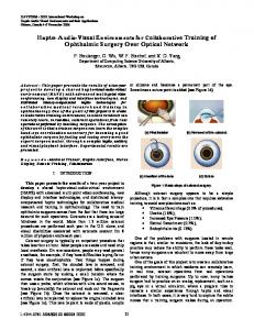
Figure 1: Hardware set-up of the GRID system. The two expansion boards shown are placed inside the personal computer. The video camera is normally attached to a microscope.
sively Loral Fairchild CCD-5000/1 cameras (Loral Fairchild, Sunnyvale, CA, USA). This is an industrial charge coupled device (CCD) camera, with the CCD chip cooled to 20 K below ambient temperature by a Peltier element. These cameras have a special purpose connector (user I/O connector) which allows access to the camera's video timing circuitry. A special expansion board designed by M.H.F. Wilkinson (Wilkinson et al., 1992) is connected to this connector, and allows software exposure time control. The exposure time of the camera can be increased from a single video frame (=33 ms) to any integer number of frames. This feature is used extensively in faint light detection (e.g. fluorescence). Each system has two video monitors: one is connected to the standard video output of the computer and is used for the normal dialogue, and one is connected to the frame grabber, to display the current video 120
image, the progress of the image processing, etc. Each video camera is connected to a (fluorescence) microscope. Several types of microscopes are in use: a Leitz Orthoplan, two Olympus BH2s and a Zeiss fluorescence microscope. The Leitz Orthoplan microscope has electronically controlled shutters in front of both standard and ultra-violet light sources. These can be software controlled using an extension of the exposure control board. A diagram of the hardware is shown in Figure 1. Software General design considerations Many image processing packages, such as GIPSY and AIPS in astronomy (Allen and Terlouw, 1981), and a plethora of commercial packages (e.g. Groen et al., 1988), require quite a bit of specific image processing knowledge from the end user. Usually, they are menu driven, or use an interpreter of
some kind to access image processing functions. Though these packages are extremely flexible and easy to use for image processing and computer specialists, they leave much to be desired for medics, biologists, and many other scientist who frequently use image processing systems. Most of these systems do allow the user to store a sequence of commands as a "macro", so it can be played back using a single command, and other systems are fully programmable, but the knowledge needed to customise the systems this way is often underestimated. It was decided that GRID should be designed as a set of programs, each capable of performing a fairly limited set of tasks, capable of running with as little user involvement as possible, and asking only those questions which all those not fully versed in the inner workings of image processing could make sense of. Making each program perform a limited set of tasks greatly increases user friendliness, by not offering too many options at once, at the expense of some flexibility. Vital in this approach was the continuous involvement of one or more programmers, with experience in image processing. The task of these programmers was to analyse the requirements of the microbiologists, and then to write programs that perform precisely the tasks required. In our view, it is a tribute to this design philosophy, that many scientists and students with little or no previous experience with computers, have collected and processed many thousands of images on our systems. Usually they could work with the system and get their data on paper within a few days. Apart from user-friendliness, the system should be easily portable to other platforms, as hardware obsolescence proceeds at an exponential rate. Therefore, the software has been written in a layered fashion, the bottom layer
shielding the hardware specific functions from the top. In this way only a relatively small set of procedures need be rewritten when moving to different hardware. The last port of the system to different hardware took only about three weeks, for a single programmer. To ensure software durability, documentation of the inner workings is essential, and guidelines for documentation have been drawn up, though when writing quick patches for problems they are not always adhered to. This is often a problem in a single programmer environment. Too often, the short-term operation of the system takes precedence over long-term objectives, and writing documentation gets shifted to a low priority. Application programs As has been described, the GRID system consists of a number of programs, which perform specific image processing tasks. These programs fall into three broad categories. The first category consists of interactive image acquisition programs. Each such program is geared to a specific type of experiment. They are highly automated, and do not require much (if any) knowledge of image processing to operate them. The oldest, simply dubbed RECORD, acquires grey-scale images of bacterial slides, stained in a variety of ways, segments them automatically into objects (bacteria) and background, and stores the resulting binary images for further analysis. Alternatively, it can store grey-level information as well to allow analysis programs to measure the density of the stain in each object. The automated detection of object boundaries is the key feature of the program, which allows objective measurement of the morphology of the bacteria. The method, due to Kittler et al. (1985), uses the gradients in the image to determine which grey levels lie precisely
121
Figure 2: Nigrosin counterstained bacteria acquired by GRID system program MEDEYES. Background noise has been subtracted. Neither background nor bacteria have a uniform brightness (A). Gradient of image "A", showing edges of bacteria. Note that certain edges are much brighter than others, indicating differences in the brightness of the bacteria (B). Image "A" segmented into objects (white) and background (black), using local thresholds determined from image "A" and "B". Note that bacteria of different brightness have all been segmented properly (C).
on object boundaries in each region of the image. Pixels darker than these local thresholds (or lighter, depending on the stain) are considered part of an object, others are part of the background. Figure 2 shows the grey scale image, gradient image, and resulting binary image, with white representing objects, and black background. A somewhat more complicated program, called PHASECON, geared to phase contrast combined with immunofluorescence acquisition also exists. It allows the acquisition of phase contrast and immunofluorescence images of a single field of view, at user selectable exposure times. The phase contrast image is segmented into objects 122
and background in the same way as in RECORD. Only parts of the fluorescence image which coincide with an object in the phase contrast image, or lie within a certain distance from an object, are stored for analysis. A second group of programs is rather simpler. They perform analysis on the images acquired by the first set of programs in a batch-oriented way. All the user must do is specify which image files must be analysed, and the programs will analyse the entire batch. This analysis is generally carried out at night. A typical system can routinely analyse 40,000 to 60,000 bacteria per day. The fastest system has done more than 100,000 in a single batch at night.
Figure 3: Modified scatterplot (Bart-O-Gram) of surface area vs. perimeter of nigrosin counterstained faecal bacteria. An approximately quadratic relationship between the two parameters is visible.
The analysis can be broken down into three phases. In the first phase, simple morphological parameters such as width, surface area, and perimeter are measured, with or without fluorescence brightness or stain density. In second phase the raw morphological data are converted to three more or less independent shape factors dubbed F1, F2 and F3, or principle components. This
method was pioneered by Meijer et al. (1990) in our laboratory. It has been shown that these shape factors, which are weighted averages of the logarithms of the raw data, explain more than 99% of the variability of bacterial shapes within human faecal flora, and are not highly correlated, as the raw data are (Meijer et al., 1991b). The difference is clearly shown in Figures 3 and 4,
Figure 4: Bart-O-Gram of optimum shape factors F1 vs. F2. Interdependence has clearly decreased. Not all bacteria are plotted to prevent cluttering of image. 123
Figure 5: Surface plot of number of bacteria as a function of both F1 and F2. Omission of bacterial shapes allows more quantitative information to be shown (A). Contour plot showing number of bacteria as a function of F1 and F2. Contours represent isodensity lines at 2log intervals (B).
which show plots of perimeter vs. surface area, and F1 vs. F2. In the third phase of analysis, the processed morphological data are used to compute statistics of either the distribution of the number of bacteria or their fluorescence as a function of the shape factors. This allows quick assessment of changes in the bacterial population as a whole, either purely morphological, or in fluo-
124
rescence distribution. Surface and contour plots of both number of bacteria and mean fluorescence as a function of F1 and F2 are shown in Figures 5 and 6. The last category currently contains just one program: DATAPLOT. It is an interactive graphical data representation program, specially adapted to our image processing needs. Though our pro-
Figure 6: Surface plot of mean fluorescence (in arbitrary units) as a function of both F1 and F2 (A). Contour plot showing mean fluorescence (in arbitrary units) as a function of F1 and F2. Contours show iso-fluorescence lines at 1/2 2log intervals (B).
grams generate files which could be read by any graphical package available commercially, the number of display options found in these packages was found inadequate. DATAPLOT allows users to create a multitude of different graphs, displaying the relationships between and distributions of any two or three parameters in the data set. The
simplest form of plot is the scatter plot, which can be made by most graphical packages. A modified version of these plots is also available in DATAPLOT. Instead of printing a star or other symbol at the appropriate location in the graph, the actual bacterial shapes are plotted. This type of graph (dubbed Bart-O-Gram, after the inventor Bart
125
Meijer [1990]) is shown in Figures 3 and 4. DATAPLOT can also compute and plot the distributions as shown in Figures 5 and 6, optionally adding a Bart-O-Gram as an overlay, to get an idea of the bacterial shapes. The GRID system contains one more program: MEDEYES (Meijer, 1991c). It is radically different from all others in the system in that it does require image processing knowledge to utilise it to its full capability. It is an easily expandable interpreter, which is used mainly by the programmers of the system to experiment with new image processing procedures. It uses a Pascal-like language with full-blown flow control with IFTHEN-ELSE constructs, REPEATUNTIL, WHILE-DO and FOR loops. Any new image processing function or procedure can be added to the program, and can be tested thoroughly, in a variety of conditions, without the need for a special test program. MEDEYES is also useful when one simply wants to take a couple of snapshots of a slide, and wants to see what various image processing functions do to it. All programs in the system have been developed in our lab, using mainly Microsoft Pascal 4.0, C 5.1, and occasionally assembly language (Microsoft Macro Assembler) as programming languages. The frame grabber functions are accessed through libraries supplied by the manufacturer (MATROX). To improve portability, these functions are never called directly, but are reached through an interface library. Two interface libraries exist at this moment: One for the MVP-AT and one for the PIP1024. These interface libraries shield the main program modules from differences between the frame grabber boards, allowing easy upgrading of each PIP1024 to MVP-AT systems. Frame grabbers supplied by other manufacturers could also be linked to the GRID system in this way.
126
The user interface All interactive programs, except for MEDEYES, have the same user interface. This improves the user friendliness of the system, since all programs have the same "look-and-feel". The user interface, dubbed SCREENIO, is basically keyword driven, and only slightly more sophisticated than the glass teletype interface supported by standard Pascal and C. It is, however, far more robust than the standard interface of these languages, and it provides a standard screen layout. The robustness of the interface stems from the fact that the input of the user is considered to be a simple stream of characters, which is parsed for meaningful information. If the string is not considered meaningful to the program, it alerts the user to this with an error message, and re-states the question. By contrast, if the Pascal READLN statement were to be used to get a number from the user, and the user entered a character other than a digit, the program would crash immediately, and the user would, justifiably, be upset. Whenever possible, SCREENIO offers default values, which are chosen when the user only presses "enter". These values are chosen in such a way that the user can almost always just press "enter" to proceed. SCREENIO also supports menus, function keys, and special commands, and "hidden" questions. Hidden questions are questions that are never put on the screen. They are more or less internal (or advanced) features of programs, the settings of which should not be altered by inexperienced users, but which should be accessible by certain experienced users. The screen layout is fairly basic. The screen is divided into five areas. At the top of the screen there is the INFO area, a sort of status bar. It is a single line of information on the general status of the program. The second and largest area
by far, is the display area, which is used by all programs to display any kind of data or instructions to the user. Below this is the user-type-in (UTI) area. This is the area in which all questions appear, and in which the user types his answer. Error messages appear below this in a two-line error message area. At the very bottom of the display there is a function key display. In the most recent additions to the programs some window-oriented features have been added, to improve user friendliness even further. These windows are simple forms, which pop up on the display, and let the user edit the value of each item. Future extensions of the system are bound to be even more window oriented, as users place higher demands on the ease of use of programs, but for relatively simple programs such as RECORD and PHASECON, the simple SCREENIO system is quite sufficient. Applications and results The GRID-system has been operational as a research tool for about 3-4 years now, and a growing number of publications using results obtained by it have been produced. During the first stage of its use, the system had to be validated, and its strengths and weaknesses evaluated. Meijer et al. (1990) first showed that the system could distinguish pure cultures of bacteria purely on the basis of the morphological distribution of the constituent bacteria. Single bacteria could not be assigned to a particular species on the basis of shape. It was also shown (Meijer et al., 1991b) that subtle changes in time in morphological composition of the faecal flora healthy volunteers could be detected by the system. These same fluctuations pose a lower limit to the magnitude of changes in morphology as a result of treatment or disease that can be detected. Later the system was compared to
classical microbiological methods, in a study of the effect of ceftriaxone on the intestinal flora (Meijer et al. 1991a; de Vries-Hospers et al., 1991). The results of the image processing were obtained much faster than anaerobic culturing, and correlated well with the other methods. At this time the statistical processing (phase 3 of the analysis) was standardised for the morphometric part of the GRID system. In 1989 the fluorescence package (PHASECON) became operational, and it too had to be validated first. It was first shown by Apperloo-Renkema et al. (1991) that the system could measure titres of circulating antibodies directed against Enterobacteriaceae with greater accuracy than the human observer. Later this computerised immunofluorescence technique was combined with the morphometry (Apperloo-Renkema et al. 1992), assessing antibody titres as a function of shape in healthy volunteers (Apperloo-Renkema et al. 1990a, 1990b). Though the interpretation of the results was (and is) not straightforward, the system proved to be a good tool in investigating the interaction between immune system and intestinal flora, and allowed rapid monitoring of changes in that interaction. Both hardware and software have since been improved, and each improvement had to be validated in its turn. The addition of exposure control has boosted the sensitivity dramatically (more than a hundred fold) (Wilkinson et al., 1992). It has been shown that the accuracy of titre measurement has been improved (Jansen et al., 1993a), and titres were found to be reproducible to within 10% on a linear scale. This corresponds to an accuracy of better than 0.15 titre step on a 2log dilution sequence. Impressive though this may seem, a fluorescence activated cell sorter (FACS) could probably do the same, and faster. The addition of mor-
127
phological information is in fact the most interesting option offered by this system, and this is the field we are currently working on. As ever, the first
stage of such work is the validation of the system on healthy volunteers, and this has recently been done (Jansen et al., 1993b).
DISCUSSION The current status of GRID Image processing has proved a useful technique in many fields of science, and the GRID system is just one of many image processing systems allowing scientists to quantify what they see. The main difference with many other systems is that it has been designed for use by medics and biologists, not computer scientists. This 'push-button', rather than menu oriented, approach means that the scientists can work with programs tailor made for specific problems. They can concentrate on biological or medical issues, not computer science. The disadvantage of our approach is that it takes rather longer to get started in the first place. A commercially available package can simply be ordered, plugged in, and run. Adapting the existing system to new problems requires more work too, though we are currently streamlining our software to cope with this. The modular approach we (and many others) use means that we now have a large library of ready-for-use building blocks, that can be assembled in a variety of ways to produce different programs. The major problem in this approach is the need for a certain critical mass of programmers and other computer scientists to keep the system viable in the long run. Until now, the development has largely been a single programmer effort. In practice this means that it is difficult to keep up with new developments, and keep the software well documented and readable to others. Each programmer has his own style of programming, and what seems perfectly simple and readable code to
128
one programmer, who has grown into a specific field of work, may be completely unclear to others. To alleviate this problem we have now contacted the department of computing science of the University of Groningen, to assist us in our work. Scientific results and prospects GRID has been used successfully as a tool in the research of the intestinal flora, both purely morphologically (Meijer et al., 1990, 1991a, 1991b), and in its interaction with the immune system (Apperloo-Renkema, 1991, 1990a, 1990b). It has been shown that shifts in the morphological distribution of bacteria in a population can be correlated with classical microbiological measurements. Others (Bacquero et al., 1988) have argued that the morphological diversity of bacteria is a measure of the biological diversity and therefore the health of the ecosystem. This morphological diversity has been quantified using the GRID system (Meijer, 1991a, 1991b) in a fast (within one day), reliable, and reproducible way. What morphometry cannot do is identify bacterial species. Immunofluorescence measurement using this system is both faster and more accurate than naked eye observation, with an added advantage that the measurement need not be done in a dark room. Using the system purely for immunofluorescence does not utilise the system fully. Though more expensive, a FACS is probably a better tool, if fluorescence information is all that is required.
Adding (immuno-)fluorescence to the morphological technique is probably the most powerful option of our system. The interpretation of the results does still pose something of a problem, but inter-individual differences, or shifts in time, in the interaction between immune system and gut flora can be detected and quantified. Future developments At this moment we are working on multicolour fluorescence measurement, allowing such measurements as simultaneous IgA, IgG and IgM measurement on bacteria. Aside from immunofluorescence, any other fluorescent probe or monoclonal antibody
could be used in this technique. This, and the simultaneous measurement of Ig and any combination of probes could open up new areas of research, though the interpretation of the data would certainly require some thought. Work is also in progress on the file and project management. In future it should be possible to compile image databases, which the scientists could browse through at will. All data pertaining to slide, patient, and experiment should be available to the scientist immediately. Such sets of data and images could also allow computer programs to perform systematic searches for those features, which most accurately distinguish between different diseases.
CONCLUDING REMARKS Using the GRID system, we have started to measure a number of interesting effects, which could probably not have been detected in any other way. As ever, new data pose at least as many questions as they answer, so more work, both on the theoretical and experimental side, is needed to interpret what is seen. Whatever the problems in the interpretation, image processing has greatly increased the value of our microscopes. GRID is an effective research tool,
but as with many tools it must be honed to proper sharpness to maximise its usefulness. A system developed in a laboratory requires on-site knowledge of computer programming techniques to keep it operational. A single programmer is not enough in the long run: Three or four computer experts is probably a minimum "critical mass" necessary. This is not always affordable. To ensure continuity, it is best to collaborate with a department of computing science, and such collaborations can be very fruitful.
ACKNOWLEDGEMENTS We would like to thank the Institute for Microecology in Herborn (Germany), for their financial support and encouragement of our research, throughout the development of GRID, and B. Deddens for the use of his data in Figures 5 and 6. LITERATURE Aikens, R.S., Agard, D.A., and Sedat, J.W.: Solid-state imagers for microscopy. In: Methods in cell biology, Vol. 29: Fluorescence microscopy of living cells in culture,
Part A (Eds.: Taylor, L.S. and Wang, Y.L.). Academic Press, San Diego, 291313 (1989). Allen, R.J. and Terlouw, J.P.: A multi-tasking
129
operating system for interactive data reduction. In: Proceedings of the Workshop on IUE Data Reduction (Ed.: Wiess, W.). Observatory of Vienna, 193-203 (1981). Apperloo-Renkema, H.Z., Jagt, T.G., Wilkinson, M.H.F, and van der Waaij, D.: Colonization resistance and antibodies against the indigenous intestinal microflora in 10 healthy volunteers: A relationship? Microecol. Ther. 20, 489-494 (1990a). Apperloo-Renkema, H.Z., Jagt, T.G., Wilkinson, M.H.F, Meijer, B.C., and van der Waaij, D.: Characterization of the interindividual diversity of the serum antibacterial antibody response against the indigenous intestinal microflora of healthy volunteers. Microecol. Ther. 20, 495-499 (1990b). Apperloo-Renkema, H.Z., Wilkinson, M.H.F, Oenema, D.G., and van der Waaij, D.: Objective quantitation of serum antibody titres against Enterobacteriaceae using indirect immunofluorescence, read by videocamera and image processing system. Med. Microbiol. Immunol. 180, 93-100 (1991). Apperloo-Renkema, H.Z., Wilkinson, M.H.F, and van der Waaij, D.: Circulating antibodies against faecal bacteria assessed by immunomorphometry: Combining quantitative immunofluorescence and image analysis. Epidemiol. Infect. 109, 497-506 (1992). Bacquero, F., Fernandez-Jorge, A., Vicente, M.F., Alós, J.I., and Reig, M.: Diversity analysis of the human intestinal flora: A simple method based on bacterial morphotypes. Microb. Ecol. Health Dis. 1, 101108 (1988). de Vries-Hospers, H.G., Tonk, R.H.J., and van der Waaij, D.: Effect of intramuscular ceftriaxone on aerobic oral and faecal flora of 11 healthy volunteers. Scand. J. Infect. Dis. 23, 625-633 (1991). Evans-Hurrell, J.A., Adler, J., Denyer, S., Rogers, T.G., and Williams, P.: A method for the enumeration of bacterial adhesion to epithelial cells using image analysis. FEMS Microbiol. Lett. 107, 77-82 (1993). Froehling, P.E.: A comparison of some methods for measuring the size distribution of two-dimensionally clustered latex particles in TEM images using mathematical morphology. J. Microsc. 164, 81-87 (1991). Groen, F.C.A., Ekkers, R.J., and de Vries, R.J.: Image processing on personal com-
130
puters. Signal Processing 15, 279-291 (1988). Gross, D. and Loew, M.L.:, Fluorescent indicators of membrane potential: Microspectrofluorimetry and imaging. In: Methods in cell biology, Vol. 30: Fluorescence microscopy of living cells in culture, Part B (eds.: Taylor, L.S. and Wang, Y.L.). Academic Press, San Diego, 193-219 (1989). Hewison, C., Hedley, G.P., Brittain, M., and Doughty, R.W.: A blood grouping and antibody screening system using image analysis. Transfusion Medicine 3, 29-34 (1993). Hiraoka, Y., Sedat, J.W., and Agard, D.A.: The use of a charge-coupled device for quantitative optical microscopy of biological structures. Science 238, 36-41 (1987). Jansen, G.J., Wilkinson, M.H.F., Deddens, B., and van der Waaij, D.: Statistical evaluation of an improved quantitative immunofluorescence method of measuring serum antibody levels directed against the intestinal flora. J. Microbiol. Meth. 17, 137-144 (1993a). Jansen, G.J., Wilkinson, M.H.F., Deddens, B., and van der Waaij, D.: Characterization of human faecal flora by means of an improved fluoro-morphometrical method. Epidemiol. Infect. 111, 265-272 (1993b). Kittler, J., Illingworth, J., and Föglein, J.: Threshold selection based on a simple image statistic. Comp. Vision Graph. Image Proc. 30, 125-147 (1985). Lamaziere, J.D., Lavallee, J., Zunino, C., and Larrue, J.: Semiquantitative study of the distribution of two cellular antigens by computer directed color analysis. Lab. Invest. 68, 248-252 (1993). Meijer, B.C., Kootstra, G.J., and Wilkinson, M.H.F.: A theoretical and practical investigation into the characterisation of bacterial species by image analysis. Binary Comp. Microbiol. 2, 21-31 (1990). Meijer, B.C., Kootstra, G.J., Geertsma, D.G., and Wilkinson, M.H.F.: Effects of ceftriaxone on faecal flora: Analysis by micromorphometry. Epidemiol. Infect. 106, 513-521 (1991a). Meijer, B.C., Kootstra, G.J., and Wilkinson, M.H.F., Morphometrical parameters of gut microflora in human volunteers. Epidemiol. Infect. 107, 383-391 (1991b). Meijer, B.C.: Medeyes: An extensible language for image analysis. Binary Comp. Micro-
biol. 3, 57-60 (1991c). Singh, A., Pyle, B.H., and McFeters, G.A.: Rapid enumeration of viable bacteria by image analysis. J. Microbiol. Meth. 10, 91-101 (1989). Swanson, J.: Fluorescent labeling of endocytic compartments: Microspectrofluorimetry and imaging. In: Methods in cell biology, Vol 29: Fluorescence microscopy of living cells in culture, Part A (Eds.: Taylor, L.S. and Wang, Y.L.). Academic Press, San Diego, 137-151 (1989). Tanke, H.J.: Does light microscopy have a future. J. Microscopy 155, 405-418 (1989). Taylor, C.E.D. and Heimer, G.V.: Quantitive immunofluorescence studies. Ann. N.Y. Acad. Sci. 254, 151-156 (1975). Tsien, R.Y.: Fluorescent indicators of ion concentrations. In: Methods in cell biology, Vol. 30: Fluorescence microscopy of living cells in culture, Part B (Eds.: Taylor, L.S.
and Wang, Y.L.). Academic Press, San Diego, 127-156 (1989). Vardi, E. and Grover, N.B.: Aggregation of Escherichia coli B/r A during agar filtration: Effect on morphometric measurements. Cytometry 13, 540-544 (1992). Waterhouse, R.N., Silcock, D.J., and Glover, L.A.: Use of charge coupled device (CCD) image-enhancement for rapid screening and monitoring of prokaryotic promoter expression. Let. App. Microbiol. 16, 136-141 (1993). Wilkinson, M.H.F., Jansen, G.J., and van der Waaij, D: Very low level fluorescence detection and imaging using a long exposure charge coupled device system. In: Biotechnology applications of microinjection, microscopic imaging, and fluorescence (Eds.: Bach, P.H., Reynolds C.H., Clark, J.M., Poole, P.L., and Mottley, J.). Pergamom Press, London, 221-231 (1992).
131