Review
PDZ-containing proteins: alternative splicing as a source of functional diversity Jimena Sierralta *, Carolina Mendoza Program of Physiology and Biophysics, Institute of Biomedical Sciences, Faculty of Medicine, Universidad de Chile, and Centro de Neurociencias Integradas, Independencia 1027, Santiago, Chile
Abstract Scaffold proteins allow specific protein complexes to be assembled in particular regions of the cell at which they organize subcellular structures and signal transduction complexes. This characteristic is especially important for neurons, which are highly polarized cells. Among the domains contained by scaffold proteins, the PSD-95, Discs-large, ZO-1 (PDZ) domains are of particular relevance in signal transduction processes and maintenance of neuronal and epithelial polarity. These domains are specialized in the binding of the carboxyl termini of proteins allowing membrane proteins to be localized by the anchoring to the cytoskeleton mediated by PDZ-containing scaffold proteins. In vivo studies carried out in Drosophila have taught that the role of many scaffold proteins is not limited to a single process; thus, in many cases the same genes are expressed in different tissues and participate in apparently very diverse processes. In addition to the differential expression of interactors of scaffold proteins, the expression of variants of these molecular scaffolds as the result of the alternative processing of the genes that encode them is proving to be a very important source of variability and complexity on a main theme. Alternative splicing in the nervous system is well documented, where specific isoforms play roles in neurotransmission, ion channel function, neuronal cell recognition, and are developmentally regulated making it a major mechanism of functional diversity. Here we review the current state of knowledge about the diversity and the known function of PDZ-containing proteins in Drosophila with emphasis in the role played by alternatively processed forms in the diversity of functions attributed to this family of proteins.
Theme: Neurotransmitters, modulators, transporters, and receptors Topic: Regional localization of receptors and transmitters Keywords: PDZ domains; Drosophila melanogaster; Cell polarity; Receptors localization
Contents 1. General . . . . . . . . . . . . . . . . . . . . . . . . . . . . . . 2. PDZ Domains . . . . . . . . . . . . . . . . . . . . . . . . . . 3. PDZ-containing proteins in Drosophila . . . . . . . . . . . . . 4. PDZ-containing proteins that participate in cell polarity . . . . . 5. PDZ-containing proteins in signal transduction processes . . . . 6. Alternative gene processing as a source of functional variability 7. Future perspectives . . . . . . . . . . . . . . . . . . . . . . . . Acknowledgements . . . . . . . . . . . . . . . . . . . . . . . . . . References . . . . . . . . . . . . . . . . . . . . . . . . . . . . . . .
* Corresponding author. Tel.: +56-2-678-6708; fax: +56-2-777-6916. E-mail address:
[email protected] (J. Sierralta).
. . . . . . . . .
. . . . . . . . .
. . . . . . . . .
. . . . . . . . .
. . . . . . . . .
. . . . . . . . .
. . . . . . . . .
. . . . . . . . .
. . . . . . . . .
. . . . . . . . .
. . . . . . . . .
. . . . . . . . .
. . . . . . . . .
. . . . . . . . .
. . . . . . . . .
. . . . . . . . .
. . . . . . . . .
. . . . . . . . .
. . . . . . . . .
. . . . . . . . .
. . . . . . . . .
. . . . . . . . .
. . . . . . . . .
. . . . . . . . .
. . . . . . . . .
. . . . . . . . .
. . . . . . . . .
. . . . . . . . .
. . . . . . . . .
. . . . . . . . .
. . . . . . . . .
. . . . . . . . .
106 106 108 108 109 109 111 112 112
1. General Scaffold proteins allow specific protein complexes to be assembled in particular regions of the cell where they are needed for signal transduction or to define subcellular domains. The scaffold proteins are conformed by modular structures such as PSD-95, Discs-large, ZO-1 (PDZ) domains, phosphotyrosine binding (PTB) domains, Pleckstrin homology (PH) domains, SH2 and SH3 (Src homology 2 and 3) domains, among others [66]. All these domains can act as sites for specific protein – protein interactions, and the presence of more than one of these domains within a single polypeptide is consistent with its function as a scaffolding protein. Scaffold proteins have been identified from yeast to humans through their ability to bind a variety of membrane and cytoplasmic proteins. Among the domains contained by scaffold proteins, the PDZ domains are of special relevance in the maintenance of cell polarity and in signal transduction processes. The ability of the PDZ domains to attach to the carboxyl termini of proteins is important for the binding of membrane proteins, which can be anchored or localized to particular regions of the cell through linkage to the cytoskeleton. Thus, many PDZ-containing proteins are involved in the generation and maintenance of polarity in epithelial cells and in the assembly of transduction complexes in particular membrane domains, including the synapse. Reviews about the role of Drosophila PDZ-containing proteins in the maintenance of polarity in epithelial cells [10] and of PDZ-containing proteins in the structure of the synapse [27,77,78] have been published recently. Together with localizing proteins in specific membrane domains, PDZ domains have a role in the destination and recycling of these proteins that is beginning to be explored. In vertebrates, PDZ-containing proteins have been implicated in the endocytic recycling of the NMDA receptors [49] and h2-adrenergic receptors [107], targeting of proteins to intracellular organelles [99], and trafficking [84]. In Drosophila, a role in targeting has been demonstrated for the photoreceptor scaffold protein InaD where the partial preassembly of the InaD complex in the Golgi allows the correct targeting of the proteins PLCh and PKC [97] to the rhabdomeric compartment. Studies carried out in Drosophila mutants have contributed to the understanding of the in vivo function of scaffold proteins. From these studies it has been learned that the role of many scaffold proteins is not limited to a single process; thus, in many cases the same proteins or products of the same gene are expressed in different tissues and participate in apparently very different processes. For example, products of the tumor suppressor discs large (dlg) gene are components of septate junctions in epithelial cells and participate in the maintenance of this structure [105]. They also participate early in the development, establishing the polarity of the epithelia [12,92]. On the other hand, dlg products are necessary for the basal localization of cell fate determinants and the size of the spindle in the asymmetric
cell division of neuroblasts [3,65,68]. Moreover, in the larval neuromuscular junction (NMJ), dlg products are required for normal synaptic bouton structure [47]. In this synapse, dlg products containing PDZ domains are expressed both presynaptically and postsynaptically, and they determine the localization of the Shaker potassium channel [90] and the adhesion molecule Fasciclin II [93]. Further, the pre and postsynaptic functions do not appear to be equivalent since some of the defects observed in dlg mutants can be rescued with just the presynaptic targeting [16,93,94]. Another epithelial-expressed gene important for polarity known to participate in multiple functions is scribble (scrib), which also appears to be important in the olfactory behavior [26], the synapse [71], and the polarity of follicle cells [12]. The diversity of functions observed for genes encoding scaffold proteins can be explained by the differential expression of the proteins that interact directly with these molecules. In addition, the expression of alternative transcripts of these molecular scaffold genes can also play a role in this diversity.
2. PDZ Domains Like many other protein – protein interaction domains, PDZ domains are relatively small (around 100 amino acids) and fold into a compact globular shape, displaying N- and C-termini close to one another in the folded structure [33,78]. Three types of PDZ sequences have been characterized based on homology and the consensus sequence they bind [8,33]. The binding site resides in the carboxyl tail of proteins; the last three amino acids are the most important ones for the interaction. Known exceptions of this rule are the binding site of the TRP channel to the third PDZ of the photoreceptor protein InaD and the interaction between nNOS and the second PDZ domain of PSD95. TRP has a canonical binding site for the type 2 PDZ (SXV) but is located 32 amino acids from the carboxy terminus [80]. On the other hand, nNOS binds to the second PDZ of PSD95 via a PDZ – PDZ interaction [15]. PDZ domains are highly modular and could easily be integrated into existing proteins without significant structural disruption through the course of evolution. The modularity of the PDZ domains has been demonstrated by many in vivo and in vitro experiments. For instance, the Drosophila InaD protein, which contains five PDZ domains, may harbor mutations in a particular PDZ domain that impair the interaction with a specific partner without disturbing the interactions of other partners with their corresponding PDZ [96]. Thus, the inad2 allele, which carries a point mutation in the PDZ5 of InaD, shows a delocalization of the effector protein Phospholipase C, which binds both PDZ1 and 5 [81,96], without mislocalization of the light-activated channel TRP (which binds the third PDZ domain) or the eyespecific PKC (which binds the PDZs 2 and 4) [1,96].
Table 1 The table displays the domain composition, known function, and the reported alternative transcripts of PDZ-containing proteins in Drosophila Gene
Protein – protein interaction domains
Other domains
Reported function
Reported splice variants
arc (a) bazooka (baz)
2 PDZ 3 PDZ, 1 Proline rich, Protein kinase C binding 2 L27, 1 PDZ, SH3, GUK
No No
No No
connector enhancer of ksr (kinase suppressor of ras) (cnk) discs large (dlg)
1PDZ, 1PH, 1SAM
No
3 PDZ, SH3, HOOK, GUK
No
discs lost (dlt)
1 L27, 4 PDZ
No
dishevelled (dsh)
PDZ, DAX, DEP
No
dizzy/gef26 dmpar-6
1 PDZ 1 PDZ
GEF activity No
dgrip
7 PDZ
No
inactivation no after potential (inad) lap1 lim kinase (limk1) locomotion defects (loco)
5 PDZ
No
9 LRR, 1 PDZ 2 LIM, 1 PDZ 1 PDZ, Raf like binding domain, PH domain-like 4 PDZ, 2 WW 1 PDZ 3 PDZ, SH3, GUK, Proline rich region 1 PDZ, 1 SH3, 6 ANK Band 4.1, 1 PDZ 1 PDZ 1 PDZ, 1 C2 1 PDZ 1 PDZ, 1 PH, cysteine rich region 1 PDZ, 2 C2 16 LRR, 4 PDZ
No No GTPase activator GoLoco/LGN
Wing and eye development Mitotic spindle orientation/ asymmetric protein localization Regulation of CamKII/ locomotor behavior Actin binding protein/eye development/JunK cascade regulation Eye and wing development/Positive regulator of the RAS/MAP kinase pathway Maintenance of epithelial polarity/ synapse structure and plasticity/tumor suppressor Maintenance of epithelial polarity/Tumor suppressor Wnt signal/maintenance of cell polarity Axon target recognition Epithelial polarity/asymmetric division in neuroblasts Participates in muscle development during embryogenesis Integrity and speed of phototransduction cascade Not known Actin cytoskeletal reorganization Regulator of G-protein signaling activity
No Myosin ATPases No
Scaffold in larval NMJ Not known Segregation and specification of SOPs
No Tyr phosphatase GTPase activator GTPase activator No GEF
Scaffold in larval NMJ Not known Not known Not known Not known Cell shape changes in gastrulation Not known Cell proliferation/epithelial polarity/tumor suppressor Establishment and/or maintenance of epithelial cell polarity Synaptic transmission/ synaptic differentiation Ecdysone-regulated gene
calcium/calmodulin-dependent protein kinase (caki/camguk) canoe (cno)
magi myosin-heavy chain like (mhcl) polychaetoid (pyd) prosap ptpmeg rhogap19d rhogap100f rhophilin (rhp) rhogef2 rim scribble (scrib) stardust (sdt) still life (sif ) spinophilin (spn) sry interacting protein (sip1) syntrophin-like 1 (syn1) syntrophin-like 2 (syn2) tungus (tun) veli/dlin7 x11l/mint
1 PDZ, SMAD/FHA
Ser/thr kinase activity No
1 L27, 1 PDZ, 1 SH3, 1 GUK 2 PH, 1 PDZ 1PDZ, 1SAM, 1 Coiled-coil 1 PDZ 1 PDZ, 2 PH 1 PDZ 1 PDZ, 4 LIM 1 PDZ 1 PTB, 2 PDZ, PH like
No No No GEF No No No No No No No
Na/H antiporter regulator Structural constituent of muscle Cytoskeletal protein binding Cell motility/cytoskeleton organization and biogenesis/muscle contraction Vesicle trafficking in the synapse Postsynaptic organization
Yes [20,25,53] Yes [25,60] No
Yes [25,58,103,104] Yes [9,25] Yes [25] No No No No No Yes [25,64] Yes [25,31]
Yes [25] Yes [25] Yes [25,86,100] Yes [25] Yes [25] Yes [25] Yes [25] Yes [25] Yes [6] Yes [25] Yes [12,25,26,52] Yes [4,35] Yes [25,82] Yes [25] No Yes [25] Yes [25,32] Yes [25] Yes [25] No
ANK = ankyrin repeats, C2 = protein kinase C conserved region 2, DAX = domain present in Dishevelled and Axin, DEP = for Dishevelled, Egl-10, Pleckstrin, FHA = forkhead-associated domain, GAP = GTPase activating protein, GEF = GTP exchange factor, GoLoco = GEF specific for Gai proteins, GUK = guanylate kinase, L27 = for LIN2 and LIN7, LIM = for LIN-11, ISL-1, MEC-3, LRR = leucine rich repeat, PDZ = for PSD95, Discs large, ZO1, PH = pleckstrin homology, PTB = phosphotyrosine-binding domain, SAM = sterile alpha motif, SH3 = Src homology 3, SMAD = Sma and Mad (mother against dpp) homology domain, WW = domain with two conserved Tryptophane (W) residues.
Consequently, a much slower response to light is observed. On the other hand, in vitro pull down experiments show specificity of interaction of isolated domains with the specific partner [96] and transfections of individual PDZs localize and bind the appropriate partner without loss of specificity [44,90]. The modularity of the PDZ and other protein – protein domains raises the possibility of the selection of scaffold proteins that can organize different kinds of complexes by assembling in a single polypeptide the domains necessary to recruit the different sets of proteins. This can be accomplished in the course of evolution by gene duplication [57] and exon shuffling that generates multiple genes encoding PDZ domains as well as genes encoding proteins with PDZ domains and other protein-interacting domains [89]. As shown in Table 1, PDZ-containing proteins in Drosophila display a remarkable diversity not only in the number of PDZ domains they contain but also especially in the kind of other domains that are put together with the PDZs, reflecting the modularity of the protein-interacting domains in general. In addition of the diversity in the protein-interacting domains, most of the genes can be alternatively processed, an additional manner to increase the diversity of proteins by means of adding or deleting protein interaction domains from a basic structure. Alternative splicing is a key mechanism for generating protein diversity, and it appears to be of central importance for neuronal genes [30,50], including a role in some human diseases [22]. Taking into account that splice variants are not easy to predict from genomic sequences, and therefore their annotation is most probably underestimated, we could expect that the known repertoire of scaffold proteins will greatly increase in the near future.
3. PDZ-containing proteins in Drosophila Table 1 shows a partial list of Drosophila genes encoding proteins that contain PDZ domains with a studied function or display homology to known genes. The names of the respective Drosophila genes listed in the table either refer to mutant phenotypes, homology with known genes in other organisms, or homology to known catalytic domains. In addition to the genes listed in Table 1, there are at least 30 other Drosophila genes that encode PDZ-containing proteins of unknown function and without any homology to known genes [25,61]; 25 of them contain only one PDZ domain (three of them contain other known protein –protein interaction domains) while five contain multiple (2 –6) PDZ domains; for many of these genes, ESTs [25] have been reported with differences in their length or exon composition, showing that they could encode more than one protein. Among the genes listed in the table, the ones comprised by the MAGUK family (dlg, sdt, caki, and pyd) are particularly well conserved between Drosophila, vertebrates, and Caenorhabditis elegans. Nevertheless, in many
cases, a single Drosophila gene corresponds to several homologous vertebrate counterparts, probably reflecting events of gene duplication during the evolution of vertebrate genomes. For instance, the Drosophila gene sdt has at least two homologous genes, PALS1 and PALS 2, while dlg gene has four homologues (SAP102, PSD95, SAP97, and PSD93). The pyd gene has at least three homologous genes, zo1, zo2, and zo3. This fact makes difficult to obtain data about the function of these genes through the use of knockout or transgenic models in vertebrates because of the possible functional redundancy. For instance, transgenic mice that express truncated forms of the synaptic proteins PSD95 and SAP97, which recruit glutamate receptors and adhesion and regulatory proteins to the synapse, do not exhibit obvious structural defects in the synapses [45,59].
4. PDZ-containing proteins that participate in cell polarity Among the genes for which a function is better understood, there are eight genes encoding proteins with PDZ domains involved in apico-basal polarity in epithelia. discs large (dlg), discs lost (dlt), stardust (sdt), scribble (scrib), bazooka (baz), and dm-par6 participate in the generation and maintenance of the epithelial polarity in the embryo [12,92]. The other two, polychaetoid (pyd) and canoe (cno), participate in specific events during eye development, sensory organ patterning, and dorsal closure of embryo, and are part of the Jun kinase signal transduction cascade [63,85], but apparently they are not involved in epithelial integrity. All of these gene products are distributed in one of three defined zones in the Drosophila epithelia. Dlt together with Baz, Dm-Par6, and Sdt is located in embryonic epithelia in the marginal zone, the most apical region of the epithelial cells. Dlt and Sdt bind to the transmembrane protein Crumbs [87,91], while Baz is part of a complex together with dm-Par6 and the atypical PKC [102]. Pyd and Cno are located in the adherens junctions, the area just basal to the marginal zone, while Dlg and Scrib are located in the embryonic and larval epithelia in the septate junctions, the most basal of the structures [10,92]. The genetic interactions between these genes allow to postulate a model in which the maturation of the cell polarity in the epithelia is given by the coordinated and sequential assembly of PDZ-containing protein complexes, where Baz and the Baz interactor proteins would be the initial components and apical organizers, then the scrib complex would allow the basolateral development repressing the Bazooka complex that gives apical characteristics. Crumbs would play the role of antagonize the Scribble repressive activity to Baz, thus stabilizing the apical domain [11,13,40,87]. There is evidence that polarity complexes of the same type exist in vertebrates and that they are involved in cell polarity in neurons [79], non-neuronal cells [70,72], and in epithelial tight junction formation [37].
Some of the mentioned genes are also necessary for establishing the polarity of adult epithelial tissue and in the localization of proteins in neurons. For example, dlg and scrib are important for the plasticity and maintenance of the structure of the neuromuscular junction in the larvae [16,54,71] and for the follicule cells polarity [12,88]. They are also present in the adult central nervous system where their function is beginning to be studied [26,35,73]. Recently, scrib has been described to play a role in the olfactory avoidance behavior in a sex-specific manner in the adult [26]. In addition, one member of the apical complex dmpar6 is involved in the formation of the cellular borders of photoreceptor cells [62]. The processes just mentioned, namely epithelial polarity formation and maintenance, synaptic localization of proteins, and asymmetric cell division, require the maintenance of asymmetric localization of proteins, but each process has its own distinctiveness. dlg, baz, and dm-par6 genes are involved in the asymmetric division of neuroblasts and in the planar asymmetric division in sensory organ precursors [7,39]. During the neuroblast asymmetric division, the protein Dlg localizes apically with Pins (Partner of Inscuteable), Insc (Inscuteable), Baz, and dm-Par-6 [39,46,65, 68,69]. Dlg, despite its apical localization, is important for the basal localization of cell fate determinants Prospero and Numb [65,68], while Baz defines the spindle orientation and localizes apical proteins [76]. As it was already mention, during neuroblast asymmetric division Dlg and the fate determinant Numb are localized in opposite sides [68]; however, in the planar division of sensory organ precursors, Dlg is localized in the same pole as Numb [7]. Actually, the proteins that direct the asymmetric division process in neuroblasts and in sensory organ precursors are not the same. Thus, in planar asymmetric division, the process is directed by the Frz/Dsh complex, which transduces an unconventional Wnt signal that involves the activation of the Jun kinase without the participation of Inscuteable [2,7,63]; in neuroblasts the asymmetric division is dependent on the Insc/Baz complex [39]. Therefore, although some of the proteins involved in the determination of epithelial polarity and neuroblasts and planar asymmetric division of sensory organ precursor are the same, the relationships among them are not completely conserved in all these processes.
5. PDZ-containing proteins in signal transduction processes PDZ-containing proteins localize proteins involved in the signal transduction pathway, improving the specificity and speed of the pathways in which they participate. This is the role of InaD, which binds and localizes proteins of the phototransduction cascade in the rhabdomeric subcompartment [95,96]. Another gene dsh coordinates the Wnt signal transduction from the Frizzled (Frz) receptor. Dsh is a
master protein that directs the Wnt signal to one of two pathways (or may be to both in parallel): the canonical Wnt signal through the armadillo/beta catenin pathway or the JunK cascade that involves regulation of the cytoskeleton (reviewed in Ref. [67]). This bipolar activity of Dsh is related to the modular nature of this protein. Dsh has three identified domains, a DIX or DAX (present in Dishevelled and Axin) domain, a PDZ, and a Dishevelled, EGL-10, and Pleckstrin (DEP) domain. Deletion experiments have shown that the DEP domain is dispensable for the canonical Wnt signal but is necessary for the unconventional Wnt pathway that is activated for the planar asymmetric division of the sensory organ precursors [14]. Besides the functions described previously, Dlg has also been suggested to be part of a signal transduction pathway that regulates cell growth, since dlg mutants develop tumors of the imaginal discs with loss of the planar epithelial layers architecture. Studies using constructs lacking specific domains suggest that the loss of growth control can be separated from the loss of apico-basal polarity [36]. In mammals, the Dlg homologue hDlg can bind the viral protein APC, and it is necessary to transduce the cell cycle blocking signal mediated by APC [38,55]. Recently, it has been reported that epithelial Dlg is part of a complex that includes the fly homologues of Cask (Caki), Veli (dLIN7), Mint (XIIL), and EGFR (Egfr) (LIN2, LIN7, LIN10, and LET23 in C. elegans, respectively), suggesting that the loss of growth control could be due to the mislocalization of the EGF receptor in the epithelia [19]. The proteins Cask, Veli, and Mint are known to be part of a tripartite complex both in C. elegans organism in which it localizes the EGF receptor (LET-23) [41] and in mammalian neurons where it is involved in the exocytosis of synaptic vesicles [17]. A more direct role of PDZ-containing proteins in signal transduction can be predicted for proteins that also contain catalytic domains. This is the case for RhoGAP19D, RhoGEF26, and Caki among others (Table 1); these proteins can potentially modulate the transduction cascade of receptors or proteins to which they are attached by the PDZ domain they contain.
6. Alternative gene processing as a source of functional variability Alternative processing is a versatile form of genetic control whereby a gene is processed into multiple mRNA molecules differing in the precise combination of exon sequences. Comparing with other posttranscriptional mechanisms (like posttranslational modifications and RNA editing), alternative splicing is the most important one for generating protein diversity. From genome-wide analysis comes the notion that the human brain is a tissue with about three times the number of tissue-specific alternative splice forms as compared with other human tissues [108]. The best-studied examples of regulated and specific alternative
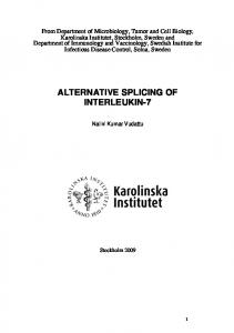
splicing occur in neurons. This correlates with the complex ability of neurons to respond differentially to electrical and chemical signals. The study of splicing aberrations in neuronal mRNAs in mammals and in their homologues in Drosophila has allowed to relate them with several neurological diseases [22,29,43]. An extreme example of alternatively splicing products in Drosophila is the dDscam gene, the Drosophila homologue of the human Down syndrome cell adhesion molecule (Dscam) [75], a protein member of the immunoglobulin superfamily. dDscam has been demonstrated to participate in axonal guidance during nervous system development. Recently, it was reported that the dDscam gene can potentially generate about 38,000 different isoforms through the alternative use of exons in four different sites. The potential diversity of the products of this gene was demonstrated by sequencing 50 transcripts, which resulted in the identification of 48 different forms [75]. The immense variety of gene products may account for the specificity in the axon guidance clues that are suspected to be of great variability [98]. In most cases, although the occurrence of alternative transcripts for a particular gene is known, it has been difficult to determine the role of individual splice variants or to determine differences in the expression patterns. Recently, several reports have described genes encoding scaffold proteins that undergo alternative splicing. For the mammalian scaffold protein ERBIN (16 LRR in the Nterminal, 1 PDZ in the carboxyl terminus), there are seven variants some with a truncated PDZ domain [24]. Although there are no studies about their function, it can be speculated that these truncated forms can serve as dominant negative regulators as it has been demonstrated for the Drosophila gene nitric oxide synthetase (nos) [83]. The mouse atypical MAGUK magi-1 (GUK in the N-terminal, two WW domains, four PDZs) has three transcripts that are expressed in a tissue-specific form and encode proteins with unique COOH termini [21]. Two of the forms (Magi-1a and Magi1b) are found mainly in nonnuclear fractions. The third variant Magi-1c is found primarily in the nucleus. Thus, the use of alternative exons is not only defining tissue specificity but also cell localization [48]. On the other hand, zo-1, a mammalian gene that encodes a MAGUK, was originally described as a component of the tight junction and also undergoes alternative splicing, resulting in two proteins that are distinguished by the presence of an 80-amino acid segment [5,101]. The two isoforms are expressed in different cell types in a pattern that correlates with junctional plasticity [5]. Interestingly, the Drosophila homologue of zo-1, the gene pyd also displays splice variants that show the presence or absence of a 78 -amino acids stretch (exon 6) in the same position where the splicing of zo-1 occurs, although both domains display low homology [100]. The two pyd isoforms (Fig. 1) have a differential subcellular localization in the epithelia where the 6+ isoform is localized at the adherens junctions and the 6 isoform is dispersed
through the basolateral membrane and is not present in the adherens junctions [100]. Another Drosophila PDZ-containing gene scrib exhibits splice variants that differ in their carboxy terminal end (Fig. 1); thus, while the Scrib1 form has four PDZ domains, the other Scrib2 is shorter and has only two PDZ domains and a specific carboxyl tail. The two forms display different expression patterns: Scrib1 is expressed in the ventral cord and the apical and lateral membrane of the ectoderm of the embryo while Scrib2 is expressed in the basal membrane of the ectoderm and in the peripheral nervous system [52]. More recently, adult isoforms of scribble have been described; seven different transcripts were isolated and one of them appears to be expressed only in females [26]. This genetic study suggests that some of these transcripts are involved in the olfactory behavior in the adult without perturbing other developmental and structural functions of the products of this gene. The gene sdt also has three splice variants; Sdt A is the longest [4,34], Sdt B lacks a domain at the amino terminal end [34], and StdC [4] has a different transcription start point and lacks the PDZ and the SH3 domains. This short form is unable to rescue the defects in the null mutant [4]. The expression patterns and functional roles of these isoforms are not known. The dlg gene exhibits alternative processing both in Drosophila and in humans. hdlg originates splice variants that differ in the use of alternative exons in three different sites [56]. Two of the three sites are localized in the amino terminal part of the protein, proximal to the first PDZ domain, and the third is localized between the SH3 and the GUK domain (HOOK domain). They define the presence of 10 –30 amino acid inserts. The HOOK domain has been predicted as a site of intramolecular interaction with the GUK domain. This interaction would be able to block the binding capability of the GUK domain with its partners [106]. Regions of the amino terminal domain compete with the GUK domain for the interaction with HOOK, and in this form they release the GUK domain to freely interact with other proteins [106]. The alternative splicing within the regions encoding the HOOK domain and the amino terminal domain could interfere with these intramolecular interactions [106]. A more direct function for the insertion I3 in the region encoding the HOOK domain has been recently described [74]; this insertion confers the ability to target SAP97 (the rat homologue of hDlg) to the synapse, leading to an increase of AMPA receptor delivery to this site (AMPA receptors are known partners for the PDZ1 and two of SAP97). Moreover, a small insert in the region encoding an amino terminal domain, insert I1A, has been involved in the ability to cluster potassium channels and thus to increase the potassium current carried by Kir 2.2 and Kv 1.5 channels in rat heart [28]. Drosophila DlgA (Dlg1, Ref. [103]) was the first gene product of the discs large gene reported, and with PSD95 and ZO-1, one of the first proteins where the PDZ domains were described. It has recently been found that dlg undergoes alternative processing that accounts for more products than DlgA
Fig. 1. Domain organization of Drosophila PDZ proteins. Baz and Dlt are examples of multiple PDZ proteins. dlg, pyd, scrib, and sdt genes encode several isoforms, some of them are expressed in different tissue. SdtA is referred as Sdt-MAGUK1 in Ref. [4], SdtC isoforms do not contain PDZ domains and is referred as Sdt-GUK1 in Ref. [4]. Sdt3 domain denotes the translation of exon 3 of sdt gene and Pyd6 domain indicates the translation of exon 6 of pyd gene.
[58]. The described alternative products are more complex than the ones described for hdlg. The gene has different transcription start codons and alternative splicing of exons, which define at least five different products. The longest of the novel products involves the replacement of the short region before the PDZ1 in DlgA (exon 8) by three exons defining a different start codon and a domain (S97N) homologous to the amino terminal region of mammalian SAP97, which contains another protein interaction domain, an L27 domain. Forms comprising the S97N domain are expressed in the ventral cord, peripheral nervous system, and muscle in the embryo and in the brain and neuromuscular synapse in the larvae. In spite of this, they are absent in the ectoderm epithelia in the embryo and in the imaginal discs epithelia in the larvae where, as it was already mentioned dlg products are expressed [58]. These results could explain the pleiotropic phenotype of dlg mutants and the multiple functions described for this gene. Recently, a new splice variant for the vertebrate protein PSD95 [18], another close homologue of Dlg, has been described. The splice site is in an equivalent site to the one described for
Dlg, just proximal to the first PDZ. The a isoform of PSD95 (the originally described) is defined by a short amino terminal domain (similar in length to the one in DlgA) that can be palmitoylated. The h isoform has a longer amino terminal domain, which has an L27 domain and cannot be palmitoylated. The different amino terminal domains define specific functions for these isoforms; thus, in the a form, it allows the regulated palmitoylation that in turn modulates the synaptic expression of AMPA receptors, in the h form, it appears to participate in the endocytic traffic of AMPA receptors.
7. Future perspectives The potential source of variability generated by the use of alternative exons in the nervous system is just starting to be unveiled. The identification of tissue-specific splice variants is a first step in the discovery of novel interaction domains and specific features that can account for the use of genes in different processes, developmental stages, and tissues.
The functional genomic, genetic, cellular, and molecular techniques available in the Drosophila model make possible to analyze the function of specific splice forms in vivo. For example, it is possible to study mutant alleles that affect just specific splice variants or to perform transgenic rescue experiments involving these variants. This analysis can be done in a temporally and tissue-specific regulated fashion; moreover, it is also possible to carry out loss of function experiments for specific splice variants in a defined set of cells in distinct developmental stages using RNAi approaches coupled to the UAS-GAL4 system [23,42] or using the MARCM technique [51]. The regulation of the expression of specific splice variants constitutes another major field to be explored. Although regulatory elements and RNA binding proteins involved in the splicing process have been identified, little is known about the mechanisms that control the specificity of expression in a cell or in a particular stage of development of a particular splice variant. The study of genes encoding scaffold proteins in Drosophila may prove specially suitable to address these general questions by combining the power of the model system with the characteristics of scaffold proteins, in which domain addition, subtraction, and shuffling are easy to recognize and the functional results of these rearrangements can be predicted on the basis of known functions of the domains involved. In particular, the study of genes encoding PDZ-containing proteins may provide general answers relevant to how the diversity and specificity of synapses formation and modulation arise.
[8] [9]
[10] [11]
[12]
[13]
[14]
[15]
[16]
[17]
[18]
Acknowledgements
[19]
We thank Dr. M. Kukuljan for critical reading and support. Supported by Fondecyt 1000824 and 1030795 and ICM-P01007.
[20]
[21]
References [1] F.M. Adamski, M.-Y. Zhu, F. Bahiraei, B.-H. Shieh, Interaction of eye protein Kinase C and Inad in Drosophila localization of binding domains and electrophysiological characterization of a loss of association in transgenic flies, J. Biol. Chem. 273 (1998) 17713 – 17719. [2] P.N. Adler, J. Taylor, Asymmetric cell division: plane but not simple, Curr. Biol. 11 (2001) R233 – R236. [3] R. Albertson, C.Q. Doe, Dlg, Scrib and Lgl regulate neuroblast cell size and mitotic spindle asymmetry, Nat. Cell Biol. 5 (2003) 166 – 170. [4] K. Bachmann, M. Schneider, E. Theilenberg, F. Grawe, E. Knust, Drosophila Stardust is a partner of Crumbs in the control of epithelial cell polarity, Nature 414 (2001) 638 – 643. [5] M.S. Balda, J.M. Anderson, Two classes of tight junctions are revealed by ZO-1 isoforms, Am. J. Physiol., Cell Physiol. 264 (1993) C918 – C924. [6] K. Barrett, M. Leptin, J. Settleman, The Rho GTPase and a putative RhoGEF mediate a signaling pathway for the cell shape changes in Drosophila gastrulation, Cell 91 (1997) 905 – 915. [7] Y. Bellaiche, A. Radovic, D.F. Woods, C.D. Hough, M.L. Parmentier,
[22]
[23] [24]
[25]
[26]
[27]
C.J. O’Kane, P.J. Bryant, F. Schweisguth, The partner of inscuteable/ Discs-large complex is required to establish planar polarity during asymmetric cell division in Drosophila, Cell 106 (2001) 355 – 366. I. Bezprozvanny, A. Maximov, Classification of PDZ domains, FEBS Lett. 509 (2001) 457 – 462. M.A. Bhat, S. Izaddoost, Y. Lu, K.-O. Cho, K.-W. Choi, H.J. Bellen, Discs lost, a novel multi-PDZ domain protein, establishes and maintains epithelial polarity, Cell 96 (1999) 833 – 845. D. Bilder, PDZ proteins and polarity: functions from the fly, Trends Genet. 17 (2001) 511 – 519. D. Bilder, N. Perrimon, Localization of apical epithelial determinants by the basolateral PDZ protein Scribble, Nature 403 (2000) 676 – 680. D. Bilder, M. Li, N. Perrimon, Cooperative regulation of cell polarity and growth by Drosophila tumor suppressors, Science 289 (2000) 113 – 116. D. Bilder, M. Schober, N. Perrimon, Integrated activity of PDZ protein complexes regulates epithelial polarity, Nat. Cell Biol. 5 (2003) 53 – 58. M. Boutros, N. Paricio, D.I. Strutt, M. Mlodzik, Dishevelled activates JNK and discriminates between JNK pathways in planar polarity and Wingless signaling, Cell 94 (1998) 109 – 118. J.E. Brenman, D.S. Chao, S.H. Gee, A.W. McGee, S.E. Craven, D.R. Santillano, Z. Wu, F. Huang, H. Xia, M.F. Peters, et al., Interaction of nitric oxide synthase with the postsynaptic density protein PSD-95 and alpha1-syntrophin mediated by PDZ domains, Cell 84 (1996) 757 – 767. V. Budnik, Y.H. Koh, B. Guan, B. Hartmann, C. Hough, D. Woods, M. Gorczyca, Regulation of synapse structure and function by the Drosophila tumor suppressor gene dlg, Neuron 17 (1996) 627 – 640. S. Butz, M. Okamoto, T.C. Sudhof, A tripartite protein complex with the potential to couple synaptic vesicle exocytosis to cell adhesion in brain, Cell 94 (1998) 773 – 782. D.M. Chetkovich, R.C. Bunn, S.-H. Kuo, Y. Kawasaki, M. Kohwi, D. Bredt, Postsynaptic targeting of alternative postsynaptic density-95 isoforms by distinct mechanisms, J. Neurosci. 22 (2002) 6415 – 6425. C.M. De Lorenzo, A.W. Huwe, M. Spillane, P.J. Bryant, The Dlg multimeric complex and its function in cell proliferation control, Dros. Res. Conf. 42 (2001) 442A. S.D. Dimitratos, D.F. Woods, P.J. Bryant, Camguk, lin-2, and CASK: novel membrane-associated guanylate kinase homologs that also contain CaM kinase domains, Mech. Dev. 63 (1997) 127 – 130. I. Dobrosotskaya, R.K. Guy, G.L. James, MAGI-1, a membraneassociated guanylate kinase with a unique arrangement of protein – protein interaction domains, J. Biol. Chem. 272 (1997) 31589 – 31597. B.K. Dredge, A.D. Polydorides, R.B. Darnell, The splice of life: alternative splicing and neurological disease, Nat. Rev., Neurosci. 2 (2001) 43 – 50. J.B. Duffy, GAL4 system in Drosophila: a fly geneticist’s Swiss army knife, Genesis 34 (2002) 1 – 15. B. Favre, L. Fontao, J. Koster, R. Shafaatian, F. Jaunin, J.H. Saurat, A. Sonnenberg, L. Borradori, The hemidesmosomal protein bullous pemphigoid antigen 1 and the integrin beta 4 subunit bind to ERBIN. Molecular cloning of multiple alternative splice variants of ERBIN and analysis of their tissue expression, J. Biol. Chem. 276 (2001) 32427 – 32436. The FlyBase Consortium, The FlyBase database of the Drosophila genome projects and community literature, Nucleic Acids Res. 31 (2003) 172 – 175. http://.flybase.org. I. Ganguly, T.F.C. Mackay, R.R.H. Anholt, Scribble: a PDZ domain protein that contributes to sexually dimorphic olfactory avoidance behavior in Drosophila, Genetics 164 (2003) 1447 – 1457. C.C. Garner, J. Nash, R.L. Huganir, PDZ domains in synapse assembly and signalling, Trends Cell Biol. 10 (2000) 274 – 280.
[28] D. Godreau, R. Vranckx, A. Maguy, C. Goyenvalle, S.N. Hatem, Different isoforms of synapse-associated protein, SAP97, are expressed in the heart and have distinct effects on the voltage-gated K + channel Kv1.5, J. Biol. Chem. 278 (2003) 47046 – 47052. [29] P.J. Grabowski, Splicing regulation in neurons: tinkering with cellspecific control, Cell 92 (1998) 709 – 712. [30] P.J Grabowski, D.L. Black, Alternative RNA splicing in the nervous system, Prog. Neurobiol. 65 (2001) 289 – 308. [31] S. Granderath, A. Stollewerk, S. Greig, C.S. Goodman, C.J. O’Kane, C. Klaembt, Loco encodes an RGS protein required for Drosophila glial differentiation, Development 126 (1999) 1781 – 1791. [32] M.J. Greener, R.G. Roberts, Conservation of components of the dystrophin complex in Drosophila (1), FEBS Lett. 482 (2000) 13 – 18. [33] B.Z. Harris, W.A. Lim, Mechanism and role of PDZ domains in signaling complex assembly, J. Cell Sci. 114 (2001) 3219 – 3231. [34] Y. Hong, B. Stronach, N. Perrimon, L.Y. Jan, Y.N. Jan, Drosophila Stardust interacts with Crumbs to control polarity of epithelia but not neuroblasts, Nature 414 (2001) 634 – 638. [35] Y. Hong, L. Ackerman, L.Y. Jan, Y.N. Jan, Distinct roles of Bazooka and Stardust in the specification of Drosophila photoreceptor membrane architecture, Proc. Natl. Acad. Sci. U. S. A. 100 (2003) 12712 – 12717. [36] C.D. Hough, D.F. Woods, S. Park, P.J. Bryant, Organizing a functional junctional complex requires specific domains of the Drosophila MAGUK Discs large, Genes Dev. 11 (1997) 3242 – 3253. [37] T.W. Hurd, L. Gao, M.H. Roh, I.G. Macara, B. Margolis, Direct interaction of two polarity complexes implicated in epithelial tight junction assembly, Nat. Cell Biol. 5 (2003) 137 – 142. [38] T. Ishidate, A. Matsumine, K. Toyoshima, T. Akiyama, The APC – hDLG complex negatively regulates cell cycle progression from the G0/G1 to S phase, Oncogene 19 (2000) 365 – 372. [39] Y.N. Jan, L.Y. Jan, Asymmetric cell division in the Drosophila nervous system, Nat. Rev., Neurosci. 11 (2001) 772 – 779. [40] K. Johnson, A. Wodarz, A genetic hierarchy controlling cell polarity, Nat. Cell Biol. 5 (2003) 12 – 14. [41] S.M. Kaech, C.W. Whitfield, S.K. Kim, The LIN-2/LIN-7/LIN-10 complex mediates basolateral membrane localization of the C. elegans EGF receptor LET-23 in vulval epithelial cells, Cell 94 (1998) 761 – 771. [42] S. Kalidas, D.P. Smith, Novel genomic cDNA hybrids produce effective RNA interference in adult Drosophila, Neuron 33 (2002) 177 – 184. [43] M.D. Kaytor, H.T. Orr, RNA targets of the fragile X protein, Cell 107 (2001) 555 – 557. [44] E. Kim, M. Niethammer, A. Rothschild, Y.N. Jan, M. Sheng, Clustering of Shaker-type K+1 channels by interaction with a family of membrane-associated guanylate kinases, Nature 378 (1995) 85 – 88. [45] N. Klocker, R.C. Bunn, E. Schnell, G. Caruana, A. Bernstein, R.A. Nicoll, D.S. Bredt, Synaptic glutamate receptor clustering in mice lacking the SH3 and GK domains of SAP97, Eur. J. Neurosci. 16 (2002) 1517 – 1522. [46] R. Kraut, W. Chia, L.Y. Jan, Y.N. Jan, J.A. Knoblich, Role of inscuteable in orienting asymmetric cell divisions in Drosophila, Nature 383 (1996) 50 – 55. [47] T. Lahey, M. Gorczyca, X.X. Jia, V. Budnik, The Drosophila tumor suppressor gene dlg is required for normal synaptic bouton structure, Neuron 13 (1994) 823 – 835. [48] R.P. Laura, S. Ross, H. Koeppen, L.A. Lasky, MAGI-1: a widely expressed, alternatively spliced tight junction protein, Exp. Cell Res. 275 (2002) 155 – 170. [49] G. Lavezzari, J. McCallum, R. Lee, K.W. Roche, Differential binding of the AP-2 adaptor complex and PSD-95 to the C-terminus of the NMDA receptor subunit NR2B regulates surface expression, Neuropharmacology 45 (2003) 729 – 737. [50] C.J. Lee, K. Irizarry, Alternative splicing in the nervous system: an
[51]
[52]
[53]
[54]
[55]
[56]
[57]
[58]
[59]
[60]
[61]
[62]
[63] [64]
[65]
[66] [67] [68]
emerging source of diversity and regulation, Biol. Psychiatry 54 (2003) 771 – 776. T. Lee, L. Luo, Mosaic analysis with a repressible cell marker for studies of gene function in neuronal morphogenesis, Neuron 22 (1999) 451 – 461. M. Li, J. Marhold, A. Gatos, I. Torok, B.M. Mechler, Differential expression of two scribble isoforms during Drosophila embryogenesis, Mech. Dev. 108 (2001) 185 – 190. J.R. Martin, R. Ollo, A new Drosophila Ca2+/calmodulin-dependent protein kinase (Caki) is localized in the central nervous system and implicated in walking speed, EMBO J. 15 (1996) 1865 – 1876. D. Mathew, L.S. Gramates, M. Packard, U. Thomas, D. Bilder, N. Perrimon, M. Gorczyca, V. Budnik, Recruitment of Scribble to the synaptic scaffolding complex requires GUK-holder, a novel Dlg binding protein, Curr. Biol. 12 (2002) 531 – 539. A. Matsumine, A. Ogai, T. Senda, N. Okumura, K. Satoh, G.H. Baeg, T. Kawahara, S. Kobayashi, M. Okada, K. Toyoshima, T. Akiyama, Binding of APC to the human homolog of the Drosophila discs large tumor suppressor protein, Science 272 (1996) 1020 – 1023. M. McLaughlin, R. Hale, D. Ellston, S. Gaudet, R.A. Lue, A. Viel, The distribution and function of alternatively spliced insertions in hDlg, J. Biol. Chem. 277 (2002) 6406 – 6412. A. McLysaght, K. Hokamp, K.H. Wolfe, Extensive genomic duplication during early chordate evolution, Nat. Genet. 31 (2002) 200 – 204. C. Mendoza, P. Olguı´n, G. Lafferte, U. Thomas, S. Ebitsch, E.D. Gundelfinger, M. Kukuljan, J. Sierralta, Novel isoforms of Dlg are fundamental for neuronal development in Drosophila, J. Neurosci. 23 (2003) 2093 – 2101. M. Migaud, P. Charlesworth, M. Dempster, L.C. Webster, A.M. Watabe, M. Makhinson, Y. He, M.F. Ramsay, R.G.M. Morris, J.H. Morrison, T.J. O’Dell, S.G. Grant, Enhanced long-term potentiation and impaired learning in mice with mutant postsynaptic density-95 protein, Nature 39 (1998) 433 – 439. H. Miyamoto, I. Nihonmatsu, S. Kondo, R. Ueda, S. Togashi, K. Hirata, Y. Ikegami, D. Yamamoto, Canoe encodes a novel protein containing a GLGF/DHR motif and functions with Notch and scabrous in common developmental pathways in Drosophila, Genes Dev. 9 (1995) 612 – 625. E.W. Myers, G.G. Sutton, A.L. Delcher, I.M. Dew, D.P. Fasulo, M.J. Flanigan, S.A. Kravitz, C.M. Mobarry, K.H. Reinert, K.A. Remington, E.L. Anson, R.A. Bolanos, H.H. Chou, C.M. Jordan, A.L. Halpern, S. Lonardi, E.M. Beasley, R.C. Brandon, L. Chen, P.J. Dunn, Z. Lai, Y. Liang, D.R. Nusskern, M. Zhan, Q. Zhang, X. Zheng, G.M. Rubin, M.D. Adams, J.C. Venter, A whole-genome assembly of Drosophila, Science 287 (2000) 2196 – 2204. S.C. Nam, K.W. Choi, Interaction of Par-6 and Crumbs complexes is essential for photoreceptor morphogenesis in Drosophila, Development 130 (2003) 4363 – 4372. S. Noselli, F. Agnes, Roles of the JNK signaling pathway in Drosophila morphogenesis, Curr. Opin. Genet. Dev. 9 (1999) 466 – 472. K. Ohashi, T. Hosoya, K. Takahashi, H. Hing, K. Mizuno, A Drosophila homolog of LIM-kinase phosphorylates cofilin and induces actin cytoskeletal reorganization, Biochem. Biophys. Res. Commun. 276 (2000) 1178 – 1185. T. Ohshiro, T. Yagami, C. Zhang, F. Matsuzaki, Role of cortical tumor-suppressor proteins in asymmetric division of Drosophila neuroblast, Nature 408 (2000) 593 – 596. T. Pawson, J.D. Scott, Signaling through scaffold, anchoring, and adaptor proteins, Science 278 (1997) 2075 – 2080. M. Peifer, P. Polakis, Wnt signaling in oncogenesis and embryogenesis a look outside the nucleus, Science 287 (2000) 1606 – 1609. C. Peng, L. Manning, R. Albertson, C.Q. Doe, The tumor suppressor genes lgl and dlg regulate basal protein targeting in Drosophila neuroblasts, Nature 408 (2000) 596 – 600.
[69] M. Petronczki, J.A. Knoblich, DmPAR-6 directs epithelial polarity and asymmetric cell division of neuroblasts in Drosophila, Nat. Cell Biol. 3 (2001) 43 – 49. [70] P.J. Plant, J.P. Fawcett, D.C. Lin, A.D. Holdorf, K. Binns, S. Kulkarni, T. Pawson, A polarity complex of mPar-6 and atypical PKC binds, phosphorylates and regulates mammalian Lgl, Nat. Cell Biol. 5 (2003) 301 – 308. [71] J.P. Roche, M.C. Packard, S. Moeckel-Cole, V. Budnik, Regulation of synaptic plasticity and synaptic vesicle dynamics by the PDZ protein Scribble, J. Neurosci. 22 (2002) 6471 – 6479. [72] M.H. Roh, B. Margolis, Composition and function of PDZ protein complexes during cell polarization, Am. J. Physiol., Renal Physiol. 285 (2003) F377 – 387. [73] C. Ruiz-Canada, Y.H. Koh, V. Budnik, F.J. Tejedor, DLG differentially localizes Shaker K+-channels in the central nervous system and retina of Drosophila, J. Neurochem. 82 (2002) 1490 – 1501. [74] G. Rumbaugh, G-M. Sia, C.C. Garner, R.L. Huganir, Synapse-associated protein-97 isoform-specific regulation of surface AMPA receptors and synaptic function in cultured neurons, J. Neurosci. 23 (2003) 4567 – 4576. [75] D. Schmucker, J.C. Clemens, H. Shu, C.A. Worby, J. Xiao, M. Muda, J.E. Dixon, S.L. Zipursky, Drosophila Dscam is an axon guidance receptor exhibiting extraordinary molecular diversity, Cell 101 (2000) 671 – 684. [76] M. Schober, M. Schaefer, J.A. Knoblich, Bazooka recruits Inscuteable to orient asymmetric cell divisions in Drosophila neuroblasts, Nature 402 (1999) 548 – 551. [77] M. Sheng, Molecular organization of the postsynaptic specialization, Proc. Natl. Acad. Sci. U. S. A. 98 (2001) 7058 – 7061. [78] M. Sheng, C. Sala, PDZ domains and the organization of supramolecular complexes, Annu. Rev. Neurosci. 24 (2001) 1 – 29. [79] S.H. Shi, L.Y. Jan, Y.N. Jan, Hippocampal neuronal polarity specified by spatially localized mPar3/mPar6 and PI 3-kinase activity, Cell 112 (2003) 63 – 75. [80] B.-H. Shieh, M.-Y. Zhu, Regulation of the TRP Ca21 channel by INAD in Drosophila photoreceptors, Neuron 16 (1996) 991 – 998. [81] B.-H. Shieh, M.-Y. Zhu, J.K. Lee, I.M. Kelly, F. Bahiraei, Association of INAD with NORPA is essential for controlled activation and deactivation of Drosophila phototransduction in vivo, Proc. Natl. Acad. Sci. U. S. A. 94 (1997) 12682 – 12687. [82] M. Sone, M. Hoshino, E. Suzuki, S. Kuroda, K. Kaibuchi, H. Nakagoshi, K. Saigo, Y. Nabeshima, C. Hama, Still life, a protein in synaptic terminals of Drosophila homologous to GDP-GTP exchangers, Science 275 (1997) 543 – 547. [83] Y. Stasiv, M. Regulski, B. Kuzin, T. Tully, G. Enikolopov, The Drosophila nitric-oxide synthase gene (dNOS) encodes a family of proteins that can modulate NOS activity by acting as dominant negative regulators, J. Biol. Chem. 276 (2001) 42241 – 42251. [84] N.L. Stricker, R.L. Huganir, The PDZ domains of mLin-10 regulate its trans-Golgi network targeting and the surface expression of AMPA receptors, Neuropharmacology 45 (2003) 837 – 848. [85] K. Takahashi, T. Matsuo, T. Katsube, R. Ueda, D. Yamamoto, Direct binding between two PDZ domain proteins Canoe and ZO-1 and their roles in regulation of the jun N-terminal kinase pathway in Drosophila morphogenesis, Mech. Dev. 78 (1998) 97 – 111. [86] M. Takahisa, S. Togashi, T. Suzuki, M. Kobayashi, A. Murayama, K. Kondo, T. Miyake, R. Ueda, The Drosophila tamou gene, a component of the activating pathway of extramacrochaetae expression, encodes a protein homologous to mammalian cell – cell junctionassociated protein ZO-1, Genes Dev. 10 (1996) 1783 – 1795. [87] G. Tanentzapf, U. Tepass, Interactions between the crumbs, lethal giant larvae and bazooka pathways in epithelial polarization, Nat. Cell Biol. 5 (2003) 46 – 52. [88] G. Tanentzapf, Ch. Smith, J. McGlade, U. Tepass, Apical, lateral,
[89]
[90]
[91]
[92]
[93]
[94]
[95]
[96]
[97]
[98]
[99]
[100]
[101]
[102]
[103]
[104]
[105]
and basal polarization cues contribute to the development of the follicular epithelium during Drosophila oogenesis, J. Cell Biol. 151 (2000) 891 – 904. S.A. Teichmann, A.G. Murzin, C. Chothia, Determination of protein function, evolution and interactions by structural genomics, Curr. Opin. Struct. Biol. 11 (2001) 354 – 363. F.J. Tejedor, A. Bokhari, O. Rogero, M. Gorczyca, J. Zhang, E. Kim, M. Sheng, V. Budnik, Essential role for dlg in synaptic clustering of Shaker K+ channels in vivo, J. Neurosci. 17 (1997) 152 – 159. U. Tepass, C. Theres, E. Knust, Crumbs encodes an EGF-like protein expressed on apical membranes of Drosophila epithelial cells and required for organization of epithelia, Cell 61 (1990) 787 – 799. U. Tepass, G. Tanentzapf, R. Ward, R. Fehon, Epithelial cell polarity and cell junctions in Drosophila, Annu. Rev. Genet. 35 (2001) 747 – 784. U. Thomas, E. Kim, S. Kuhlendahl, Y.H. Koh, E.D. Gundelfinger, M. Sheng, C.C. Garner, V. Budnik, Synaptic clustering of the cell adhesion molecule fasciclin II by discs-large and its role in the regulation of presynaptic structure, Neuron 19 (1997) 787 – 799. U. Thomas, B. Phannavong, B. Muller, C.C. Garner, E.D. Gundelfinger, Functional expression of rat synapse associated proteins SAP97 and SAP102 in Drosophila dlg-1 mutants: effects on tumor suppression and synaptic bouton structure, Mech. Dev. 62 (1997) 161 – 174. S. Tsunoda, C.S. Zuker, The organization of INAD-signaling complexes by a multivalent PDZ domain protein in Drosophila photoreceptor cells ensures sensitivity and speed of signaling, Cell Calcium 26 (1999) 165 – 171. S. Tsunoda, J. Sierralta, Y. Sun, R. Bodner, E. Suzuki, A. Becker, M. Socolich, C.S. Zuker, A multivalent PDZ domain protein assembles signalling complexes in a G-protein-coupled signalling cascade, Nature 388 (1997) 243 – 249. S. Tsunoda, Y. Sun, E. Suzuki, C.S. Zuker, Independent anchoring and assembly mechanisms of INAD signaling complexes in Drosophila photoreceptors, J. Neurosci. 21 (2001) 150 – 158. J. Wang, C.T. Zugates, I.H. Liang, Ch-H.J. Lee, T. Lee, Droso phila Dscam is required for divergent segregation of sister branches and suppresses ectopic bifurcation of axons, Neuron 33 (2002) 559 – 571. W.-L. Wang, S.-F. Yeh, Y.-I. Chang, S.-F. Hsiao, W.-N. Lian, C.-H. Lin, C.-Y.F. Huang, W.-J. Lin, PICK1, an anchoring protein that specifically targets protein kinase C to mitochondria selectively upon serum stimulation in NIH 3T3 Cells, J. Biol. Chem. 278 (2003) 37705 – 37712. X. Wei, H.M. Ellis, Localization of Drosophila MAGUK protein Polychaetoid is controlled by alternative splicing, Mech. Dev. 100 (2001) 217 – 231. E. Willott, M.S. Balda, A.S. Fanning, B. Jameson, C. Van Itallie, J.M. Anderson, The tight junction protein ZO-1 is homologous to the Drosophila discs-large tumor suppressor protein of septate junctions, Proc. Natl. Acad. Sci. U. S. A. 90 (1993) 7834 – 7838. A. Wodarz, A. Ramrath, A. Grimm, E. Knust, Drosophila aty pical protein kinase C associates with bazooka and controls polarity of epithelia and neuroblasts, J. Cell Biol. 150 (2000) 1361 – 1374. D.F. Woods, P.J. Bryant, Molecular cloning of the lethal(1)discs large-1 oncogene of Drosophila, Dev. Biol. 134 (1989) 222 – 235. D.F. Woods, P.J. Bryant, The discs-large tumor suppressor gene of Drosophila encodes a guanylate kinase homolog localized at septate junctions, Cell 66 (1991) 451 – 464. D.F. Woods, C. Hough, D. Peel, G. Callaini, P.J. Bryant, Dlg protein is required for junction structure, cell polarity, and proliferation control in Drosophila epithelia, J. Cell Biol. 134 (1996) 1469 – 1482.
[106] H. Wu, C. Reissner, S. Kuhlendahl, B. Coblentz, S. Reuver, S. Kindler, E.D. Gundelfinger, C.C. Garner, Intramolecular interactions regulate SAP97 binding to GKAP, EMBO J. 19 (2000) 5740 – 5751. [107] Y. Xiang, B. Kobilka, The PDZ-binding motif of the h2-adrenoceptor is essential for physiologic signaling and trafficking in
cardiac myocytes, Proc. Natl. Acad. Sci. U. S. A. 100 (2003) 10776 – 10781. [108] Q. Xu, B. Modrek, C. Lee, Genome-wide detection of tissue-specific alternative splicing in the human transcriptome, Nucleic Acids Res. 30 (2002) 3754 – 3766.