Case Report
Periampullary Carcinoma with Skull Metastasis: A rare case report Deepti Sharma1,*, Garima Singh2 1,2Dept.
of Radiation Oncology, VMMC & Safdarjung Hospital, New Delhi
*Corresponding Author: Email:
[email protected]
Abstract Periampullary cancer consist of pancreatic cancer, carcinoma of ampulla of vater, distal common bile duct and duodenum. With the use of multimodality treatment, the prognosis of periampullary carcinoma has been improved. Skull (calvarium) metastasis is uncommon presentation. Only few cases of periampullary carcinoma with skull metastasis are available in English literature. Although rare, metastatic periampullary adenocarcinoma should be considered as a differential diagnosis in patients presenting with abnormal scalp swelling and tenderness. We reported a case of 48 year old female who presented with lytic expansile bony lesion with associated soft tissue mass in left parietal region with no neurological defecit 18 months after the whipples procedure. Keywords: Periampullary Carcinom; Scalp Metastasis; Skull Metastasis
Access this article online Website: www.innovativepublication.com DOI:
10.5958/2455-6793.2016.00020.1 Introduction The global annual incidence rate for carcinoma of pancreas is about 8/100,000 persons1 and is currently the fourth leading cause of cancer mortality in the United States It is anticipated that it become the second by 2020.2 Skull (calvarium) metastasis is common from malignancies like breast, lung colon, prostate, kidney and ovary.3 Gastrointestinal and pancreatic cancer rarely metastasized to brain and skull.4 However there are only few anecdotal reports in which ampulla of vater adenocarcinoma had metastasis to skull.5,6 Here we are presenting a case of carcinoma head of pancreas with expansile osteolytic calvarial metastasis. Case Presentation A 48 years old post-menopausal female was diagnosed with periampullary carcinoma, underwent whipple surgery and feeding jejunostomy.[Fig. 1] Histopathology was suggestive of moderately differentiated adenocarcinoma, R0 resection with node positive. Case was discussed in multidispilanry clinic and planned for adjuvant chemotherapy. She had received adjuvant chemotherapy with Gemcitabine and Cisplatin till Sept 2014 and was kept on follow up. After 11 months of follow up, she developed swelling on left side of forehead, which was progressive increasing in size with mild pain. No complain of discharge from swelling, vomiting, headache, seizures, decrease of vision. On examination 5x5 hard fixed
swelling present on left side of forehead with tense shiny skin, non-tender, temperature over the swelling not raised. Biopsy from the swelling was suggestive of metastatic carcinoma. CECT Scan of skull showed permeative lytic expansile bony lesion with associated soft tissue mass approximately 4.6X3.5 cm in left parietal region possibly metastasis. CECT thorax and abdomen was reveaed heterogenous enhancing irregular soft tissue mass esion in the retroperitoneum closely abutting right renal vessels approximately 2.5X2.2 cm suggestive of recurrent lesion with norma choedochojejunal, gastro-jejunal and pancreatico-jejuna anastomosis. Patient then received palliative radiotherapy to the skull lesion and is now on palliative chemotherapy. Good palliation was achieved.
Fig. 1: CECT abdomen showed heterogenous enhancing irregular soft tissue mass lesion in the retroperitoneum
International Journal of Medical Pediatrics and Oncology, July-September, 2016:2(3):120-122
120
Deepti Sharma et al.
Periampullary Carcinoma with Skull Metastasis: A rare case report
Table 1: Studies showing Metastatic periampullary carcinoma Study Miyahara et al12 Ambro et al.13 Bhat W et al.14 Bdeiri K et al15 Aydin MV et al16 Hopf S et al6 Jeon JY et al5 Present study
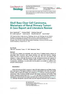
Fig. 2: CECT Scan of skull showed permeative lytic expansile bony lesion with associated soft tissue in left parietal region Discussion Periampullary cancer consist of pancreatic cancer, carcinoma of ampulla of vater, distal common bile duct and duodenum. Periampullary cancer are usually managed by radical operative procedures in early stages. However 80% of patients present with disease that cannot be cured with radical surgery.7 In a study by Lee and Tatter, patients with carcinoma pancreas and periampullary cancer invariably present with metastasis to abdominal lymph node, liver and lung.8 Cutaneous metastases is present in 0.7% to 9% of all patients with cancer, and is common in breast, lung, and colon cancer but are uncommon in pancreatic cancer.9 In pancreatic carcinoma cutaneous metastasis are usually multiple and are confined to periumbilical region.10 Isolated non umbilical metastasis are uncommon.11 Pancreatic cancer with cutaneous metastasis to the scalp is rare. So far to the best of our knowledge, there are only 7 cases of pancreatic cancer with scalp metastasis which are documented in literature.5,6,12,13,14,15,16 Out of these there are only 5 cases including present study, in which skull is involved. In a study by Miyahara et al, 20 patients out of 22 reported with cutaneous metastasis prior to diagnosis of pancreatic cancer. In 11 of these, skin involvement was the first presentation. Hopf S et al reported a case of cancer of ampulla of vater with right frontal skull metastasis 5 years after pylorus preserving pancreaticoduodenectomy. 1 In another study, by Jeon JY et al 65 year old Koren man presented with parietal scalp swelling after whipple procedure.10 Aydin et al also reported a case of a 65year-old woman, presented with painless frontoparietal scalp swelling which developed within three months and is the first presenting symptom of pancreatic adenocarcinoma.26 (Table 1)
No. of patients 43 65 59 70 65
Sex
Site
M M F F F
Uncus Ductal Tail Tail Tail
54 65 48
F M F
Ampulla of vater Ampulla of vater Periampullary carcinoma
In the present study, patient of periampullary carcinoma developed skull metastasis 18 months after the curative whipples procedure, with no neurological deficits other than mild headache and scalp swelling despite adjacent dura is involved and underlying cortex is compressed. Conclusion With the use of multimodality treatment, the prognosis of periampullary carcinoma has improved, although rare, metastatic periampullaryl adenocarcinoma should be considered as a differential diagnosis in patients presenting with abnormal scalp swelling and tenderness. References 1.
Raimondi S, Maisonneuve P, Lowenfels AB. Epidemiology of pancreatic cancer: an overview. Nat Rev Gastroenterol Hepatol.2009;6:699–708. 2. The Alarming Rise of Pancreatic Cancer Deaths in the United States, Pancreatic Cancer Action Network, 2012. 3. Osborn AG: Miscellaneous tumors, cysts and metastasis, In: Osborn AG (ed) Diagnostic Neuroradiology. Mosby, St Louis, 1994,pp 626-670. 4. Sabo RA, Kalyan –Raman UP: Multiple intracerebral metastasis from an islet cell carcinoma of the pancreas: case report, Neurosurgery.1995;37:326-328. 5. Jeon JY, Yi HJ, Lee SR, Paik SS, Lee SS. Skull metastasis from ampulla of vater adenocarcinoma: case report. J Neurooncol. 2004;67:107-113. 6. Hopf S, Rudiger B Scheil F, Heusermann U, Borm W. Skull metastasis of ampulla of vater adenocarcinoma 5 years after Whipple operation: case report and literature review. J Neurooncol 2009;95:141-145. 7. Li D, Xie K, Wolff R et al. Pancreatic Cancer. Lancet.2004;363:1049-1057. 8. Lee YT, Tatter D. Carcinoma of the pancreas and periampullary structures. Pattern of metastasis at autopsy. Arch Pathol Lab Med .1984;108:584-587. 9. Lookingbill DP, Spangler N, Helm KF: Cutaneous metastases in patients with metastatic carcinoma: a retrospective study of 4020 patients. J Am Acad Dermatol.1993;29:228–236. 10. Yendluri V, Centeno B, Springett GM: Pancreatic cancer presenting as a Sister Mary Joseph’s nodule: case report and update of the literature. Pancreas2007;34:161–164.
International Journal of Medical Pediatrics and Oncology, July-September, 2016:2(3):120-122
121
Deepti Sharma et al.
Periampullary Carcinoma with Skull Metastasis: A rare case report
11. Abdel-Hafez HZ: Cutaneous pancreatic metastasis: a case report and review of literature. Indian J Dermatol 2008;53: 206 –209. 12. Miyahara M, Hamanaka Y, Kawabata A, et al: Cutaneous metastasis from pancreatic cancer. Int J Pancreatol.1996;20:127–130. 13. Ambro CM, Humphreys TR, Lee JB: Epidermotropically metastatic pancreatic adenocarcinoma. Am J Dermatopathol.2006; 28:60–62. 14. Bhat W, Abood A, Maraveyas A, et al: Cutaneous metastasis from pancreatic carcinoma: a case report and review. J Clin Exp Dermatol Res 1:206–111,2010. 15. Bdeiri K, Kamar FG. Cutaneous Metastasis of Pancreatic Adenocarcinoma as a First Clinical Manifestation: A Case Report and Review of the Literature. Case reports in GI oncology.2013:61-63. 16. Aydin MV et al. Unusual case of skull metastasis secondary to pancreatic adenocarcinoma . Pathology & Oncology Research .Sep 2005;11(3):182-183.
International Journal of Medical Pediatrics and Oncology, July-September, 2016:2(3):120-122
122