regarding the field of dermatology (250-1000 words). CLINICAL DERMATOLOGY
. CME articles. Content should be specific to the field of dermatology following.
Jul 31, 2012 - with granulomatous rosacea, as well as rosacea fulminans, with reductions in both the rhinophyma and the associated multiple sebaceous ...
Jul 31, 2012 - for Category I (Preapproved) CME credit by the American Academy of ...... surgery. He is director of a Mohs Surgery Fellowship program.
ent RBC half-lives of 5â10, 12â25, and. 25â40 days ... Cardiac Troponin T in Serum of a ... diac Troponin T and normal cardiac troponin I. Clin Chem 1999 ...
We shall first show that the fluid proper time is finite as the solution approaches its ..... Wandelt, [astro-ph/0407196]; S. Das, P.S. Corasanuti and J. Khoury,. [astro-ph/0510628]. [24] T. R. Jaffe, A. J. Banday, H. K. Eriksen, K. M. Gorski and F.
cosmological models with a tilting, but otherwise conventional, perfect fluid, .... no hair theorem, but since the tilt does not die away, isotropization of the.
At the hip (Hip disarticulation). 1 (6.7). 0. 0. Total. 5 (33.3). 9 (60.0). 1 (6.7). Table 2. The intensity of pain in their residual limbs in landmine explosion survivors ...
Jul 20, 2011 - Gabriele Cipriani1, Lucia Picchi1, Marcella Vedovello1, Angelo ... for Biological Sciences, CAS and Springer-Verlag Berlin Heidelberg 2011.
phase with high precision and determine its critical exponents. KEY WORDS: ... drop has long been a challenge; and very diverse methods have been used, some of them ... approximating the shape by spline functionsâ) and, in a more refined way, ... t
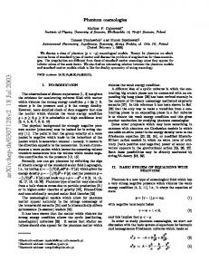
Jul 18, 2003 - The physical background for strongly negative pres- sure matter (phantom) ... matter appear naturally in the mirage cosmology of the braneworld ..... y â ̺ â â) and the second describes a cycle in y and ̺ from zero to a ... sti
Valves for heating, ventilation, air conditioning – for industrial and power plant
use. 0508 / 112966 / HORA / GB. PA-N160+280_GB_0508. Operating Manual.
automotive tuning and accessories importer based out of Burlingame, CA. Ads were ... website. Distributed at local dance
the demand for hospital care, outpatient medical care, care related to acute and chronic episodes of sickness, dental care, and well-care (a label used for ...
The program promotes the wise management and use of our forest resources to ..... between the BOF Communications Section
an astonishingly different cosmic end game. Here, we explore the consequences that follow if the dark energy is phantom energy, in which the sum of the ...
deploy by land, sea, or air to conduct mobile-armed offensive and defensive
operations worldwide. .... During World War II the Corps earned the nickname. “
Phantom ..... Pattern (OCP) Uniform is designed for wear in Afghanistan only with
the ...
High Signal Intensity in Dentate Nucleus and Globus Pallidus on Unenhanced ... Laura Kate Young1, Shona Matthew1, Stephen Gandy1,2, Lukasz Priba2, John ...
the US Forest Service after the passage of the 1990 Farm Bill and continues to be successful today .... Quality Deer Man
Field Staff ⦠.... Jobs: Pennsylvania's forest industry supports nearly. 60,000 jobs ..... between the BOF Communicati
rently gravitationally bound, such as the Milky Way and perhaps the Local Group, .... âSome say the world will end in fire, Some say in iceâ. [36] â for a new fate ...
Nov 6, 2008 - in certain alternative theories to general relativity [31]. ...... recently, static models with two interacting phantom and ghost scalar fields were.
ediTorial missioN: The JDPA is the official clinical journal of the Society ...
dermatological patient care by publishing the most innovative, timely, practice-.
May 4, 2017 - Introduction: Pericytes (PCs) located on the outside of capillaries play a pivotal role in formation and stabilization of blood vessels and are ...
Annual Meeting SVGO / SBMS Thursday May 4th, 2017 Cellularized Perfusable Microvessels for the Study of Human Pericytes Response to Paracrine Signals AR Pereira 1, L Barbe 2, M Herrmann 1, M Alini 1, S Verrier 1. 1
AO Research Institute, Davos, Switzerland; 2 CSEM, Landquart, Switzerland.
Introduction: Pericytes (PCs) located on the outside of capillaries play a pivotal role in formation and stabilization of blood vessels and are suggested to contribute to regenerative processes [1]. However, the complex interplay between endothelial cells and pericytes is still incompletely understood. To access the response of pericytes to paracrine signaling (e.g. inflammation), here we developed a microfluidic perfusable 3D co-culture system for pericytes and human umbilical vein endothelial cells (HUVECs). Methods: The microfluidic platform comprises three different parts: a glass slide stage, a polycarbonate chamber including two capillary guides and a PDMS lid (Figure 1A). Type I-collagen (2.5 mg/ml) is poured in the chamber and polymerized at 37ºC for one hour. Two parallel microvascular channels are generated by gently retraction of microcapillaries. Each channel is individually connected to a reservoir of endothelial growth medium perfused using a piezoelectric micro-pump. GFP-HUVECs are injected into the channels and left to adhere for 16h. Cell-seeded microchannels are perfused under physiological conditions (≤10 μl/min) and observed using a time-lapse microscope. Seeding and co-culture protocols of PCs and HUVECs were optimized using a Vena8 Endothelial+™ biochip (Cellix). Results: The created channels showed regular and stable shape (diameter 150 μm) either in static or perfusion conditions (Figure 1B). The seeding procedure and perfusion conditions allowed for good cell viability and efficient endothelialization of the channel (Figure 1C). Using the cellix biochip, the coculture cell seeding protocol was optimized with the use of 4% dextran. In addition, compared to sequential cell seeding, direct co-seeding of both cell types in a 4:1 ratio (HUVECs:PCs) showed superior channel endothelialization (Figure 1D). Conclusion: We successfully produced on-chip perfusable micro capillary-like structures comprising endothelial cells and pericytes. Next, pericyte behavior and migration will be monitored in response to perfused paracrine factors. Acknowledgement: This work is supported by the AO Foundation and the 3R Research Foundation Switzerland (#139-14). [1] Crisan M. et al, Cell Stem Cell 2008. 3:301-313.