Microbiological Research 211 (2018) 47–56
Contents lists available at ScienceDirect
Microbiological Research journal homepage: www.elsevier.com/locate/micres
Phenotypic and genotypic characterisation of an unique indigenous hypersaline unicellular cyanobacterium, Euhalothece sp.nov
T
Trisha Moganya, Feroz M. Swalahab, Mushal Allamc, Phillip Senzo Mtshalic, Arshad Ismailc, ⁎ Sheena Kumaria, Faizal Buxa, a
Institute for Water and Wastewater Technology, Durban University of Technology, Durban, 4001, South Africa Department of Biotechnology and Food Technology, Durban University of Technology, Durban, 4001, South Africa c Sequencing Core Facility, National Institute for Communicable Diseases, National Health Laboratory Service, Sandringham, 2192, Johannesburg, South Africa b
A R T I C LE I N FO
A B S T R A C T
Keywords: Cyanobacteria Euhalothece Hypersaline polyphasic taxonomy Molecular phylogeny Whole genome sequencing
A novel halotolerant species of cyanobacterium of the order Chroococcales was isolated from hypersaline estuary in Kwa-Zulu Natal, South Africa. A comprehensive polyphasic approach viz., cell morphology, pigment composition and complete genome sequence analysis was conducted to elucidate the taxonomic position of the isolated strain. The blue-green oval to rod-shaped cells were 14–18 μm in size, and contained a high amount of phycocyanin pigments. The strain was moderate thermotolerant/alkalitolerant halophile with the optimum conditions for growth at 35 °C, pH 8.5 and 120 g/l of NaCl. Based on 16S rRNA gene sequence phylogeny, the strain was related to members of the ‘Euhalothece’ subcluster (99%). The whole genome sequence was determined, and the annotated genes showed a 90% sequence similarity to the gas-vacuolate, spindle-shaped Dactylococcopsis salina PCC 8305. The size of the genome was determined to be 5,113,178 bp and contained 4332 protein-coding genes and 69 RNA genes with a G + C content of 46.7%. Genes encoding osmoregulation, oxidative stress, heat shock, persister cells, and UV-absorbing secondary metabolites, among others, were identified. Based on the phylogenetic analysis of the 16S rRNA gene sequences, physiological data, pigment compositions and genomic data, the strain is considered to represent a novel species of Euhalothece.
1. Introduction Cyanobacteria, also known as blue-green algae, are a group of photosynthetic microorganisms with a wide morphological and metabolic diversity (Coutinho et al., 2016). These microorganisms form a major component of the biota of hypersaline environments including salt lakes, soda lakes, solar salterns, hypersaline lagoons, and hypersaline sulphur springs (Oren, 2008). Due to their abundance in such environments, cyanobacteria play a major role in almost all biochemical cycles on Earth. They are involved in the primary production of biomass, global oxygen supply, carbon dioxide (CO2) sequestration, as well as nitrogen (N2) fixation (Prabha et al., 2016). Cyanobacteria are also considered as a promising resource for third generation bio-energy. The extraordinary adaptive ability of these organisms has allowed them to survive in a wide range of ecosystems (Brito et al., 2017). They have developed mechanisms to maintain osmotic balance with the external environment through the accumulation of solutes internally (Pfeffer et al., 2016). The utilisation of atmospheric CO2 and sunlight for growth has triggered renewed interest in the organization of
⁎
Corresponding author. E-mail address:
[email protected] (F. Bux).
https://doi.org/10.1016/j.micres.2018.04.001 Received 4 October 2017; Received in revised form 16 February 2018; Accepted 2 April 2018 Available online 03 April 2018 0944-5013/ © 2018 Elsevier GmbH. All rights reserved.
cyanobacterial metabolism. Cyanobacteria also have great commercial potential owing to their fairly simple structure, minimal nutritional requirements, and an ability to synthesize a wide variety of metabolites and high-value chemicals (Kothari et al., 2013; Lau et al., 2015). Furthermore, enzymes and protein complexes encoded by halophiles often display better stability compared to non-halophilic cyanobacteria (Roberts, 2005) and are thus, highly valuable for applications in industrial biotechnology. Classification of cyanobacterial taxa has traditionally been done by comparison of phenotypic features (i.e. cell morphology), which does not reflect the evolution and the degree of relationship among different taxa (Kim et al., 2014; Komárek et al., 2011). Recently, the use of molecular taxonomy based on nucleotide sequence homology of marker genes (usually 16S, 23S or 16S–23S intergenic spacer regions) has been studied (Bravakos et al., 2016; Rajendhran and Gunasekaran, 2011). However, phylogenies derived from single gene comparison showed inconsistency with each other due to different factors such as horizontal gene transfer (HGT), paralogy and highly variable rates of evolution (Snel et al., 2002). Therefore, whole genome sequencing (WGS) has
Microbiological Research 211 (2018) 47–56
T. Mogany et al.
505 nm). The images were captured and analysed using a software (Axiovision imaging v 8.0).
emerged as a powerful tool to identify and resolve the functional characteristics of cyanobacterial species (Tan et al., 2016). Recent advancements in cyanobacterial genome sequencing have enabled largescale multi-gene phylogenetic analyses that have provided a robust framework to resolve deep branching relationships, understand the evolutionary history of these organisms, (Chrismas et al., 2015) and identify the different factors such as vertical descent, gene transfer, gene and genome duplication, gene invention, gene loss and degradation that affect the tree lineage (Eisen, 2000). Genome sequencing also provides opportunities for understanding microbial diversity and metabolic organization in diverse environments (Lee et al., 2005). Despite the rapidly developing technology, there are currently only two known complete genomes of cyanobacterium belonging to the genus Cyanothece subcluster Euhalothece. Since these cyanobacteria are widespread in extremely mineralised waters, they are expected to have higher genome diversity between strains. This study focused on the morphological and phenotypical identification of an indigenous halotolerant cyanobacterium. Thereafter, the genomic information from Euhalothece sp. nov was obtained in order to gain insights into adaptations allowing the cyanobacterium to dominate a hypersaline, nutrient-limited estuary in Kwa-Zulu Natal, South Africa. The draft genome sequence of Euhalothece sp. nov helped us to understand the cyanobacteria’s physiological capabilities and revealed the differences from other strains. Furthermore, this information can potentially aid in the discovery of novel metabolites and genes that are useful for engineering the cyanobacterium for various biotechnological applications.
2.2.3. Scanning electron microscopy (SEM) Samples were fixed in 2.5% glutaraldehyde in 0.1 M sodium phosphate buffer (pH 7.2) overnight, followed by a post-fixation step, whereby the sample was immersed in 0.5% OsO4 for 1 h and thereafter, rinsed twice with sodium phosphate buffer (pH 7.2). This was followed by series of dehydration steps using 30, 50, 75, 85, 95 and 100% ethanol for 5 min and dried at a critical point for 3 h under CO2 atmosphere. The samples were subsequently mounted onto standard stubs (Ø12.7 mm) and sputter coated with gold, using a Quorum (Q 150R ES sputter coater). The coated samples were then examined with a ZEISS LEO 1450 scanning electron microscope operated at 10 or 20 kV and with a working distances of 7–9 mm. 2.2.4. Transmission electron microscopy (TEM) Ultrastructure micrographs of cyanobacterial cells were analysed using transmission electron microscopy (TEM). Cells from 10 mL culture was collected by centrifugation at room temperature (112g, 5 min) and washed with DdH2O, and transferred into 2% bacteriological agar in 0.1 M phosphate buffer (pH 7.2). Thereafter, cells were fixed in 3% glutaraldehyde in phosphate buffer and incubated for 24 h at 4 °C. Postfixation was done on 1–2 mm agar blocks with osmium tetroxide in phosphate buffer for 2 h at 4 °C. Samples were then dehydrated in ethanol, embedded in Spurr’s resin, sectioned, stained with uranyl acetate and lead citrate (Spurr, 1969). Visualization and photography were performed using a JEOL 1010 transmission electron microscope (Jeol, MA, USA) operated at 100 kV.
2. Materials and methods 2.1. Cyanobacterium isolation
2.3. Effect of salinity, temperature, and pH on growth and C-Phycocyanin production
Water samples (100 mL) were collected from False Bay region at Lister's Point (Lake St. Lucia, South Africa, 27.976S 32.362E), the largest hypersaline estuarine lake in Africa. The physiochemical parameters viz., temperature (40 °C), pH (8.5), dissolved oxygen and salinity (120 g/L) were measured using a potable multiparameter YSI 556 MPS system (Yellow Spring Systems, USA) during the time of sample collection. Isolation and purification of the isolate was carried out using serial dilutions and repeated streak-plating on 1% Blue-Green (BG11) agar medium (Stanier et al., 1971) medium, pH 7. Wet mounts were prepared and examined using light microscopy (Zeiss Axio microscope) to ensure purity of the culture. Single colonies were picked from agar plates and inoculated into liquid BG 11 medium (100 mL) and were incubated under Sylvania® Gro-Lux® illumination (100 μmol photon m−2 s−1), for 16 h light: 8 h dark cycles at 27 ± 2 °C for two weeks, following sub-culturing.
Cells were harvested by centrifugation (112 ×g, 10 min) in the exponential growth phase and re-suspended in fresh medium containing different concentrations of NaCl (30–150g/L), which was prepared from sea water with the addition of natural sea salt. To observe the influence of temperature on growth and phycocyanin (PC) accumulation, the culture was grown at different temperatures i.e. 20, 25, 30, 35, 40 and 45 °C. Similarly, the effect of pH on growth and phycocyanin (PC) was investigated at pH 5, 6, 7, 8, 8.5, 9 and 10. The pH was adjusted with 10 M NaOH and 1 N HCl solution. For each set of experiments, 500 mL Erlenmeyer flask containing 200 mL BG11 medium (autoclaved) were inoculated with a 10% v/v of culture in the exponential growth phase. Thereafter, the flasks were incubated in an orbital shaking incubator at 90 rpm at different salinities/temperature ranges/pH at a constant light intensity of 100 μmol photons per m2 per−s with a 16:8 light: dark cycle. Biomass analysis was done according to Leema et al. (2010). Phycocyanin was extracted in 10 mM sodium phosphate buffer (pH 7.0), and subjected to repeated freeze-thaw cycles of −20 °C and 4 °C, and measured at 620 and 652 nm according to Soni et al. (2008).
2.2. Phenotypic characterisation 2.2.1. Light microscopy Cells were examined using a Zeiss Axio microscope equipped with transmitted light and phase contrast illumination (Axio Imager A1; Carl Zeiss, Germany). Photomicrographs were taken using a digital camera and Zen software. The morphological features were compared to the already described taxa following published literature (Komárek and Anagnostidis, 1998; Komárek et al., 2014)).
2.4. Genotypic identification 2.4.1. DNA isolation Genomic DNA extraction was done by using a FastDNA™ SPIN Kit for soil according to the manufacturer's instruction. (MP Biomedicals, Thermo Fisher Scientific) The extracted genomic DNA was quantified using a Qubit® 2.0 Fluorometer (Thermo Fisher Scientific) and the integrity of the extracted genomic DNA was determined using 0.8% agarose-gel electrophoresis.
2.2.2. Epifluorescent microscopy The nucleic acids were stained with 4′, 6-diamidino-2-phenylindole (DAPI) modified method according to Mukherjee and Ray (2015). Cells were harvested and washed with phosphate buffer and stained with 100 uL of 20 μg/mL of DAPI for 30 min. The stained cells were examined with an epifluorescence microscope (Axiolab, Carl Zeiss, Germany) applying a mercury source (HBO 50) interfaced with a Zeiss Filter set 49 (excitation 365, beam splitter FT 395 and emission 445/
2.4.2. Genome sequencing and assembly Multiplexed paired-end libraries (2 × 300 bp) were prepared using the Nextera XT DNA sample preparation kit (Illumina, San Diego, CA, USA). Sequencing was performed on an Illumina MiSeq instrument at 48
Microbiological Research 211 (2018) 47–56
T. Mogany et al.
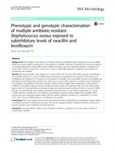
time of isolation, the cells were 8–9 μm. However, after the inoculation and incubation in BG 11 medium for ± 5 weeks, the cells reached stationary phase and had a width of > 14 μm and length of > 18 μm. (Fig. 1A). Cells were found to possess a thin, fine slime layer around them, and this layer was found to be loosely associated with the cell surface (Fig. 1E). Cell division was always perpendicular to the longer axis of oval cells. Actively diving younger cell appeared to be smooth and had a very thin mucous layer around them. Older cells had irregular net-like granule appearance and were buoyant due to the presence of internal gas vesicles (G) (Fig. 1F). Furthermore, the cyanobacterium was found to adjust buoyancy, accordingly to achieve access to re-mineralised nutrients and float to obtain light energy for photosynthesis. The presence of nucleic acids in cells, when stained with DAPI, was observed as a blue colour, with a mosaic of red (chlorophyll), whereas polyphosphate bodies were observed as greenish-yellow dense granules in the cells (Fig. 1D) (Cepák and Komárek, 2010). The DNA was spread unevenly across the entire cell cytoplasm, with no distinct nucleus. The nucleoid has a netlike structure − numerous skeins of DNA without visible DNA threads. Transmission electron micrographs showed the presence of a thick outer cell wall. The external layer (EL) surrounded the cell walls, consisting of thin fibres that emanated from the outer membrane and were arranged perpendicular to it (Fig. 1H). The photosynthetic machinery of the cyanobacterium was made up of numerous irregular thylakoids (th) radiating all over the cell, partly in fascicles (Fig. 1F). Light-harvesting pigments, phycobilisomes (PBS) with a blackberry-like appearance can be seen between the thylakoids (Fig. 1H). In agreement with methylene blue stain (Fig. 1C), the cell also contained numerous polyphosphate (p) granules that store phosphate (Fig. 1G). Cyanophycin (cy) granules that store nitrogen were also present (Fig. 1H); these appeared to be bodies of globular-like appearance with varied size. The cell also contained various other cellular inclusions, including and glycogen granules (g), which store carbon and carboxysomes (cb), which are nitrogen storage compounds that are membrane bound.
the NICD Sequencing Core Facility. The resulting paired-end reads were quality trimmed and de novo assembled using CLC Genomics Workbench version 8 (CLC Bio-Qiagen, Aarhus, Denmark). All resultant contigs were then submitted to GenBank, where gene annotation was implemented using the NCBI Prokaryotic Genome Annotation Pipeline (PGAP) (Tatusova et al., 2016). The annotation was further uploaded to Rapid Annotation using Subsystem Technology (RAST) for subsystemsbased annotation (Aziz et al., 2008; Brettin et al., 2015; Overbeek et al., 2014). The 16S rRNA gene sequence of cyanobacterial isolate was extracted from genome data was compared against the sequences from GenBank, National Center for Biotechnology Information (NCBI) (www. ncbi.nlm.nih.gov) using Basic Local Alignment Search Tool (BLAST). Closely related sequences from GenBank were retrieved and used for phylogenetic analysis. Nucleotide sequences obtained were edited and aligned using BioEdit V7.26. The phylogenetic tree was constructed using the Neighbor-joining method (Saitou and Nei, 1987) with 1000 bootstrapand in MEGA 7 software (Kumar et al., 2008; Tamura et al., 2011). A similarity matrix was built comparing the 16S rRNA sequences using BioEdit. Genome relatedness indices (OGRI) used in this study were DNA–DNA hybridization (DDH) and average nucleotide identity (ANI). The in-silico DDH estimate and the difference in the average GC content were calculated using the Genome-to-Genome Distance Calculator 3 (GGDC V 2.0) http://ggdc.dsmz.de/distcalc2.php (MeierKolthoff et al., 2013). The average Nucleotide Identity by Orthology (OrthoANIu) between the pair of genome sequences was done online using http://www.ezbiocloud.net/tools/ani (Yoon et al., 2017). By using antibiotics & Secondary Metabolite Analysis SHell (antiSMASH) software https://antismash.secondarymetabolites.org secondary metabolite biosynthesis gene clusters in the genome were identified. The program aligns the identified regions at the gene cluster level to their nearest relatives from a database containing all other known gene clusters (Medema et al., 2011). 3. Results 3.1. Morphological description of Euhalothece sp. nov
3.2. Effect of selected cultivation conditions (salt, temperature, and pH) on biomass and pigment production
The classification based on cellular morphology and general features of the isolate are summarised in Table 1. The cyanobacterial cells were unicellular, blue green, oval with cylindrical shape. The strain contained a high quantity of C-Phycocyanin (C-PC) (Fig. S1). At the
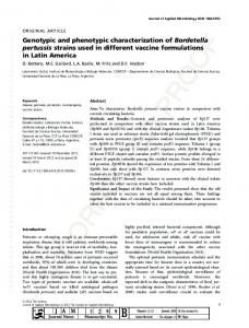
The effect of abiotic factors such as pH, salinity, and temperature on the growth of the isolate was determined by biomass and C-PC production. The response of Euhalothece sp. nov under different salt concentrations was also compared by examining the differences in cell size and content. The strain was found to tolerate a range of NaCl concentrations from 30 to 150 g/L, with the optimum being at 120 g/L (Fig. 2A). A significant increase (p < 0.05) in both biomass and C-PC yield was observed with an increase in salinity. Furthermore, salt concentration was found to influence cell morphology. At salinities > 60 g/L, large green/blue- green cells were observed. However, at a low salinity i.e. 30 g/L, majority of Euhalothece sp. nov cells turned brown and the cell wall began to disintegrate. At a higher salinity (150 g/L), the cells were longer in size. Among the different pH regimes investigated, the highest C-PC production was observed at pH 8 and the least C-PC yield was seen at pH 10. A 14% increase in C-PC was observed when pH was increased from the control (i.e. pH 7) to pH 8.5, and a 40% decrease in PC resulted from pH 8.5 to pH 10 (Fig. 2B). These results revealed that the Euhalothece sp. nov exhibited optimum growth at pH 8.5 with biomass production of 1.13 g/L. A further increase in pH (> 9) resulted in a lower biomass yield. By varying temperature (20–45 °C), it was ascertained that the Euhalothece sp. nov can grew effectively up to 40 °C (Fig. 2C). An increase in temperature from 25 to 35 °C resulted in a significant increase (p < 0.05) in the final biomass (1.6–2.1 g/L). A similar trend was also observed for PC production whereby an increase in C-PC production was observed with an increase in temperature. Among the different
Table 1 Morphological features and characterisation of isolated Euhalothece sp. nov. Classification Domain Phylum Class Order Family Genus Cluster Habitat Salinity Water depth Optimum temperature pH range
Bacteria Cyanobacteria Cyanophyceae Chroococcales Cyanobacteriaceae Cyanothece Cluster 3: Halothece Sub cluster: Euhalothece Marine (hypersaline) 120 g/L ± 40 cm 35 °C 7–9
Cell description Cell shape Cell size (stationary phase of growth) Cell content Gram stain Pigment
Cylindrical- oval cells 16 μm Granular Gram-variable Phycocyanin and chlorophyll
Growth characteristics Biomass Initially growth rate
2–4 g/L 0.76 gday−1
49
Microbiological Research 211 (2018) 47–56
T. Mogany et al.
Fig. 1. Micrographs of Euhalothece sp. nov. (A) Light microscopy showing spherical to ovoid (14–18 μm) cells in singles and undergoing binary fission (1000×), (B) methylene blue stain showing polyphosphate products, scale bars = 15 μm. (C) Eipfluorescent microscopy showing red auto fluorescence of pigments, chlorophyll and phycobiliproteins, (D) showing DAPI stained cells, whereby DNA fluoresces blue and cytoplasma is red, scale bars = 10 μm. (E) SEM showing slime surrounding cell aggregates (micrograph 7.26 kx), (F) TEM ultrathin cross section showing the irregular thylakoids (th) arrangement, inter thylakoid spaces (inter.sp) and (G) gas vesicles, scale bars = 100 nm (G) TEM showing longitude section of cells. (H) TEM enlargement of micrograph G, showing glycogen (g), phycobilisomes (pbs), carboxysomes (cs), cyanophycin granules (cy) and serrated external layer (EL); scale bars = 100 nm (For interpretation of the references to colour in this figure legend, the reader is referred to the web version of this article.)
were annotated as protein-coding genes and 69 for RNA genes (9 for rRNA, 4 for tRNA and 4 other nc RNA). There were 5 clustered regularly interspaced short palindromic repeat (CRISPR) arrays. General genome features and assembly statistics of the cyanobacterium are provided in Table 3.
temperatures tested, 35 and 40 °C supported the highest C-PC yield of 17.5 and 16 mg/g respectively. A decrease in biomass was observed when the temperature was further increased to 45 °C. 3.3. 16S sequence analysis The phylogenetic relationships of the isolate and other reported cyanobacteria based on 16S rRNA gene is presented in a tree calculated applying the maximum likelihood method (Fig. 3). The results indicated that the isolate is closely related (99% similarity) to the Euhalothece sp. and distantly related to other cyanobacterial sequences (91% or lower similarity) available in the NCBI database. Similarity matrix calculations of the 16S rRNA sequences of the selected strains are shown in Table S1. The sequences were aligned using the multiplesequence alignment tool in the ClustaW package in BioEdit (Supplementary data Fig. S2).
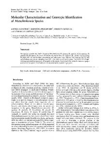
3.6. Insights from genome sequence and secondary metabolism gene clusters Using RAST algorithm, the subsystem distribution, representing a collection of functionally related protein families in the genome of Euhalothece sp. nov revealed a total of 381 subsystems. Amongst these, cofactors, vitamins, prosthetic groups, and pigments subsystem featured the largest number (331) of the assigned CDS. Other major subsystems annotated were amino acids and derivatives (299), carbohydrates (293), protein metabolism (293), DNA metabolism (139) and RNA metabolism (158) (Fig. 4). By using antiSMASH, the secondary metabolites gene clusters from the Halothece sp. nov genome were predicted. Seven biosynthetic gene cluster (BGC) were identified, of which 2 were non ribosomal peptides (NPRS), 3 were terpenes, 1 polyketide synthase (PKS) and 1 ladderane were found in the genome.
3.4. Phenotypic comparison between Halothece strains Even though all strains belonging to the Halothece genus were unicellular, there were noticeable differences amongst them especially with regards to cell size and shape (Table 2). ‘Dactylococcopsis' (Myxobakton salium) PCC 8305 cells a fusiform forms (4–8 × 35–80 μm) with many gas vacuoles at the periphery of cells. Strain PCC 7418 is rodshaped (3 μm) with low number of gas vesicles. Euhalothece sp. nov cells were large cells (8–9 μm) with densely packed gas vesicles. The thylakoid arrangement amongst the strains were also different. Strain PCC 8305 have enlarged thylakoids, whereas numerous irregular thylakoids were present in Euhalothece sp. nov cells. PCC 7418 had numerous radially oriented thylakoids.
4. Discussion 4.1. Morphological identification of Euhalothece sp. Based on the phenotypic and morphological characteristics, the isolated strain resembled members of Subsection 1 (formerly order Chroococcales). Members of Subsection 1 are unicellular, spherical, ellipsoidal or rod-shaped cyanobacteria that reproduce by binary fission or budding (Boone et al., 2001). Furthermore, the cytomorphology i.e. cell morphology, type of cell division and cell structure of the isolate conforms to descriptions of the genus Cyanothece, subcluster Euhalothece as established by Komarek (1976) and elaborated by Waterbury and Rippka (1979). The genus Cyanothece was suggested by Komarek (1976) to accommodate the cyanobacteria that was
3.5. Genome sequencing and properties The draft genome of Euhalothece sp. nov with a total of 5.11 Mbp from 1408 contigs, contains 46.7% G + C content. A total of 5247 genes were predicted. Of these 846 were pseudogenes. The remaining 4332 50
Microbiological Research 211 (2018) 47–56
T. Mogany et al.
et al., 2001). The common features of strains belonging to the Halothece cluster are extreme halotolerance (optimal growth at salinities between 5 and 20%), moderate thermophiles, and only contains PC pigment (Garcia-Pichel et al., 1998). Garcia-Pichel et al. (1998) described the characteristics of 13 extremely halotolerant strains of unicellular cyanobacteria, isolated from various hypersaline environments, including hypersaline and solar evaporation ponds. Two distinct subclusters of Cyanothece were acknowledged: (1) ‘Euhalothece', comprising 12 closely related strains of similar morphology and were originally identified as members of three traditional genera Aphanothece, ‘Dactylococcopsis' (Myxobakton) and Cyanothece (Komárek et al., 2014), and (2) ‘true Halothece', consisting only a single strain, MPI 96P605, which was isolated from a benthic gypsum crust in a solar evaporation pond (Eilat, Israel). Cepák and Komárek (2010), reported that strains belonging to Halothece can be further divided into two groups based on cytomorphological and ultrastructural characteristics. a) Euhalothece cells divide only by binary fission, forming identical daughter cells and they do not form any chains, whereas b) true Halothece’ strains form chains or pseudofilaments under selected environmental conditions. 4.2. Effect of physiochemical parameters on Euhalothece sp. nov The isolated strain, Euhalothece sp. nov was found to tolerate NaCl concentrations up to 150 g/L, with the optimum NaCl concentration being at 120 g/L (Fig. 2A). At a low salinity i.e 30 g/L, the cell wall began to disintegrate and a loss in C-PC was observed. At higher salinities (60–120 g/L), the cells were intense blue-green. Similarly, Walsby et al. (1983) reported that PCC 8305, was not able to grow in seawater (30 g/L salinity). The optimum NaCl concentration for Halothece sp. PCC 7418 (originally called Synechococcus PCC 7418) was 100 g/L, the strain was able to tolerate higher salinity up to 200 g/L however, a decrease in growth was observed (Garcia-Pichel et al., 1998). Brock (1976) and Borowitzka (1981) reported that Aphanothece halophytica isolated from the Great Salt Lake, Utah required a minimum salt concentration of 30 g/L with the optima in the range of 60–150 g/L. Temperature optima for the isolated strain was between 30 and 40 °C (Fig. 2C) and is regarded as moderately thermophilic. A similar thermotolerance characteristic were also reported for other cyanobacterial strains in Halothece cluster (Garcia-Pichel et al., 1998). Tolerance to high temperatures might be an adaptation of the these cyanobacteria, since the low heat capacity of hypersaline waters allows them to easily reach high temperatures when heated by sunlight (Nubel et al., 2000). Brauer et al. (2015) reported that the growth of Halothece sp. was temperature-dependent and was found to grow best at 35 °C. Similarly, Dechatiwongse et al. (2014) found that the maximum biomass productivity (0.1 g L−1 h−1) of Cyanothece 511 was observed when the temperature was maintained between 31 and 35 °C. The cyanobacteria possess different mechanisms for maintenance of pH homeostasis depending their natural habitat (Babu et al., 2015). Usually hypersaline environments have alkaline pH range of 9–11.5 (Chakraborty et al., 2017). The results from this study (Fig. 2B) also confirmed that Euhalothece sp. nov were able to tolerate a range of pH from 6.5 to 10. Therefore, the strain may be considered to be alkalitolerant whose growth is optimum at pH 8.5. The pH is known to influence the solubility of carbon dioxide and minerals (Nagle et al., 2010). Cyanobacteria species isolated from Lake Magadi, a soda lake in Kenya had higher growth rates at pH 7. (Muruga et al., 2010). Touloupakis et al. (2016) reported Synechocystis PCC 6803 grew well between pH 7.5 and 10, and was the culture was still viable when the pH was maintained above 11.
Fig. 2. Biomass and phycocyanin yield at varying (A) salinities fro m 30 to 150 g/L, micrographs depict the change in pigmentation, cell size and shape (B) pH range 5–10 and, (C) temperatures ranging from 20–45 °C of Euhalothece sp. nov. SEM = 3.
inaccurately placed in genus Synechococcus. The major features to distinguishthese two genus were Cyanothece are present in single cells or pairs, and able to fix nitrogen whereas Synechococcus can be found in chains and unable to fix nitrogen (Komárek and Cepák, 1998; Reddy et al., 1993; Turner et al., 2001). Subsequently, Waterbury and Rippka (1979), proposed that the coccoid to rod-shaped cyanobacteria with cells larger than 3 μm in diameter dividing by binary fission and lacking a sheath to be provisionally place in the Cyanothece group. Further subdivisions of the Cyanothcece genus were used to separate strains based on their original habitats such as freshwater, marine or hypersaline environments etc (Rippka and Herdman, 1992). Cyanothece is divided into 3 clusters based on their characteristics such as salt tolerance, cell size, and DNA base compositions. Generally, Cluster 1 and 2 comprises of strains isolated from freshwater environments, whereas strains belonging to cluster 3 ‘Halothece’ grow at higher salinities > 3% (Boone
4.3. Morphological and phylogenetic comparisons of Euhalothece strains The 16S ribosomal genes has all the characteristics of a phylogenetic marker genes due to the universal distribution in prokaryotes, presence 51
Microbiological Research 211 (2018) 47–56
T. Mogany et al.
Fig. 3. Reconstructed phylogenetic tree of Euhalothece sp. nov and 13 related cyanobacteria that were aligned based on 16S rRNA sequences using the maximum likelihood (ML) method (with 1000 bootstrap replicates). All sequences were extracted from NCBI database. GenBank accession numbers for each strain are shown in parentheses. Multiple sequence alignments were performed using the BioEdit sequence alignment editor. The evolutionary distances were computed using the Kimura 2-parameter method. Sequence alignment as well as tree building were performed using MEGA7. The tree is drawn to scale, with branch lengths in the same units as those of the evolutionary distances used to infer the phylogenetic tree. All positions containing gaps and missing data were eliminated. There were a total of 1238 positions in the final dataset.
of variable and conserved regions and high information content (Sarma, 2013). Thus, this region has been used extensively for phylogenetic studies in prokaryotes including cyanobacteria (Nubel et al., 1997). Based on the phylogenetic analysis (Fig. 4) and pairwise nucleotide similarity matrix of the 16S rRNA genes (Table S1), the isolated strain examined in this study appeared to be closely related to representatives of the not yet legitimate but widely studied genus Halothece (Euhalothece sub-cluster). The NCBI BLAST analysis showed that among all the matches for the 16S rRNA gene sequences from Genbank, the strain showed the highest similarity to Dactylococcopsis PCC 8305 (AJ000711) and Cyanothece 115 (DQ243690.1) isolated from sea water off China’s coast (Zhang et al., 2007). These two sequences shared 99% identity, with 100% coverage to the isolate and also formed a stable clade. Strain PCC 8305 was originally wrongly designated as Dactylococcopsis salina (Walsby et al., 1983), which is a type species of green alga, which was reclassified to the genus Myxobactron salinum (Komárek and Anagnostidis, 1998). The isolate also shared a 98% identity (99% coverage) to Cyanothece sp. GSL007 (FJ546715.1) isolated from Great Salt Lake, Utah. Among the other cultured representatives of the genus, Euhalothece, strains Z-M001 isolated from Lake Magadi and Euhalothece sp. BAA002 (DQ457013), isolated from Libyan hypersaline lake, QabarOnn were the closet (99% identity, 96% coverage). Our isolate also
Table 3 Genome features of Euhalothece sp. nov draft genome. Attributes
Values
NCBI Bioproject NCBI Biosample ID NCBI genome accession number Assembly size (bp) Total number of contigs G+C content (%) Genes (total) Number of coding sequences Number of rRNAs Number of tRNAs Number of ncRNAS Number of Pseudogenes CRISPR Arrays
PRJNA335957 SAMN05467874 MDVL00000000 5,113,178 1408 46.7 5247 4332 3,3,3 (5S, 16S, 23S) 56 4 846 5
shared identities to sequences of uncultured bacteria (KU309088.1) from Salt flats United Arab Emirates (McKay et al., 2016). The isolate was related (showing 96% similarity) to Halothece sp. PCC 7418 isolated from Solar Lake on the eastern shore of the Sinai Peninsula in 1972. The Euhalothece sp. nov was distantly related (showing > 90% similarity) to freshwater Cyanothece sp belonging to cluster 2. The low
Table 2 Comparison of the characteristics (morphology and ultrastructural features) of Euhalothece sp. nov with related strains. Euhalothece sp.nov
Dactylococcopsis salina PCC 8305
Cyanothece 115
Euhalothece sp. Z-M001
Halothece PCC 7418
Site of isolation Tolerance
Lake St Lucia, South Africa Halophile
Solar Lake, Sinai, Egypt Halophile
Sea water off China’s coast Marine
Solar Lake, Sinai, Egypt Halotolerant
Salinity (%) Temperature (°C) Gas vesicles Thylakoid organisation Cell morphology Cell size (μm) Cell division
6–12 25–45 yes irregular arrangement
5–20 30–34 yes irregular position of widened thylakoids long, spindle-shaped 4−8 × 35−80 divides in one plane
1–5 28 no –
Soda Lake Magadi Extreme natronophile (180 g/l Na2CO3) 3–10 35 – thylakoid membranes densely packed at the cell periphery round 2.7–4 divides into two daughter cells
3–6 25–37 yes numerous, radially oriented thylakoids rod-round 5–7.4 single plane
Mikhodiuk et al. (2008)
Cohen et al. 1975
Isolated by
oval −cylindrical 8–14 binary fission divides into two identical cells In this study
Walsby et al. (1983)
rod shaped 2.5−5 × 7.5−10 binary fission in a single plane. Zhang et al. (2007)
52
Microbiological Research 211 (2018) 47–56
T. Mogany et al.
Fig. 4. Genes connected to subsystems and their distribution in different categories. 34% of genes belong to subsystem, and remaining 66% are not in the subsystem.
Somaliland, with close similarity to the isolates; Cyanothece halobia and Aphanothece shiloi (now called Cyanothece shiloi) from the heliothermal salt works in Greece and Solar (Roussomoustakaki and Anagnostidis, 1991). Due to its too low taxonomic resolution, sometimes cyanobacteria linages are not easily discriminated by the 16S rRNA gene marker. The other useful markers widely used for phylogenetic analysis of cyanobacteria include RNA polymerase gamma subunit (rpoC1), the 16S-23S internal transcribed spacer region (ITS) (nifH) and the phycocyanin operon (cpcBA-IGS) (Dall'agnol et al., 2012; Dyble et al., 2002; Robertson et al., 2001). However, the sequence data available for some of these genes are rather limited (Nubel et al., 1997), and inconsistency of phylogenetic results when using single genes have been reported (Tan et al., 2016). Therefore, WGS are expected to give more reliable phylogeny and identification, since it allows the comparison of entire genome content (Das et al., 2014).
salt tolerance, among the other properties, distinguishes these strains from members of the Halothece genus. Currently the isolates belonging to genus Euhalothece have not been assigned to species level, therefore, the interpretation of the similarity levels of the 16S rRNA gene sequences could not reveal the intra-species and inter-species relatedness (Mikhodiuk et al., 2008). Thus the branches of the phylogenetic tree inside the Euhalothece cluster indicate the high genetic heterogeneity within the Euhalothece. Characteristics of the Halothece strains that showed similarity based on 16S rRNA genes are shown in Table 2. PCC 8305 has distinct spindle shaped cells and a different ultrastructure from the other Euhalothece sp. However, PCC 8305 shared similarities to Euhlaothece sp. nov both strains sustained growth from standard seawater and preferred higher salinity (15%), therefore is considered a true halophile, whereas the other stains were considered halotolerant. Furthermore PCC 8305 and Euhalothece sp. nov cells were filled with densely gathered gas vesicles that determined their strong buoyancy ability. Strain PCC 7418 was reported to have a low number of gas vesicles, whilst the other two strains (Z-M001 and 115) were not reported to produce gas (Margheri et al., 1999). In 2003, Margheri et al. confirmed by 16S rRNA sequence analysis of 13 strains, that these cyanobacteria were different from any other known marine or freshwater unicellular cyanobacteria, forming a monophyletic cluster, for which the authors proposed the name ‘Halothece’. However, the establishment of the new genus of Halothece subsequently stirred up opposition mainly due to the viewpoint of phycological taxonomy on the grounds that classification of Halothece did not follow the International Code of Botanical Nomenclature. The Halothece genus was not validated owing to the absence of the diagnosis in Latin and the description of the new genus based on the difference of physiological character concerning halotolerance (Oren, 2009). Only very few names of cyanobacteria have been validly published under the bacteriological nomenclature system. Even though Halothece is not a validated geuns, to date there are approximately 45 isolates that are assigned to Halothece, subcluster Euhalothece. All known Euhalothece strains were isolated earlier from hypersaline reservoirs around the Solar Lake and the Dead Sea (Israel), Shark Bay (Australia) (Burns et al., 2004) evaporitic, hypersaline Lake 21 (Kiritimati atoll, Republic of Kiribati, Central Pacific) (Schneider et al., 2013) hypersaline evaporative ponds in Guerrero Negro (Mexico) (Green et al., 2008), Salinas del Cabo de Gata (Spain), and salt works in Greece, and Somalia, Wadi AlNatrun alkaline hypersaline lakes (Egypt) (Oren, 2008). De Philippis et al. (1998) isolated a Cyanothece sp strain from a salt pan in
4.4. Phylogenetic and genomic comparison between Euhalothece sp. nov and related strains Cyanobacterial genomes reported to date varied greatly in size and sequence diversity. The high degree of diversity between the cyanobacterial strains is likely related to its capability to adapt to different environmental niches (Tamagnini et al., 2002; Welsh et al., 2008; Kalaitzis et al., 2009). The plasticity of the Cyanothece genomes is evident from the fact that the strains have acquired many novel metabolic capabilities, which is reflected in their diverse genotypes and phenotypes (such as cell size, shape, and pigment composition) (Bandyopadhyay et al., 2011). Currently only two Halothece genomes i.e strain PCC 8305 and PCC 7418 are been sequenced. The annotated genes of Euhalothece sp. nov in this study showed sequence similarity (90%) with a planktonic gas-vacuolated cyanobacterium genome PCC 8305. However, 477 contigs (length between 1609 and 7298 bp) did not match anything in the nucleotide database. The Euhalothece sp. nov had a high GC content of ± 50% and genome size of 5.11 Mbp, similar to freshwater Cyanothece PCC 7425 (Bandyopadhyay et al., 2011). Whereas halophiles, PCC 7418 and PCC 8305 both have only 42.5% GC content, and genome size of 4.18 and 3.78 respectively (Boone et al., 2001). A total of 5247 genes were predicted. The isolate also had a high percentage (16%) of pseudogenes (846) in their genome in comparison to PCC 7418 (126 pseudogenes). Pseudogenes are presumably inactivated, non-functional genes that can accumulate in the genomes, especially those organisms undergoing processes such as niche selection 53
Microbiological Research 211 (2018) 47–56
T. Mogany et al.
during limited conditions were present. In addition, the genome of Euhalothece sp. nov contains two gene clusters encoding components of the high affinity phosphate transporter system. This transport system is comprised of the periplasmic binding protein (PstS) and membrane bound ABC transport system (PstABC). The Euhalothece sp. nov genome also encoded genes involved in synthesize of glycine betaine (GB), sucrose synthase and trehalose metabolism. These are compatible solutes that accumulate in halophilic strains to grow in saturated salt concentrations (Klähn et al., 2010). Unlike PCC 8305, Halothece sp. nov also has choline-sulfatase (3 betC genes) and a high affinity choline protein uptake protein, thus have the ability to actively take up glycine betaine from the environment. Interestingly, the Euhlaothece sp. nov was found to have phage replication (3 genes), phage packaging machinery (2 genes) and phage tail fiber proteins (1 gene), and 5 genes involved in denitrification, whereas PCC 8305 did not. The other differences between orthologs in Halothece sp. nov and PCC 8305 where the presence of 14 carotenoids genes. The genes for natural products such as the UV-absorbing compounds (mycosporine-like amino acids) and hydrocarbons were also evident in the genome. The increase in environmental temperature induced heatshock proteins (Hsps) in cyanobacteria by transcriptional activation (Rajaram et al., 2014). The HSR in Euhalothec sp. nov comprised of 21 genes, whereby the DnaK formed the chaperone machinery with cochaperones DnaJ and GrpE, Additional 25 genes encoding protection against oxidative stress were present including; manganese superoxide dismutase (sodA), superoxide dismutase (sodB), Fe-stress and redox related genes (BphO, PCh, IBP, Dps, Fr, FUR, Irr Rex, HemO), H2O2 stress response genes (PRP, OxyR, PerR), fumarate and nitrate reduction regulatory protein (Fnr), alkyl hydroperoxide reductase subunit Clike protein (AhpC), zinc uptake regulation protein (ZUR) and a non heme iron protein rubrerythrin (RTH) that protects against oxidative stress and reactive nitrogen species. Based on the above observations, it can be concluded that the novel strain of cyanobacterium (Euhlaothece sp. nov) displayed a genotypic plasticity by gaining new metabolic capabilities in comparison to other two strains while concurrently retaining the major archaic metabolic traits. Apart from the above, seven biosynthetic gene cluster (BGC) were identified, of which 2 were non ribosomal peptides (NPRS), 3 were terpenes and 1 polyketide synthase (PKS) and 1 ladderane were found in the genome. Non-ribosomal peptides are a very diverse family of natural products with an extremely broad range of pharmacological properties and biological activities. When compared with known gene cluster for secondary metabolites, Euhalothece sp.nov was predicted to synthesis puwainaphycins, a cytotoxic cyanobacterial lipopeptides, (Mareš et al., 2014). Puwainaphycins are reported to exhibit cytotoxic, anti-fungal, antibiotic, anti-proliferative activities, and a widely used immunosuppressant agent (Cheel et al., 2017). Analysis of gene cluster 5 predicted T1pks, which predicted cryptophycin and dkxanthene a yellow pigment biosynthetic gene cluster showing a 17 and 11% gene similarity respectively. Cryptophycin, is a potent cytotoxin used as a natural anticancer drug (Costa et al., 2012). The above observation indicate that isolated strain, Euhalothece sp. nov has a great potential to produce important secondary metabolites, and novel bioactive compounds with potential pharmaceutical applications.
or host specialization (Williams et al., 2009). The relatedness of our isolate, Euhalothece sp, nov and whole-genome sequences of the two other Halothece PCC 7418 and PCC 8305 were determined using ANI and DDH by pairwise comparisons. The ANI was 78.41 and 97.15%, whereas the percent DDH were 25.1 and 70.6% for comparisons of PCC 7418 and PCC 8305 respectively. The determined same-species cut-off for DDH is 70% (Goris et al., 2007; Stackebrandt and Goebel, 1994). It has been reported that values of 95–96% for ANI correspond to the 70% of the DDH analysis (Auch et al., 2010). The results from genome analysis was in agreement with the phylogenetic analyses in this study, indicating the close relationship between Euhalothece sp. nov and PCC 8305, and distinct difference from the other Halothece strains, including PCC 7418. The 16S rRNA gene phylogeny and comparative genomic analysis, displayed the same pattern, highlighting that the high salinity has contributed to the genetic relatedness of the Euhalothece sp. nov and PCC 8305. The 16S rRNA region and genome of Euhalothece sp. nov was quite unusual among the other Cyanothece strains with a shared phenotype. The study indicated that the isolated Euhalothece sp. nov has a plastic genome, incorporating new metabolic capabilities while retaining major archaic metabolic traits. 4.5. Genome analysis of Euhalothece sp.nov The Euhalothece sp. nov had the homologs of the complete sets of genes coding for photosystems I, i.e 14 genes (PsaA, -B, -C, -D, -E, -F, -J, -K, -L, -I, Ycf3, -4, -37 and BtpA) with 1 additional chlorophyll binding protein, homolog (IsiA). Photosystem II, consisted of 19 protein encoding genes (psbA, -B, -C, -D, -L, -J, -O, -P-K, -U, -V, -N, -I, -X, -Z, -27, -28), 2 cytochrome genes (psbEF), 2 genes for assembly factors (ycf39 and ycf48) and 2 additional homologs. The Euhalothece sp. nov strain also had homologs of genes coding for C-PC, including PBS core component (PCC), lyase subunits (PLabs), as well as core and rod linker polypeptides (LCM, LC LRpc and LRCpc), In addition, the genome had 4 genes for allophycocyanin subunits (APCabc), 3 phycoerythrin linkerproteins (LCpeS) and homologs for PBS degradation protein (nblB). However the strain lacked other genes required for phycoerythrocyanin and phycoerythrin biosynthesis. The complete set of genes required for the Calvin cycle (12 genes; some with additional homologs) along with the presence of key enzymes Rubisco (RbcL and Rb), phosphoribulokinase (PRK), and sedoheptulose-1,7-bisphosphatase (SBP) were present. Cyanobacteria are capable of acclimating and growing under a wide range of ambient CO2 concentrations. The Carbon-dioxide Concentrating Mechanism (CCM) enables cyanobacteria to increase the CO2 level at the carboxylating sites, i.e. carboxysomes, and thus assisting to overcome the large difference between concentration of dissolved CO2 and the Km (CO2) of the RubisCO enzyme (Burnap et al., 2015). Essential genes involved in CMM the, which included both the α and β-carboxysomes (28 genes) were evident in the genome. Circadian rhythms have intensively been studied in cyanobacteria, and genes involved in various processes of circadian timing and regulation have been identified in many species of cyanobacteria (Cerveny et al., 2013). The isolate was found to have the complete set of genes (CikA, Pex, KaiA, KaiB, KaiC, SasA, CPM and PSF) required for circadian rhythms. Within the nitrogen fixation gene cluster (16 genes) are the structural genes encoding the nitrogenase molybdenum-iron (MoFe) reductase enzyme (nifH) and Mo-Fe protein (nifDK,), genes involved in Mo-Fe cofactor biosynthesis (nifB, fdxN, nifS, nifU, nifX and nifV), Mo-Fe cofactor assembly (nifE and nifN), nitrogenase stabilizing, protective protein and Nitrogenase cofactor carrier protein (NifW and NafY) and genes of unknown function (nif T and nif Z) (Welsh et al., 2008).The cyanobacterium was found to have the main gas vesicle protein, gvpA, as well as gvp J and gvpM. The Euhalothece sp. nov also had genes encoding cobalt, zinc, cadmium, mercury and arsenic resistance, as well as resistance to chromium compounds and fluoroquinolones similar to the other Halothece strains. Gene clusters of ABCtype iron specific transporters, which are specialised for uptake of iron
5. Conclusion Overall, the evidence from the polyphasic taxonomic study based on morphological features i.e unicellular, oval/cylindrical cells with 8–14 μm diameter undergoing binary fission physiological characteristics (salinity and temperature tolerance up to 15% and 45 °C), phylogenetic analysis of 16S rRNA gene sequences, and WGS indicate that isolate should be classified as representing a novel Euhalothece species. The RAST annotation system predicted 381 subsystems, which represent only 34% of the assigned sequences. Approximately ± 600 genes are responsible for the production of secondary metabolites, 54
Microbiological Research 211 (2018) 47–56
T. Mogany et al.
which include vitamins, pigments, fatty acids, isoprenoids and UV-absorbing products. The Euhalothece sp. nov had genes for nitrogen fixation, for the uptake and assimilation of iron and phosphorus compounds, for the synthesis of compatible solutes, and for the formation of gas vesicles. Hence, the annotation and analysis of this sequence facilitated our understanding of the physiological, metabolic, and adaptive mechanisms of this cyanobacterium. Investigations into the genome of Euhalothece sp. nov also provided vital information which would aid in the detection of novel secondary metabolites, including puwainaphycins and cryptophycin as well as to engineer the cyanobacterium to over produce these valuable metabolites.
CRC Press Taylor and Francis, pp. 235–255. Cheel, J., Urajová, P., Hájek, J., Hrouzek, P., Kuzma, M., Bouju, E., Faure, K., Kopecký, J., 2017. Separation of cyclic lipopeptide puwainaphycins from cyanobacteria by countercurrent chromatography combined with polymeric resins and HPLC. Anal. Bioanal. Chem. 409, 917–930. Chrismas, N.A.M., Anesio, A.M., Sánchez-Baracaldo, P., 2015. Multiple adaptations to polar and alpine environments within cyanobacteria: a phylogenomic and Bayesian approach. Front. Microbiol. 6. Cohen, Y., Padan, E., Shilo, M., 1975. Facultative anoxygenic photosynthesis in the cyanobacterium Oscillatoria limnetica. J. Bacteriol. 123, 855–861. Costa, M., Costa-Rodrigues, J., Fernandes, M.H., Barros, P., Vasconcelos, V., Martins, R., 2012. Marine cyanobacteria compounds with anticancer properties: a review on the implication of apoptosis. Mar. Drugs 10, 2181–2207. Coutinho, F., Tschoeke, D.A., Thompson, F., Thompson, C., 2016. Comparative genomics of Synechococcus and proposal of the new genus Parasynechococcus. PeerJ 4, e1522. Dall'agnol, L.T., Ghilardi-Junior, R., McCulloch, J.A., Schneider, H., Schneider, M.P.C., Silva, A., 2012. Phylogenetic and gene trees of Synechococcus: choice of the right marker to evaluate the population diversity in the Tucuruí Hydroelectric Power Station Reservoir in Brazilian Amazonia. J. Plankton Res. 34 (3), 245–257. Das, S., Dash, H.R., Mangwani, N., Chakraborty, J., Kumari, S., 2014. Understanding molecular identification and polyphasic taxonomic approaches for genetic relatedness and phylogenetic relationships of microorganisms. J. Microbiol. Methods 103, 80–100. De Philippis, R.D., Margheri, M.C., Materassi, R., Vincenzini, M., 1998. Potential of unicellular cyanobacteria from saline environments as exopolysaccharide producers. Appl. Environ. Microbiol. 64, 1130–1132. Dechatiwongse, P., Srisamai, S., Maitland, G., Hellgardt, K., 2014. Effects of light and temperature on the photoautotrophic growth and photoinhibition of nitrogen-fixing cyanobacterium Cyanothece sp. ATCC 51142. Algal Res. 5, 103–111. Dyble, J., Paerl, H.W., Neilan, B.A., 2002. Genetic characterization of Cylindrospermopsis raciborskii (cyanobacteria) isolates from diverse geographic origins based on nifH and cpcBA-IGS nucleotide sequence analysis. Appl. Environ. Microbiol. 68, 2567–2571. Eisen, J.A., 2000. Assessing evolutionary relationships among microbes from wholegenome analysis. Curr. Opin. Microbiol. 3, 475–480. Garcia-Pichel, F., Nubel, U., Muyzer, G., 1998. The phylogeny of unicellular, extremely halotolerant cyanobacteria. Arch. Microbiol. 169, 469–482. Goris, J., Konstantinidis, K.T., Klappenbach, J.A., Coenye, T., Vandamme, P., Tiedje, J.M., 2007. DNA-DNA hybridization values and their relationship to whole-genome sequence similarities. Int. J. Syst. Evol. Microbiol. 57, 81–91. Green, S.J., Blackford, C., Bucki, P., Jahnke, L.L., Prufert-Bebout, L., 2008. A salinity and sulfate manipulation of hypersaline microbial mats reveals stasis in the cyanobacterial community structure. ISME J. 2, 457–470. Kalaitzis, J.A., Lauro, F.M., Neilan, B.A., 2009. Mining cyanobacterial genomes for genes encoding complex biosynthetic pathways. Nat. Prod. Rep. 26 (11), 1447–1465. Kim, M., Oh, H.-S., Park, S.-C., Chun, J., 2014. Towards a tax.onomic coherence between average nucleotide identity and 16S rRNA gene sequence similarity for species demarcation of prokaryotes. Int. J. Syst. Evol. Microbiol. 64, 346–351. Klähn, S., Steglich, C., Hess, W.R., Hagemann, M., 2010. Glucosylglycerate: a secondary compatible solute common to marine cyanobacteria from nitrogen-poor environments. Environmen. Microbial. 12, 83–94. Komárek, J., Cepák, V., 1998. Cytomorphological characters supporting the taxonomic validity of Cyanothece (Cyanoprokaryota). Plant Syst. Evol. 210, 25–39. Komárek, J., Kaštovský, J., Jezberová, J., 2011. Phylogenetic and taxonomic delimitation of the cyanobacterial genus Aphanothece and description of Anathece gen. nov. Eur. J. Phycol. 46, 315–326. Komárek, J., Kaštovský, J., Mareš, J., Johansen, J.R., 2014. Taxonomic classification of cyanoprokaryotes (cyanobacterial genera) 2014, using a polyphasic approach. Preslia 86, 295–335. Komarek, J., 1976. Taxonomic review of the genera Synechocystis SAUV. 1892, Synechococcus NAG. 1849, and cyanothece gen.nov. (Cyanophyceae). Archiv fir Protistenkunde 118, 119–179. Kothari, A., Vaughn, M., Garcia-Pichel, F., 2013. Comparative genomic analyses of the cyanobacterium, Lyngbya aestuarii BL J, a powerful hydrogen producer. Front. Microbiol. 4. Kumar, S., Nei, M., Dudley, J., Tamura, K., 2008. MEGA: a biologist-centric software for evolutionary analysis of DNA and protein sequences. Brief Bioinform. 9, 299–306. Lau, N.S., Matsui, M., Abdullah, A.A., 2015. Cyanobacteria: photoautotrophic microbial factories for the sustainable synthesis of industrial products. BioMed Res. Int. 2015, 754934. Lee, S.Y., Lee, D.Y., Kim, T.Y., 2005. Systems biotechnology for strain improvement. Trends Biotechnol. 23, e349–358. Leema, J.T., Kirubagaran, R., Vinithkumar, N.V., Dheenan, P.S., Karthikayulu, S., 2010. High value pigment production from Arthrospira (Spirulina) platensis cultured in seawater. Bioresour. Technol. 101, 9221–9227. Mareš, J., Hájek, J., Urajová, P., Kopecký, J., Hrouzek, P., 2014. A hybrid non-ribosomal peptide/polyketide synthetase containing Fatty-Acyl Ligase (FAAL) synthesizes the (-amino fatty acid lipopeptides puwainaphycins in the cyanobacterium Cylindrospermum alatosporum. PLoS One 9 (11), e111904. Margheri, M.C., Bosco, M., Giovannetti, L., Ventura, S., 1999. Assessment of the genetic diversity of halotolerant coccoid cyanobacteria using amplified 16S rDNA restriction analysis. FEMS Microbiol. Lett. 177 (1), 9–16. McKay, C.P., Rask, J.C., Detweiler, A.M., Bebout, B.M., Everroad, R.C., Lee, J.Z., Chanton, J.P., Mayer, M.H., Caraballo, A.A., Kapil, B., Al-Awar, M., Al-Farraj, A., 2016. An unusual inverted saline microbial mat community in an interdune sabkha in the rub' al khali (the empty quarter), United Arab Emirates. PLoS One 11 (3), e0150342. Medema, M.H., Blin, K., Cimermancic, P., Jager, V.de., Zakrzewski, P., Fischbach, M.A.,
Acknowledgements This work was supported by the National Research Foundation (NRF) for the provision of research funding. The authors would like to express their gratitude to the Microscopy & Microanalysis Unit, University of KwaZulu-Natal,Westville campus for the technical assistance with electron microscopy. Appendix A. Supplementary data Supplementary data associated with this article can be found, in the online version, at https://doi.org/10.1016/j.micres.2018.04.001. References Auch, A.F., Von Jan, M., Klenk, H.-P., Göker, M., 2010. Digital DNA-DNA hybridization for microbial species delineation by means of genome-to-genome sequence comparison. Stand. Genom. Sci. 2, 117–134. Aziz, R.K., Bartels, D., Best, A.A., DeJongh, M., Disz, T., Edwards, R.A., Formsma, K., Gerdes, S., Glass, E.M., Kubal, M., Meyer, F., Olsen, G.J., Olson, R., Osterman, A.L., Overbeek, R.A., McNeil, L.K., Paarmann, D., Paczian, T., Parrello, B., Pusch, G.D., Reich, C., Stevens, R., Vassieva, O., Vonstein, V., Wilke, A., Zagnitko, O., 2008. The RAST server: rapid annotations using subsystems technology. BMC Genom. 9, 75. Babu, P., Chandel, A.K., Singh, O.V., 2015. Survival mechanisms of extremophiles. Extremophiles and Their Applications in Medical Processes. Springer, Cham, pp. 9–23. Bandyopadhyay, A., Elvitigala, T., Welsh, E., Stockel, J., Liberton, M., Min, H., Sherman, L.A., Pakrasi, H.B., 2011. Novel metabolic attributes of the genus cyanothece, comprising a group of unicellular nitrogen-fixing Cyanothece. mBio 2, e00214–11. Boone, D.R., Garrity, G.M., Castenholz, R.W., Brenner, D.J., Krieg, N.R., Staley, J.T., 2001. 2nd ed. Bergey’s Manual of Systematic Bacteriology: The Archaea and the Deeply Branching and Phototrophic Bacteria Vol. 1. Springer, NewYork, pp. 499–504. Borowitzka, L.J., 1981. The microflora adaptations to life in extremely saline lakes. Hydrobiologia 81, 33–46. Brauer, V.S., Stomp, M., Bouvier, T., Fouilland, E., Leboulanger, C., Confurius-Guns, V., Weissing, F.J., Stal, L., Huisman, J., 2015. Competition and facilitation between the marine nitrogen-fixing cyanobacterium Cyanothece and its associated bacterial community. Front. Microbiol. 5, 795. Bravakos, P., Kotoulas, G., Skaraki, K., Pantazidou, A., Economou-Amilli, A., 2016. A polyphasic taxonomic approach in isolated strains of Cyanobacteria from thermal springs of Greece. Mol. Phylogenet. Evol. 98, 147–160. Brettin, T., Davis, J.J., Disz, T., Edwards, R.A., Gerdes, S., Olsen, G.J., Olson, R., Overbeek, R., Parrello, B., Pusch, G.D., Shukla, M., Thomason, J.A.3rd, Stevens, R., Vonstein, V., Wattam, A.R., Xia, F., 2015. RASTtk: a modular and extensible implementation of the RAST algorithm for building custom annotation pipelines and annotating batches of genomes. Sci. Rep. 5, 8365. Brito, Â., Ramos, V., Mota, R., Lima, S., Santos, A., Vieira, J., Vieira, C.P., Kaštovský, J., Vasconcelos, V.M., Tamagnini, P., 2017. Description of new genera and species of marine cyanobacteria from the Portuguese Atlantic coast. Mol. Phylogenet. Evol. 111, 18–34. Brock, T.D., 1976. Halophilic-blue-green algae. Arch. Microbiol. 107, 109–111. Burnap, R.L., Hagemann, M., Kaplan, A., 2015. Regulation of CO2 concentrating mechanism in cyanobacteria. Life 5, 348–371. Burns, B.P., Goh, F., Allen, M., Neilan, B.A., 2004. Microbial diversity of extant stromatolites in the hypersaline marine environment of Shark Bay. Aust. Environ. Microbiol. 6, 1096–1101. Cepák, V., Komárek, J., 2010. Cytomorphology of six halotolerant coccoid cyanobacteria using DAPI fluorescent and transmission electron microscopy, compared with molecular data. Fottea 10, 229–234. Cerveny, J., Sinetova, M.A., Valledor, L., Sherman, L.A., Nedbal, L., 2013. ltradian metabolic rhythm in the diazotrophic cyanobacterium Cyanothece sp: ATCC 51142. Proc. Natl. Acad. Sci. U. S. A. 110 (32), 13210–13215. Chakraborty, J., Mangwani, N., Dash, H.R., Kumari, S., Kumar, H., Das, S., 2017. Marine bacterial exopolysaccharides Functional diversity and prospects in environmental restoration. In: Kim, S.-K. (Ed.), Marine Glycobiology: Principles and Applications.
55
Microbiological Research 211 (2018) 47–56
T. Mogany et al.
Schneider, D., Arp, G., Reimer, A., Reitner, J., Daniel, R., 2013. Phylogenetic analysis of a microbialite-forming microbial mat from a hypersaline lake of the Kiritimati Atoll, Central Pacific. PLoS One 8, e66662. Snel, B., Bork, P., Huynen, M.A., 2002. Genomes in flux: the evolution of archaeal and proteobacterial gene content. Genom. Res. 12, 17–25. Soni, B., Trivedi, U., Madamwar, D., 2008. A novel method of single step hydrophobic interaction chromatography for the purification of phycocyanin from Phormidium fragile and its characterization for antioxidant property. Bioresour. Technol. 99, 188–194. Spurr, A.R., 1969. A low viscosity epoxy resin embedding medium for electron microscopy. J. Ultrastruct. Res. 26, 31–34. Stackebrandt, E., Goebel, B.M., 1994. Taxonomic note: a place for DNA-DNA reassociation and 16S rRNA sequence analysis in the present species definition in bacteriology. Int. J. Syst. Bacteriol. 44 (4), 846–849. Stanier, R.Y., Kunisawa, R., Mandel, M., Cohen-Bazire, G., 1971. Purification and properties of unicellular blue-green algae (order Chroococcales). Bacteriol. Rev. 35 (2), 171–205. Tamagnini, P., Leitão, E., Oliveira, P., Ferreira, D., Pinto, F., Harris, D.J., Heidorn, T., Lindblad, P., 2002. Cyanobacterial hydrogenases: diversity, regulation and applications. FEMS Microbiol. Rev. 31 (6), 692–720. Tamura, K., Peterson, D., Peterson, N., Stecher, G., Nei, M., Kumar, S., 2011. MEGA5 Molecular evolutionary genetics analysis using maximum likelihood, evolutionary distance, and maximum parsimony methods. Mol. Biol. Evol. 28, 2731–2739. Tan, B.F., Te, S.H., Boo, C.Y., Gin, K.Y.-H., Thompson, J.R., 2016. Insights from the draft genome of the subsection V (Stigonematales) cyanobacterium Hapalosiphon sp. Strain MRB220 associated with 2-MIB production. Stand. Genom. Sci. 11, 58. Tatusova, T., DiCuccio, M., Badretdin, A., Chetvernin, V., Nawrocki, E.P., Zaslavsky, L., Lomsadze, A., Pruitt, K.D., Borodovsky, M., Ostell, J., 2016. NCBI prokaryotic genome annotation pipeline. Nucleic Acids Res. 44, 6614–6624. Touloupakis, E., Cicchi, B., Benavides, A.M.S., Torzillo, G., 2016. Effect of high pH on growth of Synechocystis sp. PCC 6803 cultures and their contamination by golden algae (Poterioochromonas sp.). Appl. Microbiol. Biotechnol. 100, 1333–1341. Turner, S., Huang, T.C., Chaw, S.M., 2001. Molecular phylogeny of nitrogen-fixing unicellular cyanobacteria. Bot. Bull. Acad. Sin. 42, 181–186. Walsby, A.E., Van Rijn, J., Cohen, Y., 1983. The biology of a new gas-vacuolate cyanobacterium, Dactylococcopsis salina sp. nov. Solar Lake. Proc. Roy. Soc. Lond. B Biol. Sci. 217, 417–447. Waterbury, J.B., Rippka, R., 1979. 1989 Subsection I. Order Chlorococcales Wettstein 1924, emend. Rippka et al., 1979. In: In: Staley, J.T., Bryant, M.P., Pfennig, N., Holt, J.G. (Eds.), Bergey's Manual of Systematic Bacteriology 3. Williams and Wilkins, Baltimore, MD, pp. 1728–1746. Welsh, E.A., Liberton, M., Stockel, J., Loh, T., Elvitigala, T., Wang, C., Wollam, A., Fulton, R.S., Clifton, S.W., Jacobs, J.M., Aurora, R., Ghosh, B.K., Sherman, L.A., Smith, R.D., Wilson, R.K., Pakrasi, H.B., 2008. The genome of Cyanothece 51142, a unicellular diazotrophic cyanobacterium important in the marine nitrogen cycle. Proc. Natl. Acad. Sci. U.S.A. 105 (39), 15094–15099. Williams, D.L., Slayden, R.A., Amin, A., Martinez, A.N., Pittman, T.L., Mira, A., Mitra, A., Nagaraja, V., Morrison, N.E., Moraes, M., Gillis, T.P., 2009. Implications of high level pseudogene transcription in Mycobacterium leprae. BMC Genom. 10, 397. Yoon, S.H., Ha, S.M., Kwon, S., Lim, J., Kim, Y., Seo, H., Chun, J., 2017. Introducing EzBioCloud: a taxonomically united database of 16S rRNA and whole genome assemblies. Int. J. Syst. Evol. Microbiol. 67, 1613–1617. Zhang, Y., Chi, Z., Lu, W., 2007. Exopolysaccharide production by four cyanobacterial isolates and preliminary identification of these isolates. J. Ocean Univ. China 6, 147–152.
Weber, T., Takano, E., Breitling, R., 2011. antiSMASH: rapid identification, annotation and analysis of secondary metabolite biosynthesis gene clusters in bacterial and fungal genome sequences. Nucleic Acids Res. gkr466. Meier-Kolthoff, J.P., Auch, A.F., Klenk, H.P., Goker, M., 2013. Genome sequence-based species delimitation with confidence intervals and improved distance functions. BMC Bioinform. 14, 60. Mikhodiuk, O.S., Gerasimenko, L.M., Akimov, V.N., Ivanovskii, R.N., Zavarzin, G.A., 2008. Ecophysiology and polymorphism of the unicellular extremely natronophilic cyanobacterium Euhalothece sp. Z-M001 from Lake Magadi. Mikrobiologiia 77 (6), 805–813. Mukherjee, C., Ray, K., 2015. An Improved DAPI Staining Procedure for Visualization of Polyphosphate Granules in Cyanobacterial and Microlagal Cells. http://dx.doi.org/ 10.1038/protex.2015.066. Muruga, B.N., Wagacha, J.M., Kabaru, J.M., Amugune, N., Duboise, S.M., 2010. Effect of physicochemical conditions on growth rates of cyanobacteria species isolated from Lake Magadi, a soda lake in Kenya. JSR 2 (5), 41–50. Nagle, V.L., Mhalsekar, N.M., Jagtap, T.G., 2010. Isolation, optimization and characterization of selected Cyanophycean members. Indian J. Mar. Sci. 39 (2), 212–218. Nubel, U., Garcia-pichel, F., Muyzer, G., 1997. PCR primers to amplify 16S rRNA genes from Cyanobacteria. Appl. Environ. Microbiol. 63 (8), 3327–3332. Nubel, U., Garcia-Pichel, F., Muyzer, G., 2000. The halotolerance and phylogeny of cyanobacteria with tightly coiled trichomes (Spirulina Turpin) and the description of Halospirulina tapeticola gen. nov., sp. nov. Int. J. Syst. Evol. Microbiol. 50 (3), 1265–1277. Oren, A., 2008. Microbial life at high salt concentrations: phylogenetic and metabolic diversity. Saline Syst. 4, 2. http://dx.doi.org/10.1186/1746-1448-4-2. Oren, A., 2009. Problems associated with the taxonomic validation of the cyanobacterial Genus Halothece by Margheri et al., 2008. Phycologia 47, 477–486 Phycologia 48, 313–314. Overbeek, R., Olson, R., Pusch, G.D., Olsen, G.J., Davis, J.J., Disz, T., Edwards, R.A., Gerdes, S., Parrello, B., Shukla, M., Vonstein, V., Wattam, A.R., Xia, F., Stevens, R., 2014. The SEED and the rapid annotation of microbial genomes using subsystems technology (RAST). Nucleic Acids Res. 42, 206–214. Pfeffer, S., Sowa, S., Brown Jr., R.M., 2016. Complete genome sequence of a novel strain of cyanobacterium, Anabaena sp. Genome Announc. 4 4–3. Prabha, R., Singh, D.P., Somvanshi, P., Rai, A., 2016. Functional profiling of cyanobacterial genomes and its role in ecological adaptations. Genom. Data. 9, 89–94. Rajaram, H., Chaurasia, A.K., Apte, S.K., 2014. Cyanobacterial heat-shock response: role and regulation of molecular chaperones. Microbiology 160, 647–658. Rajendhran, J., Gunasekaran, P., 2011. Microbial phylogeny and diversity: small subunit ribosomal RNA sequence analysis and beyond. Microbiol. Res. 166, 99–110. Reddy, K.J., Haskell, J.B., Sherman, D.M., Sherman, L.A., 1993. Unicellular, aerobic nitrogen-fixing cyanobacteria of the genus Cyanothece. J. Bacteriol. 175 (5), 1284–1292. Robertson, B.R., Tezuka, N., Watanabe, M.M., 2001. Phylogenetic analyses of Synechococcus strains (cyanobacteria) using sequences of 16S rDNA and part of the phycocyanin operon reveal multiple evolutionary lines and reflect phycobilin content. Int. J. Syst. Evol. Microbiol. 51 (3), 861–871. Roberts, M.F., 2005. Organic compatible solutes of halotolerant and halophilic microorganisms. Saline Syst. 1, 5. Roussomoustakaki, M., Anagnostidis, K., 1991. Cyanothece halobia, a new planktic chroococcalean cyanophyta from hellenic heliothermal saltworks. Arch. Hydrobiol. 64, 71–95. Saitou, N., Nei, M., 1987. The neighbor-joining method: a new method for reconstructing phylogenetic trees. Mol. Biol. Evol. 4, 406–425. Sarma, T.A., 2013. Handbook of Cyanobacteria. CRC Press taylor and Francis, Florida.
56