MOLECULAR BIOLOGY/GENOMICS
Phlebotomus perfiliewi transcaucasicus, a Vector of Leishmania infantum in Northwestern Iran Y. RASSI,1,2 E. JAVADIAN,1 A. NADIM,1 S. RAFIZADEH,3 A. ZAHRAII,1 K. AZIZI,4 5 AND M. MOHEBALI
J. Med. Entomol. 46(5): 1094Ð1098 (2009)
ABSTRACT Visceral leishmaniasis is caused by Leishmania infantum, which is transmitted to humans by bites of phlebotomine sand ßies and is one of the most important public health problems in Iran. To identify the vector(s), an investigation was carried out in Germi district, an important focus of the disease in Ardebil province in northwestern Iran, during JulyÐSeptember 2004 and 2005. Using sticky papers, CDC light traps and aspirators, 3,560 sand ßies were collected and identiÞed to species. Host bloodmeal preference and Leishmania infections in female specimens were evaluated using enzymelinked immunosorbent assay (ELISA) for the former and microscopic examination followed by polymerase chain reaction (PCR) assay using species-speciÞc kinetoplast minicircle primers for the latter. Nine sand ßy species are present in the district, including Phlebotomus kandelakii Shchurenkova, Phlebotomus perfiliewi transcaucasicus PerÞlÕev, Phlebotomus major Annandale, Phlebotomus balcanicus Theodor, Phlebotomus halepensis Theodor, Phlebotomus brevis Theodor & Meshghali, Phlebotomus papatasi Scopoli, Sergentomyia dentata Sinton, and Sergentomyia sintoni Pringle, with P. p. transcaucasicus being the most prevalent representative of the genus Phlebotomus at 45%. The anthropophilic index for P. p. transcaucasicus was 36.3%, indicating a strong preference for humans. Of 905 female P. p. transcuacasicus dissected, 10 (1.1%) were found naturally infected with promastigotes. SpeciesspeciÞc ampliÞcation of promastigotes eluted from Giemsa-stained slides revealed speciÞc PCR products of L. infantum DNA. Based on its high anthropophily and natural infections with L. infantum, and the fact that it was the only species found infected with L. infantum, we conclude that P. p. transcaucasicus is the principal vector of L. infantum in northwestern Iran. KEY WORDS Phlebotomus perfiliewi, Leishmania infantum, Iran
The leishmaniases are parasitic diseases of multifaceted clinical manifestations caused by infections with species of Leishmania. These diseases are widespread in the Old World and New World, with great epidemiological diversity. At least 20 Leishmania species are transmitted to humans by bites of female sand ßies (Phlebotomus in the Old World and Lutzomyia in the New World), which cause the disease. Approximately 700 species of sand ßies are known, but only 10% of these serve as Leishmania vectors. Furthermore, only ⬇30 species are important from a public health standpoint (WHO 1990, Desjeux 2000, Sharma and Singh 2008). In general, the disease occurs in four major forms: cutaneous (CL), mucocutaneous, diffuse cutaneous, and visceral leishmaniasis (VL). More than 90% of VL 1 Department of Medical Entomology and Vector Control, School of Public Health, Tehran University of Medical Sciences, P.O. Box 6446-14155, Tehran, Iran. 2 Corresponding author, e-mail:
[email protected]. 3 Center of Disease Control, P.O. Box 6446-14155, Tehran, Iran. 4 Department of Health, Bandr Abbas University of Medical Sciences, Bandar Abbas, Iran. 5 Department of Parasitology, School of Public Health, Tehran University of Medical Sciences, P.O. Box 6446-14155, Tehran, Iran.
cases in the world were reported from Bangladesh, Brazil, India, and Sudan (WHO 1990). Visceral leishmaniasis, which is commonly caused by Leishmania infantum in the Mediterranean region, the Middle East, and Latin America, affects approximately half a million new patients each year (Lachaud et al. 2002). In the Mediterranean basin, domestic dogs (Canis familiaris) are the principle reservoir host, and some species of sand ßies belonging to the subgenus Larroussius are the primary vectors (WHO 1990). Visceral leishmaniasis is the most severe form of leishmaniasis, which is nearly always fatal if left untreated.(Shyma and Rai 2002). Sand ßy species of the genus Phlebotomus are the only known vectors in the Old World (Alexander and Maroli 2003). Although VL occurs sporadically throughout Iran, there are four important endemic foci in the country: Ardebil and East Azerbaijan in northwestern Iran and Fars and Bushehr provinces in the south. Approximately 40% of the 7,300 VL cases reported in Iran between 1949 and 2006 were from Arbedil province (CDC, Iran, unpublished). This represents the most important focus of the disease in the country. Wild and domestic carnivores are the main reservoirs, but rodents have been reported as reservoirs or accidental
0022-2585/09/1094Ð1098$04.00/0 䉷 2009 Entomological Society of America
September 2009
RASSI ET AL.: P. perfiliewi IN IRAN
1095
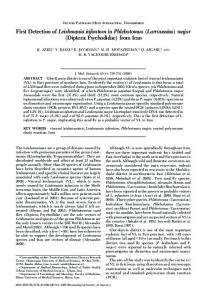
Fig. 1. Map of study area located in Ardebil province.
hosts in the Meshkin-shahr district, Ardebil province (Edrissian et al. 1999, Mohebali et al. 2005). Two sand ßy species, Phlebotomus (Paraphlebotomus) alexandri Sinton and Phlebotomus (Larroussius) kandelakii Shchurenkova have been reported as probable and proven vectors of L. infantum in Iran, respectively (Rassi etal. 2005, Azizi et al. 2006). Three other species, Phlebotomus (Larroussius) keshishiani Shchurenkova, Phlebotomus (Larrroussius) perfiliewi Parrot, and Phlebotomus (Larroussius) major Annandale, have been found naturally infected with promastigotes and are suspected vectors of VL in the country (Sahabi et al. 1992, Seyyedi-Rashti et al. 1995, Rassi et al. 1997, Rassi and Javadian 1999). This study was carried out between 2004 and 2005 in rural areas of Germi district, Ardebil province, in northwestern Iran, to identify the main vector(s) of L. infantum and determine host preferences. Materials and Methods Study Areas. The study was carried out in the city of Germi and three villages of Kalansora, Hamzeh Khanlo, and Damircholo in Germi County in northwestern Iran. The altitude is 1,100 m above sea level (Fig. 1). The total population of the Germi was ⬇91,270 in 2006. The climate is very hot (up to 40⬚C) in the summer and quite cold (⫺27⬚C) during the
winter. The summers are short, lasting from mid-May to mid-September. The main activities of the people are agriculture and animal husbandry. Sand Fly Collection. Sand ßies were collected biweekly from indoors (e.g., bedroom, guestroom, toilet, and stable) as well as outdoors (wall cracks and crevices and animal burrows) by using sticky papers (60 papers per village) and CDC light traps (three traps per village) during JulyÐSeptember 2004 and 2005. Sand ßy dissections were carried out in August and September during the peak of sand ßy activity. After removal of the terminal segments of abdomen containing the spermathecae and the heads of female sand ßies in a drop of PuriÕs medium, they were examined for the presence of Leishmania-like parasites and identiÞed to species by using keys of Theodor and Mesghali (1964). Identification of Host Preference. Bloodmeals of engorged females were blotted on Whatman No.1 Þlter paper. The ßy number, collection date, and place of collection were noted and the blotted papers were sent to the Parasitology Unit, Department of Medical Parasitology and Mycology at Tehran University of Medical Sciences for enzyme-linked immunosorbent assay (ELISA) testing (Edrissian et al. 1985). DNA Extraction. DNA was extracted as described by Motazedian et al. (2002). First, 50 l of lysis buffer (50 mM Tris-HCl, pH 7.6, 1 mM EDTA, and 1% Tween 20) was added to each slide bearing promastigotes, and after
1096
JOURNAL OF MEDICAL ENTOMOLOGY
Vol. 46, no. 5
Fig. 2. Geimsa-stained promastigotes of L. infantum from naturally infected P. perfiliewi. (Online Þgure in color.)
a few minutes the contents of each slide were transferred to a 0.5-ml microtube. Each slide was rinsed three times into the same microtube. Twelve microliters of proteinase K (19 g/ml) was then added, and the mixture was incubated at 37 C⬚ overnight before adding a 300-l solution of phenol, chloroform, and isoamyl alcohol (25: 24:1, by volume). After being shaken vigorously, the tube was centrifuged at 10,000 rpm for 10 min, and then the DNA in the supernatant solution was precipitated with 400 l of cold, pure ethanol, resuspended in 50 l of double-distilled water and stored at ⫺20⬚C before being analyzed by polymerase chain reaction (PCR). PCR. AmpliÞcation of the variable region of the minicircle Leishmania kinetoplast DNA was carried out with some modiÞcation as described by Aransay et al. (2000). Primers LINR4 (forward, 5⬘-GGG GTT GGT GTA AAA TAG GG-3⬘) and LIN17 (reverse, (5⬘-TTT GAA CGG GAT TTC TG-3⬘) were designed from the conserved region of the kinetoplast minicircle and contained conserved sequence blocks (CSBs) CSB3 and CSB2, respectively (Aransay et al. 2000). Reference strains of L. infantum (MCAN/IR/ 96/Lon49), Leishmania tropica (MHOM/IR/89/ ARD2), and Leishmania major (MHOM/IR/54/ LV39) were used as positive controls. All were obtained from the Medical Parasitology Laboratory, School of Public Health, Tehran University of Medical Sciences. The ampliÞcation reaction was carried out in a 25-l mixture, containing 250 M deoxynucleoside triphosphate, 1.5 mM MgCl2, 0.1 U of Taq polymerase (5 U/l) (CinaGene, Tehran, Iran), 1 M of each primer (LINR4 and LIN17), 1⫻ PCR buffer (Roche Diagnostics, Mannheim, Germany), and 5 l of DNA extract, overlaid with mineral oil. The DNA was am-
pliÞed in a thermocycler (CG1-96, Corbett Research, Sydney, Australia) at 94⬚C for 5 min followed by 30 cycles, each consisting of 30 s at 94⬚C, 30 s at 52⬚C, and 1 min at 72⬚C. After the last cycle, the extension was continued for 5 min. Agarose Gel Electrophoresis. Five microliters of each PCR product was resolved on a 1.5% agarose gel and visualized under an UV transilluminator with ethidium bromide. Parasites were identiÞed by comparison with positive controls of L. infantum, L. major, and L. tropica and molecular weight markers. Results In total, 3,560 female sand ßies were collected and dissected. They included P. perfiliewi (25.4%), P. kandelakii (15.6%), P. major (2%), Phlebotomus balcanicus Theodor (7%), Phlebotomus halepensis Theodor (2.5%), Phlebotomus brevis Theodor & Meshghali (0.2%), Phlebotomus papatasi Scopoli (3%), Sergentomyia dentata Sinton (35%), and Sergentomyia sintoni Pringle (9.3%). P. perfiliewi, with 45.6% prevalence, was the dominant Phlebotomus species. Upon careful examination of the genitalia of the males of this species, we determined that all were of the subspecies transcaucasus. Among the sand ßies dissected, 60% were unfed, 10% gravid, 15% semigravid, and 15% blood fed. Of 905 P. p. transcaucasicus, 10 (1.1%) were observed naturally infected with promastigotes under light microscopy at 40⫻ magniÞcation. All infected specimens were unfed and parous. Seven infected sand ßies were caught on sticky papers and three in CDC light traps. Four infected P. p. transcaucasicus were collected indoors (VL case dwellings) and six of them outdoors ⬍10-m distance from positive cases
September 2009
RASSI ET AL.: P. perfiliewi IN IRAN
1097 Discussion
Control of leishmaniasis depends on ecological and epidemiological information pertaining to the disease, such as identiÞcation of preferred hosts and detection of natural infections in the vector(s). Finding naturally infected, wild-caught specimens that are anthropophilic fulÞlls two essential requirements for incriminating a sand ßy vector (Killick-Kendrick 1990). The applicability of molecular techniques (PCR) including DNA for detection and identiÞcation of Leishmania within sand ßies by DNA hybridization have been well proven (Ready et al. 1988, Rodriguez et al. 1999, Rogers et al. 1988). One of the most suitable targets for this purpose is minicircle kinetoplast DNA because of its high number of repeats in each parasite cell (10,000) and a well-known sequence of variable region, which has high diversity in different species, facilitating differentiation (Aransay et al. 2000). In the current study, infection of P. p. transcaucasicus by L. infantum was conÞrmed using both microscopic and molecular methods. As mentioned, P. perfiliewi was Þrst found naturally infected with promastigotes from Germi district, Ardebil province, northwest of Iran, in 1997 and 1999 (Rassi et al. 1997, Rassi and Javadian 1999), but due to lack of availability of suitable molecular methods at that time, identiÞcation of the parasite was difÞcult. Therefore, P. perfiliewi was reported as the probable vector of VL in Iran (Rassi et al. 1997, Rassi and Javadian 1999). P. perfiliewi is an important vector of Leishmania infantum in Greece and is a suspected vector of VL in Serbia and of canine leishmaniasis in Tunisia (Lewis 1982). It may have transmitted VL in Emilia-Romagna in 1971 under unusual weather condition and is probably a vector of VL in Romania (Lewis 1982). Its secondary role as a vector of canine leishmaniasis in Tunisia, and its relationship to VL transmission in Yugoslavia is discussed by Maroli and Bettini (in Lewis 1982). This species also transmits L. infantum, which causes CL in Italy (Lewis 1982). The results of bloodmeal analyses indicate the P. p. transcaucasicus collected in Iran are strongly anthropophilic (36.3% with human blood), whereas in Albania P. p. perfiliewi were found fed on cow (80%) and chicken blood (20%) (Velo et al. 2005). Eduardo et al. (2005) considered this species a zoophile in Italy. The apparent secondary preference of P. p. transcaucasicus for dogs (23.5%), the main domestic reservoir of
Fig. 3. Results of PCR-based of DNA extracted from Giemsa-stained promastigote slides. The bands, shown on 1.5% agarose gel stained with ethidium bromide, correspond to molecular weight markers (lane M, 100-bp ladder), reference strains of L. tropica (lane 1), L. major (lane 2), L. infantum (lane 3), samples from two specimens of P. p. transcaucasicus (lanes 4 and 5).
houses. Twenty slides of promastigotes from infected sand ßies were prepared and stained with Giemsa (Fig. 2). The contents of 10 of these slides were analyzed by PCR for the presence of L. infantum by using standard PCR with primers LINR4 and LIN17. The resulting bands matched the L. infantum reference isolate, which was equal to 720 bp (Fig. 3). The bands of the L. major and L. tropica reference isolates were 560 bp and ⬇760 bp, respectively (Fig. 3). The results of host preference assays are presented in Table 1. Based on ELISAs of 226 bloodmeals of P. p. transcaucasicus, the proportion giving a positive reaction with alkaline phosphate antihuman conjugate ranged between 25 and 42.5%, with an average of 36.3%, indicating a strong preference for human blood. The positive proportions for dogs, the main domestic reservoir of the disease, were between 20.0 and 30.8%, with an average of 23.5%. Table 1.
Results of ELISA tests on blood meals of P. p. transcaucasicus
Name of places Germi city Kalansora Hamze-Khanlo Damircholo Total
Human
Dog
Unknown
Date of sand ßy collection (JulyÐSept.)
Total no. bloodmeal
No. (⫹)
% (⫹)
No. (⫹)
% (⫹)
No. (⫹)
% (⫹)
2004 2005 2004 2005 2004 2005 2004 2005 2004Ð2005
40 38 20 27 29 21 25 26 226
17 12 5 11 10 7 9 11 82
42.5 31.6 25.0 40.7 34.5 33.3 36.0 42.3 36.3
10 8 4 7 6 5 7 8 91
25.0 22.0 20.0 25.9 20.7 23.8 28.0 30.8 23.5
13 18 11 9 13 9 9 7 91
32.5 27.4 55.0 33.4 44.8 42.9 36.0 26.9 40.2
1098
JOURNAL OF MEDICAL ENTOMOLOGY
disease, may indicate that this species also plays an important role in transmission of VL to dogs . Based on its high degree of anthropophily and natural infections with L. infantum and the fact that it was the only species found infected with L. infantum, we conclude that P. p. transcaucasicus is the principal vector of visceral leishmaniasis in northwestern. P. kandelakii was the Þrst sand ßy incriminated as a vector of VL in Meshginshahr district in northwestern Iran (Rassi et al. 2005). The high prevalence of P. p. transcaucasus revealed by this study is consistent with the reports of Lewis (1982) on the distribution of this species in Iran and the Republic of Azerbaydzhan, which adjoins our study area. This is the Þrst report incriminating P. p. transcaucasicus as the main vector of VL due L. infantum in the region.
Acknowledgments We are grateful to Z. Zare and M. Jalali for assistance in the Þeld sampling and to M. Karamian and M. Kalantari for helping in molecular assays. We appreciate Shiraz University of Medical Sciences for providing facilities to conduct the research. This study received Þnancial support from the School of Public Health Tehran University of Medical Sciences.
References Cited Alexander, B., and M. Maroli. 2003. Control of phlebotomine sand ßies. Med. Vet. Entomol. 17: 1Ð18. Aransay, A. M., E. Scoulica, and Y. Tselentis. 2000. Detection and identiÞcation of Leishmania DNA within naturally infected sand ßies by semi-nested PCR on minicircle kinetoplast DNA. Appl. Environ. Microbiol.. 66: 1933Ð1938. Azizi, K., Y. Rassi, E. Javadian, M. H. Motazedian, S. Rafizadeh, M. R. Yaghoobi-Ershadi, and M. Mohebali. 2006. Phlebotomus (Paraphlebotomus) alexandri: a probable vector of Leishmania infantum in Iran. Ann. Trop. Med. Parasitol. 100: 63Ð 68. Desjeux, P. 2000. Leishmania/HIV coinfection, south-western Europe, 1990 Ð1998. LEISH CDS/CSR/DEC. WHO, 42.World Health Organization, Geneva, Switzerland. Edrissian, G., A. V. Manochehri, and A. Hafizi. 1985. Application of an enzyme-linked immunosorbent Assay (ELISA) for determination of the human blood index in anopheline mosquitoes collected in Iran. J. Am. Mosq. Control Assoc. 1: 349 Ð352. Edrissian, G. H., A. Nadim, A. V. Alborzi, and S. Ardehali. 1999. Visceral leishmaniasis: the Iranian experiences. Arch. Iran. Med. 1: 22Ð26. Eduardo Gomez, S., C .W. Doud, and M. Maroli. 2005. Surveillance of Leishmania sp. among sand ßies in Sicili (Italy) using a ßuorogenio real-time polymerase chain reaction. Am. J. Trop. Med. Hyg. 72: 138 Ð141. Killick-Kendrick, R. 1990. Phlebotomus vectors of visceral leishmaniasis, a review. Med. Vet. Entomol. 4: 1Ð24. Lachaud, L., S. Marchergui-Hammami, E. Chabbert, J. Dereure, J. Dedet, and P. Bastien. 2002. Comparison of six PCR methods using peripheral blood for detection of canine visceral leishmaniasis. J. Clin. Microbiol. 40: 210Ð215.
Vol. 46, no. 5
Lewis, D. J. 1982. A taxonomic review of the genus phlebotomus (Diptera: Psychodidae). Bull. Br. Mus. Hist. (Entomol.) 45: 121Ð209. Mohebali, M., H. Hajjaran, Y. Hamzavi, I. Mobedi, S. Arshi, Z. Zarei, B. Akhoundi, K. Manouchehri, R. Avizeh, and M. Fakhar. 2005. Epidemiological aspects of canine visceral leishmaniasis in the Islamic Republic of Iran. Vet. Parasitol. 129: 243Ð251. Motazedian, M. H., M. Karamian, H. A. Noyes, and S. Ardehali . 2002. DNA extraction and ampliÞcation of Leishmania from archived, Giemsa stained slides, for the diagnosis of cutaneous leishmaniasis by PCR. Ann. Trop. Med. Parasitol. 96: 31Ð34. Rassi, Y., E. Javadian, A. Nadim, A. Zahraii, H. Vatandoost, M. H. Motazedian, K. Azizi, and M. Mohebali. 2005. Phlebotomus (Larroussius) kandelakii the principle and proven vector of visceral leishmaniasis in north west of Iran. Pak. J .Biol. Sci. 8: 1802Ð1806. Rassi, Y., and E. Javadian. 1999. Study on Ph. perfiliewi, the probable vector of visceral leishmaniosis (VL) in the north west of Iran (1995Ð1998), abstract P 1. In Proceedings of the Third International symposium on phlebotomine sand-ßy, 23Ð27 August 1999, Montpellier, France. Rassi, Y., E. Javadian, and A. Nadim. 1997. Natural promastigote infection of sandßies and its Þrst occurrence in S. dentata in Ardebil province, north west of Iran. Iran. J. Public Health 6: 7Ð12. Ready, P. D., D. F. Smith, and R. Killick-kendrick. 1988. DNA hybridization on squash-blotted sand ßies to identify both Phlebotomus papatasi and infecting Leishmania major. Med. Vet. Entomol. 2: 109 Ð116. Rodriguez, N., C. M. Aguilar, M. A. Barrios, and D. C. Braker. 1999. Detection of Leishmania braziliensis in naturally infected individual sand ßies by the polymerase chain reaction. Trans. R. Soc. Trop. Med. Hyg. 93: 47Ð 49. Rogers, W. O., P. F. Burnheim, and D. F. Wirth. 1988. Detection of Leishmania within sand ßies by kinetoplast DNA hybridization. Am. J. Trop. Med. Hyg. 93: 47Ð 49. Sahabi, Z., M. A. Seyyedi-Rashti, A. Nadim, E. Javadian, M. Kazemeini, and M. R. Abai. 1992. A preliminary report on the natural leptomonad infection of Phlebotomus major in an endemic focus of V.L. in Fars province, south of Iran. Iran. J. Public Health 21: 87Ð93. Seyyedi-Rashti, M. A., Z. Sahabi, and A. Kanani-Notash. 1995. Phlebotomus (Larroussius) keshishiani Shchurenkova 1936, another vector of visceral leishmaniasis in Iran. Iran. J. Public Health 24: 23Ð30. Sharma, U., and S. Singh. 2008. Insect vectors of Leishmania: distribution, physiology and their control. J. Vector Borne Dis. 45: 255Ð272 Shyma, S., and M. Rai. 2002. Laboratory diagnosis of visceral leishmaniasis. Clin. Diag. Lab. Immunol. 9: 951Ð958. Theodor, O., and A. Mesghali. 1964. On the phlebotomine of Iran. J. Med. Entomol. 1: 285Ð300. Velo, E., A. Paparisto, G. Bongiorno, T. Dimuccio, C. Khoury, S. Bino, M. Gramiccla, L. Gradoni, and M. Maroli. 2005. Entomological and parasitological study on phlebotomine sand ßies in central and northern Albania. Parasite 12: 45Ð 49. [WHO] World Health Organization. 1990 Control of the Leishmaniases. Report of a WHO Expert Committee. Technical Report Series No. 793. World Health Organization, Geneva, Switzerland. Received 18 February 2008; accepted 13 April 2009.