Intensive Care Med (2006) 32:941–942 DOI 10.1007/s00134-006-0127-4
Matthieu Legrand Lucien Lecuyer Andry Van De Louw Stéphane Thierry
Pleural drain malposition Accepted: 22 February 2006 Published online: 24 March 2006 © Springer-Verlag 2006
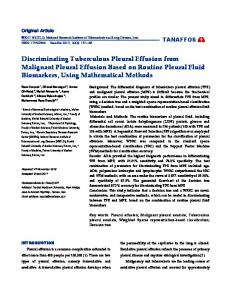
Sir: Small-bore chest drains with removable introducer needle (such as the 8-Fr Pleurocath® , Plastimed, Saint-Leu-La Fôret, France) can be used to drain liquid pleural effusions or pneumothorax [1]. We report a case of severe vascular injury due to Pleurocath® malposition, with the goal of raising awareness among physicians about this life-threatening complication, and discuss methods for a safer procedure. A 67-year-old man with a recent history of trauma and hemorrhagic shock followed by postanoxic encephalopathy was admitted to a general medicine ward for investigation of left hydropneumothorax. Thoracic computed tomography confirmed the left hydropneumothorax. A Pleurocath® was inserted on the left mid-axillary line with the patient in the 45° semi-recumbent position. Blood issued through the distal end of the catheter immediately after insertion but was mistakenly taken as evidence of hemothorax. Hemorrhagic shock occurred within a few minutes and the catheter was clamped. The patient was transferred to the intensive care unit. Catheter malposition was suspected. A chest radiograph showed the tip of the catheter in the superior mediastinum, but without an accurate location. Fluid resuscitation and blood transfusion were started. Thoracic computed tomography (CT) with contrast enhancement showed that the catheter ran through the left ventricle, as-
CORRESPONDENCE
cending aorta (Fig. 1), and right carotid. The patient was immediately transferred to a cardiothoracic surgery center, where the catheter was removed through a left thoracotomy. The patient was discharged from the unit 3 weeks later. Use of small-bore catheters may reduce morbidity compared with chest tubes [2]. Several cases of cardiac puncture have been reported with larger-bore drains over rigid inner trocars (e.g., Mallinckrodt® ) [3] and, more rarely, with small-bore drains (e.g., Pleurocath® ). However, our case report shows that small-bore catheters may be associated with life-threatening complications. In our patient, the main factors causing cardiac injury were probably insertion of the catheter low in the left chest wall, through the sixth intercostal space and the small size of the effusion. The published guidelines of the British Thoracic Society [2] recommend insertion in the “safe triangle” bordered by the anterior border of the latissimus dorsi, the lateral border of
the pectoralis major muscle, a line superior to the horizontal level of the nipple, and on the midaxillary line. Ultrasound guidance would have been particularly useful in this man with antecedent chest trauma, defining the presence of pleural symphysis. CT scan was used for location, but the patient’s position was not the same at the time of the Pleurocath® insertion. Furthermore, ultrasound guidance has been reported to decrease the incidence of complications [4]. With large-bore drains, blunt dissection through the chest wall and into the pleural space is recommended [2] because it is safer than direct insertion of the trocar. Insertion of a chest tube should never be performed with any substantial force. Because this technique is not feasible when a small-bore catheter is used, ultrasound guidance may be a good method for preventing injury. When ventricular injury during pleural catheter placement is suspected, the drain should be clamped immediately and CT performed on an
Fig. 1 The course of the catheter (arrow) as seen on thoracic CT
942
emergency basis. CT seems superior to chest radiography for establishing the diagnosis [5]. Cardiac injury requires surgical treatment. In conclusion, physicians should be aware that small-bore pleural catheters can cause vascular and cardiac injury requiring immediate clamping of the catheter, CT evaluation, and surgical repair. Simply removing the catheter might cause life-threatening bleeding, tamponade, and hemorrhagic shock. Use of ultrasonography guidance, insertion by a trained operator, and adherence to recommendations should avoid these complications.
References 1. Azoulay E (2003) Pleural effusions in the intensive care unit. Curr Opin Pulm Med 9:291–297 2. Laws D, Neville E, Duffy J, on behalf of the British Thoracic Society Pleural Disease Group, a subgroup of the British Thoracic Society Standards of Care Committee (2003) BTS guidelines for the insertion of a chest drain. Thorax 58:ii53–ii59 3. Jaillard SM, Tremblay A, Conti M, Wurtz AJ (2002) Uncommon complications during chest tube placement. Intensive Care Med 28:812–813 4. Jones PW, Moyers JP, Rogers JT, Rodriquez RM, Lee YC, Light RW (2003) Ultrasound-guided thoracentesis: is it a safer method? Chest 123:418–423 5. Baldt MM, Bankier AA, Germann PS, Poschl GP, Skrbensky GT, Herold CJ (1995) Complications after emergency tube thoracostomy: assessment with CT. Radiology 195:539–543
M. Legrand · L. Lecuyer · A. Van De Louw · S. Thierry (u) Centre Hospitalier Sud Francilien, General Intensive Care Unit, Site Evry–Quartier du Canal, 91014 Evry, France e-mail:
[email protected] Tel.: +33-1-60875225