CASE REPORT
Pleural Empyema Due to Salmonella typhi Faisal Iqbal Afridi1, Badar Jahan Farooqi1 and Arif Hussain2
ABSTRACT Salmonella serotypes most often produce gastroenteritis, enteric fever, bacteremia, vascular infection and chronic carrier state. Localized infection may occur at any site after Salmonella bacteremia. Pulmonary involvement due to Salmonella infection is rare. Empyema occurs usually in elderly patients or in patients with underlying diseases such as diabetes mellitus, malignancy, or pulmonary disease. We report the case of an 83-year-old male diabetic patient who presented with fever, productive cough, and difficulty in swallowing. The chest radiographs revealed soft shadowing mild atelactasis and pulmonary abscess on left side. CT-guided aspiration of pus was done. Salmonella enterica serotype typhi was isolated from pus sample. Pleural empyema or abscess usually requires surgical drainage in addition to antimicrobial therapy. After complete course of antimicrobial therapy, the patient improved. Key words:
Empyema. Salmonella typhi. Pulmonary abscess. Antimicrobial therapy.
INTRODUCTION Salmonellae are gram-negative, facultatively anaerobic, non-spore-forming bacilli. The wide spread distribution of Salmonella in the environment, their increasing prevalence in the global food chain, and their virulence and adaptability result in enormous medical, public health, and economic impact worldwide.1 Specific Salmonella serotypes most often produce characteristic clinical manifestations that have been given the syndrome designations gastroenteritis, enteric fever, bacteremia and vascular infection, localized infections, and chronic carrier state.1 Localized infections develop in approximately 5 – 10% cases with Salmonella bacteremia, and the presentation may be delayed.1 Extra-intestinal infectious complication especially pleural effusion or empyema due to Salmonella species, is extremely rare. Although many cases of respiratory involvement with Salmonella species have been described worldwide, only few cases with respiratory manifestations with Salmonella typhi (S. typhi) have been reported from Turkey, India, and Pakistan.2-4 Serious respiratory complications like pleural empyema usually occurs in elderly patients or in patients with severe underlying diseases. We report a case of S. typhi empyema in an 83-year-old male diabetic patient.
CASE REPORT An 83-year-old male patient was admitted in the gastrointestinal ward of Ziauddin University Hospital, Department of Microbiology1 / Pathology2 , Dr. Ziauddin University Hospital, North Nazimabad Campus, Karachi. Correspondence: Dr. Faisal Iqbal Afridi, A-79/6, Block No. 14, Gulistan-e-Jauhar, Karachi-75290. E-mail:
[email protected] Received January 19, 2012; accepted April 26, 2012.
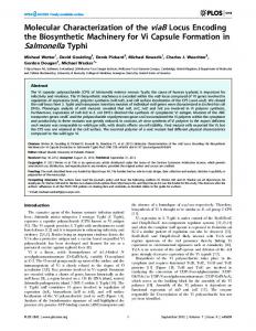
Karachi, Pakistan with the complaints of fever with rigors, productive cough and difficulty in swallowing for 1 week duration. He had experienced similar symptoms about 3 months back, but did not seek medical advice because the symptoms were self-resolved. The patient was a known case of non-insulin dependent diabetes mellitus and essential hypertension for 10 – 12 years. He smoked 40 – 50 cigarettes per day upto 40 years previously. He also had several episodes of pneumonia in the past. These pneumonic episodes were diagnosed by a general practitioner on clinical ground. He received various antimicrobials. Names, dosages, and duration of these antimicrobials are not known. The examination revealed an ill-looking man who was clinically anaemic and had signs of a left pleural effusion. The pulse rate was 80 beats per minute, blood pressure was 130/80 mmHg, temperature was 100°F, and there were no signs of heart failure. Respiratory examination revealed harsh vesicular breathing with scattered left basal crepitations. Chest radiographs posteroanterior and left lateral view (Figure 1) showed soft shadowing mild atelactasis and pleural abscess on left side. Blood samples were obtained for culture, fasting blood sugar (FBS), complete picture, malarial parasite, urea, creatinine, electrolytes, troponin-I. Troponin-I was done to rule out any cardiac pathology. His sputum sample was also taken for acid fast bacilli (AFB) smear. He was started injection piperacillin/tazobactam 4.5 gm I/V 8 hourly and amikacin 500 mg I/V OD empirically. His fever did not subside after 48 hours of antimicrobial treatment. Computed tomography (CT) guided aspiration of left parasternal lung abscess was done. Approximately, 150 ml collection of pus was aspirated and sent to laboratory for Gram stain, culture and antimicrobial sensitivities, AFB smear, and cytology. His FBS was 150 mg/dl, haemoglobin was 11.3 gm/dl, TLC 12.1 x 10⁹/L, platelet count was 571 x 109/L; differential leukocyte count was neutrophils 86%, lymphocytes 9%,
Journal of the College of Physicians and Surgeons Pakistan 2012, Vol. 22 (12): 803-805
803
Faisal Iqbal Afridi, Badar Jahan Farooqi and Arif Hussain
Figure 1: Posteroanterior and left lateral chest radiographs showing soft shadowing mild atelactasis and pleural abscess on left side.
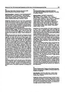
Figure 2: Non-lactose fermenting colonies on MacConkey agar.
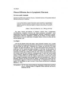
Figure 3: Biochemical reactions of Salmonella typhi with API 20E strip.
DISCUSSION
and monocytes 5%. Malarial parasite was not seen. Blood urea was 31 mg/dl, creatinine was 0.59 mg/dl, sodium was 136 mmol/L, potassium was 4.3 mmol/L, and troponin-I (quantitative) was 0.23 ng/ml. His AFB smear of sputum was negative. Cytology report of pus sample showed evidence of acute inflammation. AFB smear of pus sample was negative. The pus sample was inoculated on MacConkey (Oxoid Ltd. UK), chocolate (Oxoid Ltd. UK), sheep blood agar (Oxoid Ltd. UK), and cooked meat broth (Oxoid Ltd. UK). All these plates and cooked meat broth were incubated at 37ºC aerobically for 24 hours. After overnight 804
incubation, the sample from the bottom of the cooked meat broth was inoculated on anaerobic sheep blood agar (Oxoid Ltd. UK) plate. This inoculated anaerobic sheep blood agar plate was further incubated anaerobically for 48 hours. Gram stain of pus showed numerous pus cells and occasional Gram negative rods. After overnight incubation, the blood culture broth sample was subcultured on chocolate and MacConkey agar and incubated at 37°C aerobically for 24 hours. Blood culture yielded no growth after 7 days of incubation at 37°C. Culture of pus sample yielded pure growth of non-lactose fermenting colonies on MacConkey agar (Figure 2). The chocolate and sheep blood agar plates also showed pure growth of greyish mucoid colonies. They were Gram negative rods, catalase positive, oxidase negative, and were motile. The organism was tested biochemically using API 20E (bioMerieux). Antimicrobial susceptibility testing was carried out on the Mueller-Hinton agar (Oxoid Ltd. UK) using Clinical Laboratory Standard Institute criteria.5 The antimicrobials tested were ampicillin 10 µg (Oxoid Ltd. UK), chloramphenicol 30 µg (Oxoid Ltd. UK), cotrimoxazole 25 µg (Oxoid Ltd. UK), ceftriaxone 30 µg (Oxoid Ltd. UK), cefixime 5 µg (Oxoid Ltd. UK), and ciprofloxacin 5 µg (Oxoid Ltd. UK). Escherichia coli American type culture collection (ATCC®) 25922 used as control. Next day, the organism was identified as S. typhi (Figure 3). This was confirmed by type specific antisera (Bact-Med Diagnostics, Depto Lab Pakistan). Anaerobe was not isolated from this pus sample after 48 hours of incubation. The organism was sensitive to ampicillin, chloramphenicol, co-trimoxazole, ceftriaxone and cefixime while being resistant to ciprofloxacin. After the sensitivity report was available, injection piperacillin/ tazobactam and amikacin were stopped and injection ceftriaxone 1 gm I/V 12 hourly was started for 7 days along with supportive therapy. After 48 hours of treatment the patient was afebrile. The patient was discharged after 7 days on oral cefixime 400 mg 12 hourly for another 2 weeks. At the end of complete 3 weeks of antimicrobial treatment, the patient remained asymptomatic and afebrile.
The most common clinical presentation of Salmonella infection is acute enterocolitis, but Salmonella can cause fever, sustained bacteremia, and even extraintestinal infectious complications like pneumonia without gastrointestinal manifestations.6 The pathogenesis of extra-intestinal infectious complications of typhoid fever depends on the ingested inoculum size, virulence of the strain, host's immune response, previous exposure, and local protective factors.7 Localized infection may occur at any site after Salmonella bacteremia. Localized infection has been reported in the thyroid, meninges, bone, heart, lungs,
Journal of the College of Physicians and Surgeons Pakistan 2012, Vol. 22 (12): 803-805
Pleural empyema due to Salmonella typhi
adrenals, pancreas, spleen, liver, testes, pericardium, soft tissues, areas of necrosis or infarction, benign or malignant tumours and cysts.8 Pneumonia, empyema, and bronchopleural fistulas caused by S. typhi occur in 1 – 6% of cases.7 Saphra et al. reported 85 cases of pleural empyema with Salmonella species accounting for 1% of all cases of Salmonellosis.9 Thereafter, pleural empyema due to Salmonella species especially due to S. typhi has rarely been reported. To our knowledge, only one case of lung hydatid cyst infected with S. typhi was reported from Pakistan.4 Although few cases due to S. typhi infection with respiratory involvement has been reported from India.3,10 Pneumonia or empyema, the predominant types of serious respiratory disease occurs usually in elderly patients or in patients with underlying diseases such as diabetes mellitus, malignancy, or pulmonary disease. These underlying diseases like diabetes mellitus decreases the immune status of the host and may result in the localization of Salmonella empyema as a consequence of an inapparent Salmonella bacteremia. We could not explain whether pleural empyema was initial focus. Bacteremia might have occurred and then Salmonella settled in lung tissues. However, his blood culture was sterile, probably due to prior treatment with antimicrobials for the episodes of pneumonia before admission. As our patient also had several episodes of pneumonia before admission, it is possible that these may have predisposed to the development of empyema. In addition to this, the patient's glycemic control is not very good as indicated by his FBS report. Due to poor immune status, the organism which may have already settled in pleural cavity got the chance to multiply and produce symptoms. The diagnosis of the localized Salmonella infection can be confirmed by the culture of specimens that are normally sterile, such as blood, and pleural fluid on ordinary microbiological media. The presence of Salmonella in sputum does not necessarily imply lower respiratory tract infection because S. typhi may colonize the upper respiratory tract.8
Pleural empyema or abscess usually requires surgical drainage in addition to antimicrobial therapy. The recommended therapy for pulmonary manifestations caused by S. typhi is ceftriaxone or fluoroquinolones for 14 – 21 days.7 Alternate therapy for susceptible strains is co-trimoxazole, ampicillin, or chloramphenicol for 14 – 21 days.7 This S. typhi isolate is sensitive to all first line agents but was resistant to ciprofloxacin. This indicates the non-judicious use of fluoroquinolones in the setup.
REFERENCES 1.
Pegues DA, Ohl ME, Miller SI. Salmonella species, including Salmonella typhi. In: Mandell GL, Bennett JE, Dolin R, editors. Principles and practice of infectious diseases. 6th ed. Philadelphia: Churchill Livingstone; 2005.p. 2636-54.
2.
Kömüs N, Kilinç O, Günes J, Soytürk M. Pleural empyema due to Salmonella typhi. Tuberk Torak 2005; 53:397-400.
3.
Mishra S, Pattnaik D, Mahapatra A, Swain B. Pleural empyema due to S. typhi: a case report. Indian J Pathol Microbiol 2004; 47:75-6.
4.
Aslam F, Bhaila I, Nadeem N, Fadoo Z. Salmonella typhiinfected lung hydatid cyst. Pediatr Infect Dis J 2005; 24:270-2.
5.
Clinical and Laboratory Standards Institute. Performance standards for antimicrobial susceptibility testing: twentieth informational supplement M100-S20. Wayne: CLSI; 2010.
6.
Olutola PS, Familusi JB. Salmonella typhi pneumonia without gastrointestinal manifestations. Diagn Imaging Clin Med 1985; 54:263-7.
7.
Huang DB, DuPont HL. Problem pathogens: extra-intestinal complications of Salmonella enterica serotype typhi infection. Lancet Infect Dis 2005; 5:341-8.
8.
Rim MS, Park CM, Ko KH, Lim SC, Park KO. Pleural empyema due to Salmonella: a case report. Korean J Intern Med 2000; 15:138-41.
9.
Saphra I, Winter JW. Clinical manifestations of Salmonellosis in man: an evaluation of 7779 human infections identified at the New York Salmonella Center. N Engl J Med 1957; 256:1128-34.
10. Mohanty S, Gaind R, Paglietti B, Paul P, Rubino S, Deb M. Bacteraemia with pleural effusions complicating typhoid fever caused by high-level ciprofloxacin-resistant Salmonella enterica serotype typhi. Ann Trop Paediatr 2010; 30:233-40.
Journal of the College of Physicians and Surgeons Pakistan 2012, Vol. 22 (12): 803-805
805