REVIEW
Polynucleotide phosphorylase: Not merely an RNase but a pivotal post-transcriptional regulator Todd A. Cameron1☯‡, Lisa M. Matz1☯‡, Nicholas R. De Lay ID1,2* 1 Department of Microbiology and Molecular Genetics, McGovern Medical School, University of Texas Health Science Center, Houston, Texas, United States of America, 2 MD Anderson Cancer Center UTHealth Graduate School of Biomedical Sciences, University of Texas Health Science Center, Houston, Texas, United States of America ☯ These authors contributed equally to this work. ‡ These authors share first authorship on this work. *
[email protected]
a1111111111 a1111111111 a1111111111 a1111111111 a1111111111
OPEN ACCESS Citation: Cameron TA, Matz LM, De Lay NR (2018) Polynucleotide phosphorylase: Not merely an RNase but a pivotal post-transcriptional regulator. PLoS Genet 14(10): e1007654. https://doi.org/ 10.1371/journal.pgen.1007654
Abstract Almost 60 years ago, Severo Ochoa was awarded the Nobel Prize in Physiology or Medicine for his discovery of the enzymatic synthesis of RNA by polynucleotide phosphorylase (PNPase). Although this discovery provided an important tool for deciphering the genetic code, subsequent work revealed that the predominant function of PNPase in bacteria and eukaryotes is catalyzing the reverse reaction, i.e., the release of ribonucleotides from RNA. PNPase has a crucial role in RNA metabolism in bacteria and eukaryotes mainly through its roles in processing and degrading RNAs, but additional functions in RNA metabolism have recently been reported for this enzyme. Here, we discuss these established and noncanonical functions for PNPase and the possibility that the major impact of PNPase on cell physiology is through its unorthodox roles.
Editor: Anita K. Hopper, Ohio State University, UNITED STATES Published: October 11, 2018 Copyright: © 2018 Cameron et al. This is an open access article distributed under the terms of the Creative Commons Attribution License, which permits unrestricted use, distribution, and reproduction in any medium, provided the original author and source are credited. Funding: This work, including the efforts of Todd A. Cameron, Lisa M. Matz, and Nicholas R. De Lay, was funded by HHS | NIH | National Institute of General Medical Sciences (NIGMS; R01GM121368). The content of this publication is solely the responsibility of the authors and does not necessarily represent the official views of the National Institutes of Health. The funders had no role in the preparation of the article. Competing interests: The authors have declared that no competing interests exist.
Author summary Widely distributed among bacteria and eukaryotes, including humans, polynucleotide phosphorylase (PNPase) is a critical enzyme in RNA metabolism that functions in most organisms as a 30 to 50 exoribonuclease. In bacteria, inactivation of the gene encoding PNPase results in a wide range of consequences, including impaired growth, diminished stress responses, and loss of virulence. In mammals, PNPase has an essential role in mitochondrial function. Mutations in the gene encoding the human PNPase (hPNPase) that reduce its activity can lead to hereditary hearing loss, encephalomyopathy, severe axonal neuropathy, delayed myelination, and Leigh syndrome. In this review, we highlight both the canonical and unorthodox activities that have been reported for PNPase. Specifically, we examine its role in bacterial mRNA and rRNA decay, RNA processing, and small regulatory RNA (sRNA) degradation and stabilization. Furthermore, we explore the recently reported findings on the function of hPNPase in mitochondrial RNA import and degradation and cytoplasmic mRNA and noncoding RNA decay. Despite being discovered more
PLOS Genetics | https://doi.org/10.1371/journal.pgen.1007654 October 11, 2018
1 / 17
than six decades ago, we are still only beginning to grasp the breadth of mechanisms by which the enzymatic activities of PNPase contribute to cellular and organismal physiology.
Introduction In 1955, Grunberg-Manago and Ochoa reported that the enzyme polynucleotide phosphorylase (PNPase) from the gram-negative bacterium Azobacter vinelandii not only synthesized polynucleotides from nucleotide diphosphates but also catalyzed the reverse reaction, the phosphorolysis of polynucleotides [1, 2]. Heppel and colleagues then demonstrated that these polynucleotides were RNA [3]. PNPase was subsequently used to generate RNA of specific sequences that were employed to decipher the genetic code [4–9]. PNPase is a highly conserved enzyme that is widely distributed among bacteria and eukaryotes, except single-cell eukaryotes such as yeast and trypanosomes [10]. While PNPase can act as a polynucleotide or poly(A) polymerase in bacteria such as Escherichia coli [11] or in plant chloroplasts [12], it predominantly functions as a 30 to 50 exoribonuclease in most organisms. PNPase plays critical roles in both bacteria and mammals. Deletion of the gene encoding PNPase from bacteria typically results in a pleiotropic phenotype including increased sensitivity to stressors [13–19] and reduced virulence [14, 20–24]. Moreover, PNPase appears to have an essential function in mammals [25], and mutations that reduce its activity result in mitochondrial disorders in humans [26–29]. In this review, we examine the post-transcriptional regulatory roles of PNPase in bacteria and humans. In bacteria, these include bulk mRNA decay, rRNA degradation, and its paradoxical function in promoting small regulatory RNA (sRNA) stability and function (Table 1). These sRNAs in turn control gene expression by altering mRNA transcription, stability, or translation, placing PNPase in a pivotal position to regulate vast gene networks. We also highlight our emerging understanding of the functions that PNPase performs in humans, including transporting RNA into mitochondria, processing mitochondrial RNAs, and degrading sRNAs called microRNAs (miRNAs) that regulate mRNA translation and/or stability. Finally, we discuss how the major impacts of PNPase on cell physiology may be due to its unconventional roles in RNA metabolism. Table 1. Roles and partners of bacterial and human PNPase.
Bacteria
Humans
Context
Localization
Role
PNPase alone
Cytoplasm
• General mRNA decay • tRNA and rRNA maturation • Poly(A) and heteropolymeric tail synthesis
+ RNase E or RNase Y degradosome
Cytoplasmic membrane
• General mRNA decay • Highly structured RNA decay
+ RNA helicase (RhlB)
Cytoplasm
• rRNA decay • Highly structured RNA decay • tRNA maturation
+ Rsr and Y-RNA
Cytoplasm
• rRNA decay
+ Hfq and sRNA
Cytoplasm
• sRNA-mediated gene regulation
PNPase + other proteins?
Mitochondrial IMS
• Mitochondrial RNA import
+ RNA helicase (SUV3)
Mitochondrial matrix
• Mitochondrial RNA decay
Apoptosis, overexpression
Cytoplasm
• miRNA, mRNA and polyadenylated noncoding RNA decay
Abbreviations: Hfq, host factor for phage Qβ; hPNPase, human PNPase; IMS, inner membrance space; miRNA, microRNA; PNPase, polynucleotide phosphorylase; Rsr, Ro sixty-related protein; sRNA, small regulatory RNA; SUV3, suppressor of Var1, 3. https://doi.org/10.1371/journal.pgen.1007654.t001
PLOS Genetics | https://doi.org/10.1371/journal.pgen.1007654 October 11, 2018
2 / 17
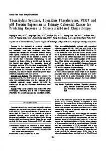
Running PNPase in reverse: Phosphorolysis of RNA by PNPase In bacteria and eukaryotes, PNPase has an important function in RNA decay. PNPase catalyzes the processive degradation of single-stranded RNA in the 30 to 50 direction using inorganic phosphate as the nucleophile to attack the 30 phosphodiester bond releasing a ribonucleoside diphosphate (NDP). A metal cofactor, Mg2+ or Mn2+, is required to stabilize the transition state of the phosphate during the reaction [30]. To degrade RNA efficiently, PNPase must bind a single-stranded stretch of RNA at least six nucleotides in length at the 30 terminus [31, 32]. PNPase subsequently degrades RNA in a stepwise motion, rapidly removing discrete segments of 6 to 7 nucleotides between short pauses [33]. A stem loop structure in an RNA substrate can act as a roadblock that halts degradation by PNPase. In vitro studies using a set of GC-rich RNA hairpins of varying length followed by single-stranded sequences demonstrated that a hairpin with a stem as short as seven base pairs inhibited degradation by PNPase [32]. However, PNPase also rapidly degrades some natural hairpins such as the Rho-independent terminators that end many sRNAs [34]. Thus, while very stable hairpins can block the exoribonucleolytic activity of PNPase, merely resulting in 30 end trimming [35], this enzyme can degrade many natural double-stranded RNAs provided that a single-stranded region is present at the 30 end to initiate degradation. This balance between degradation and inhibition is important for maturation of some tRNA transcripts, in which PNPase removes Rho-independent terminators but stops short of the CCA determinants [36–39]. Likewise, structural features of the 16S rRNA preribosomal particle are likely important for preventing excessive 30 trimming of the 16S rRNA by PNPase and other exoribonucleases during its maturation [40, 41]. Crystal structures for PNPase from Streptomyces antibioticus [42], E. coli [30, 43], Caulobacter crescentus [44], and Homo sapiens [45] have been solved, and much of the substrate specificity and activity of PNPase can be assigned to distinct structural and organizational features of this enzyme. Each PNPase monomer consists of two RNase PH-like domains, which are separated by an α-helical domain and are followed by a KH and S1 domain (Fig 1A). As a functional enzyme, PNPase is assembled into a torus-shaped trimer in which alternating RNase PH-like subunits and α-helical domains form a central ring from which the KH/S1 domains extend outward (Fig 1B and 1C) [30, 42–44]. The first RNase PH-like domain contributes to RNA and NDP binding, and the second domain additionally possesses enzymatic activity [43, 46]. The active site is positioned in a shallow groove along the inner rim of the trimer (Fig 1D, panel iii), and a constriction point formed by the FFRR loop at the entrance of the core ring creates a pore that only allows access by single-stranded RNA (Fig 1D) [43]. Catalytic activity of the central ring is facilitated by the other domains; the α-helical domain appears to regulate access of phosphate or NDP to the active site [30, 47, 48], and the KH and S1 domains each contribute to capturing and binding RNA substrates [44, 49]. Additionally, the KH domain imparts RNA directionality through the interactions of the conserved GSGG loop with the RNA backbone [44]. Finally, the processivity of PNPase is attained through its ring-like structure that retains RNA substrates via multiple RNA-binding interactions, including hydrogen binding between the GSGG loops of the KH domains and the RNA phosphate backbone (Fig 1D, panel i) and base stacking interactions between the aromatic phenylalanines in the conserved FFRR loops and a ribonucleotide base (Fig 1D, panel ii) [44].
PNPase as a component of the bacterial RNA degradosome While PNPase can function independently as a 30 to 50 exoribonuclease, in bacteria, PNPase also serves as a component of an organized RNA degradation machine (Fig 2A). Termed the degradosome, this multiprotein complex is responsible for bulk mRNA decay [50, 51]. At the core of this RNA-degrading machine in gram-negative bacteria is the endoribonuclease RNase
PLOS Genetics | https://doi.org/10.1371/journal.pgen.1007654 October 11, 2018
3 / 17
Fig 1. Domains and structure of PNPase. (A) Domain organization of PNPase, using the C. crescentus PNPase crystal structure as a reference [44]. The complete PNPase trimer viewed from the (B) top and (C) side, with domains colored the same as in (A). (D) A cut-away view of the RNA-bound trimer interior of the PNPase trimer bound to an RNA substrate (orange). Ball and stick structures depict nitrogen atoms as blue and oxygen atoms as red. Inset panels illustrate (i) the interactions between the GSGG loop of each KH domain with the RNA backbone, (ii) base stacking interactions between phenylalanine residues of the FFRR loops and several RNA bases, and (iii) the active site residues that bind phosphate and the metal cofactor. PNPase, polynucleotide phosphorylase. https://doi.org/10.1371/journal.pgen.1007654.g001
E, which initiates RNA decay. The essential N-terminal domain of RNase E contains the active site and additional features including the S1 domain and 50 sensor pocket important for binding many RNAs (reviewed in [52]). The C-terminal domain is required for formation of the
PLOS Genetics | https://doi.org/10.1371/journal.pgen.1007654 October 11, 2018
4 / 17
Fig 2. The numerous roles performed by PNPase. Schematic showing the functions performed by PNPase in bacteria and mitochondria. (A) In E. coli and other gram-negative bacteria, PNPase functions as part of the RNA degradosome in bulk mRNA decay but also operates independently of this machine to process tRNAs and rRNAs, add polynucleotide tails to RNAs, and modulate sRNA stability. PNPase binds Hfq-bound sRNAs but only degrades unbound sRNAs, which impacts both positive and negative sRNA-mediated gene regulation in E. coli. In Deinococcus radiodurans, PNPase forms a complex with Rsr mediated via Y-RNAs that degrades misfolded rRNAs. (B) The locations and functions of hPNPase are controversial, but under normal cellular conditions, hPNPase is mainly localized to the IMS where it is able to facilitate translocation of 5S rRNA, RNase P RNA, and possibly RNase MRP RNA into the mitochondrial matrix. Within the mitochondrial matrix, hPNPase degrades mitochondrial RNA and plays a role in mitochondrial DNA maintenance. Release into the cytoplasm upon overexpression or permeabilization of the mitochondrial outer membrane during apoptosis leads to the decay of mRNAs and
PLOS Genetics | https://doi.org/10.1371/journal.pgen.1007654 October 11, 2018
5 / 17
polyadenylated noncoding RNAs. hfq, host factor for phage Qβ; hPNPase, human PNPase; IMS, inner membrane space; MRP, mitochondrial RNA processing; mtRNA, mitochondrial RNA; PNPase, polynucleotide phosphorylase; Rsr, Ro sixty-related protein; sRNA, small regulatory RNA; SUV3, suppressor of Var 1, 3. https://doi.org/10.1371/journal.pgen.1007654.g002
RNA degradosome and contains binding sites for other proteins; in the canonical E. coli RNA degradosome, these proteins include the glycolytic enzyme enolase, the DEAD-box RNA helicase RhlB and PNPase [53–55]. However, the RNase E–based degradosome can vary in composition between species or even within the same organism depending on cellular conditions [56]. In C. crescentus, aconitase is exchanged for enolase, and RNase D was validated as a legitimate degradosome component [57, 58]. Gram-positive bacteria have a similar RNA degradation machine that is likewise organized around a core endoribonuclease, in this case, RNase Y. Like RNase E, RNase Y has an unstructured C-terminal region with specific binding sites for PNPase, enolase, and an RNA helicase [59]. However, despite these functional similarities, the two proteins are evolutionarily distinct, belonging to different protein superfamilies [60]. Unlike RNase E, RNase Y features an N-terminal region with an RNA-binding KH domain and an active site–containing HD domain, and its C-terminal domain additionally interacts with 50 to 30 exoribonucleases [60, 61]. PNPase and the RNA degradosome of the gram-negative bacterium E. coli have been studied in much detail. As part of the degradosome, PNPase cooperates with RNase E to degrade a specific set of mRNAs, many of which encode proteins involved in macromolecule biosynthesis or modification [50]. Additionally, within the degradosome, binding of RhlB and PNPase to the RNase E scaffold is necessary for the degradation of highly structured RNA sequences, termed repeated extragenic palindrome (REP) elements that are found in some mRNAs [62– 64]. Although PNPase participates in mRNA decay as a component of the RNA degradosome, the enzyme primarily functions independent of this machine. This is evident by the number and distribution of PNPase and RNase E molecules in E. coli. PNPase is approximately three to five times more abundant than RNase E and is mostly distributed throughout the cytoplasm (69% of PNPase; Fig 2A), whereas the majority of RNase E (91%) is located near or on the cell membrane [65]. Moreover, only a minority of E. coli PNPase trimers are bound to RNase E at any one time [53]. The independent function of PNPase is also evident by global gene expression profiling, which demonstrated that many mRNAs stabilized by the absence of PNPase are not significantly impacted by loss of the degradosome [50]. Furthermore, PNPase functions independently of the RNA degradosome in tRNA processing [37, 38, 66], rRNA degradation [40, 67], and sRNA-mediated gene regulation [68].
A specialized function for PNPase in rRNA processing and decay In bacteria, the fully assembled 70S ribosome is made up of the small 30S subunit containing the 16S rRNA and the large 50S subunit consisting of the 23S rRNA and 5S rRNA. In E. coli and presumably most gram-negative bacteria, PNPase and RNase R perform routine rRNA quality control by degrading fragments of 16S and 23S rRNAs that might otherwise compete with mature rRNAs for ribosomal proteins and impair proper ribosomal assembly [69, 70]. These rRNA fragments are generated by RNase E [70], which may cleave rRNAs that cannot be assembled into functional ribosomes due to improper processing, damage, or overabundance relative to ribosomal proteins. In E. coli, PNPase was not required for rRNA decay induced by nutrient starvation [71]. In some bacteria such as the radiation-resistant D. radiodurans, PNPase mediates rRNA degradation in response to nutrient starvation with the assistance of the Ro sixty-related protein, Rsr (Fig 2A) [72, 73]. Ro was originally identified in human cells as an autoantigen
PLOS Genetics | https://doi.org/10.1371/journal.pgen.1007654 October 11, 2018
6 / 17
recognized by antibodies from lupus erythematosus patients [74]. Ro and its homologs, which are present in many vertebrates and in roughly 5% of bacterial genome sequences [75], bind to structured noncoding RNAs called Y-RNAs [13, 72, 74, 76, 77] and are involved in rRNA decay [73, 78, 79]. In D. radiodurans, Y-RNA tethers Rsr to PNPase resulting in the formation of the “RYPER” complex (Fig 2A) that degrades structured RNA including the 5S, 16S, and 23S rRNAs [72, 73]. Y-RNAs have a highly conserved structure that consists of an extended stem generated by pairing between bases at the 30 and 50 ends and a large internal loop that in many cases is decorated with two hairpins [80, 81]. A conserved region within the Y-RNA stem binds to Rsr, whereas a region containing two hairpins, one resembling a T-arm of a tRNA, binds to the KH and S1 domains of PNPase [72, 82]. The 30 end of the misfolded rRNA appears to thread through the central pore of the toroid-shaped Rsr protein and into the central channel of PNPase, where it is degraded [72].
The opposing functions of PNPase in regulating small RNAs Although for many years it was largely assumed that PNPase only contributed to RNA processing and degradation in bacteria, it has become increasingly clear over the last decade that PNPase also plays an important role in regulating sRNA function and stability [68]. In bacteria, sRNAs range in size from 50 to 250 nucleotides and alter gene expression by sequestering regulatory proteins or by base-pairing with target mRNAs to modulate translation and transcript stability (reviewed in [83, 84]). Many sRNAs interact with RNA chaperones, such as FinO [85], ProQ [86, 87], or the host factor for phage Qβ (Hfq) [88, 89], to facilitate this process. In the latter case, Hfq protects sRNAs from degradation by occluding an endoribonuclease cleavage site [90, 91] and facilitates sRNA–mRNA annealing [92, 93]. In E. coli and its close relative Salmonella Typhimurium, binding by Hfq dictates whether an sRNA is degraded or stabilized by PNPase. For Hfq-independent sRNAs such as CopA, CsrB, and CsrC, PNPase degrades these RNAs following initial cleavage by RNase E [94, 95]. Even some Hfq-binding sRNAs such as SraL, RybB, and MicA are destabilized by PNPase [94, 96]; however, PNPase degrades only the pool of MicA that is not bound by Hfq [97, 98]. Indeed, PNPase binds and rapidly degrades several Hfq-binding sRNAs in vitro but only in the absence of Hfq [34]. In the presence of Hfq, PNPase instead forms a stable ternary complex with Hfq and sRNAs, and experimental evidence supports the existence of this complex in E. coli [34]. In vivo, PNPase stabilizes many Hfq-dependent sRNAs, and deletion of the gene encoding this RNase paradoxically results in reduced sRNA stability [34, 68, 97, 98]. What is the role of PNPase in facilitating sRNA-mediated gene regulation? In our speculative model, PNPase forms a complex with Hfq that is mediated by sRNAs (Fig 2A). Within this complex, PNPase is unable to degrade the sRNA because its 30 end is bound to Hfq. After sRNA–mRNA pairing, Hfq is released from the complex and in most cases each RNA is first cleaved by an endoribonuclease (RNase E), followed by rapid degradation of the resulting sRNA and mRNA fragments by PNPase. In the absence of PNPase, specific mRNA fragments accumulate and go on to pair with additional sRNAs resulting in their cleavage by RNase E. By this mechanism, these mRNA fragments act to deplete the pool of specific sRNAs, resulting in decreased regulation of their mRNA targets. Given that Hfq-binding sRNAs in E. coli and other gram-negative bacteria regulate many physiological processes—including DNA repair [99, 100], motility [101–104], biofilm formation [102, 105–111], and antibiotic resistance [112–114]—and that PNPase regulates sRNA stability and function, we postulate that the majority of the phenotypes associated with the loss of functional PNPase are due to its role in degrading or stabilizing sRNAs. There is already some evidence supporting this hypothesis. For example, the reduced rate of spontaneous
PLOS Genetics | https://doi.org/10.1371/journal.pgen.1007654 October 11, 2018
7 / 17
mutation observed for an E. coli pnp deletion strain [115] may originate from reduced ArcZ sRNA levels, as disruption of the negative regulation of mutS by ArcZ also reduces the spontaneous mutation rate in E. coli [100]. Similarly, the role of PNPase in promoting biofilm formation may be due to its function in stabilizing Hfq-dependent sRNAs; recent studies have collectively shown that deletion of pnp, hfq, or genes encoding the sRNAs DsrA or ArcZ from E. coli each resulted in defects in biofilm formation [108, 116].
Humans have a PNPase too Studies of the mammalian PNPase have been fraught with controversy, and many functions have been reported for the human PNPase (hPNPase), including mitochondrial RNA import, processing, and decay and miRNA and mRNA degradation (Fig 2B). Careful studies mapping the cellular location of hPNPase indicate that it mainly resides in the mitochondrial inner membrane space (IMS) located between the outer and inner membranes [117, 118]. hPNPase is guided to the IMS via a mitochondrial targeting sequence that is cleaved off when it is translocated into the IMS [118]. Upon overexpression, hPNPase accumulates in other cellular compartments such as the cytoplasm [119], but conditions in which the natively expressed hPNPase is found in this space have not been identified until recently. A newly published study revealed that natively expressed hPNPase can also be released into the cytoplasm upon mitochondrial outer membrane permeabilization during programmed cell death, whereupon hPNPase contributes to global apoptotic RNA decay by degrading mRNAs and polyadenylated noncoding RNAs (Fig 2B) [120]. Within the IMS, PNPase is a peripheral membrane protein that reportedly binds the 5S rRNA and the RNase P and RNase MRP RNAs to facilitate importation of these RNAs into the mitochondrial central space, or matrix [25]. This import function of PNPase did not appear to require its catalytic activity, and intriguingly, a 20-nucleotide stem loop structure found in the RNase P and MRP RNAs was sufficient for PNPase-mediated mitochondrial import [25]. In addition, Wang and colleagues [25] found that processing of polycistronic tRNA transcripts in vivo required the RNase P RNA. These results appear to conflict with previous work showing that the MRP RNA was undetectable [121] or at infinitesimal levels [122] in HeLa cell mitochondria, that only a very small number of RNase P RNA molecules were associated with the mitochondria of HeLa cells [122, 123], and that a reconstituted mitochondrial RNase P lacking an RNA component was functional in processing mitochondrial precursor tRNAs [124]. As argued by Wang and colleagues [25], it is possible that RNase P exists in mammalian mitochondria in both the protein-only and H1 RNA-containing forms and that the RNA-containing form of RNase P is much less abundant. Both RNases serve critical roles in mitochondria, in which RNase MRP cleaves the RNA primers used for mitochondrial DNA replication and RNase P processes the large mitochondrial polycistronic transcripts that give rise to 22 tRNAs, 12S and 16S rRNAs, as well as 13 mRNAs encoding electron transport chain (ETC) components involved in oxidative phosphorylation (reviewed in [125]), i.e., the synthesis of ATP that is powered by the transfer of electrons from NADH or FADH2 to O2. Likewise, a stable PNPase knockout in mouse embryonic fibroblasts resulted in the loss of both mitochondrial DNA and cellular respiration, supporting a role for PNPase in mitochondrial DNA maintenance [126]. The vital function of hPNPase in facilitating proper expression of the ETC components is further evidenced by the fact that knockdown of PNPase in HEK293 cells leads to impairment of the ETC and disruption of oxidative phosphorylation [117]. Additionally, several recent clinical reports demonstrate that patients suffering from hereditary hearing loss, delayed myelination, axonal neuropathy, and Leigh syndrome have mutations in PNPT1, the gene encoding hPNPase [26–
PLOS Genetics | https://doi.org/10.1371/journal.pgen.1007654 October 11, 2018
8 / 17
29, 127]. In several of these reports, the authors provided evidence that the hPNPase encoded in these patients’ genomes contributes to a defect in oxidative phosphorylation and mitochondrial RNA import [27–29]. hPNPase also catalyzes mitochondrial RNA decay with assistance from the suppressor of Var 1, 3 (SUV3) RNA helicase [128–130]. The involvement of hPNPase in this process requires that it associate with the SUV3 helicase in the mitochondrial matrix. Consistent with some hPNPase binding SUV3 in the mitochondrial matrix, PNPase coimmunoprecipitated with SUV3 from mitochondrial cell extracts and foci of exogenously produced hPNPase and SUV3 colocalized with mitochondrial DNA and RNA [128, 130]. Furthermore, knockdown of hPNPase in HeLa or T-Rex 293 cells resulted in stabilization of mitochondrial mRNAs [128, 129], and depletion of hPNPase or SUV3 led to accumulation of mitochondrial doublestranded RNA [131]. hPNPase also facilitates degradation of the c-myc mRNA [132, 133] and miRNAs including miR-221, miR-222, and miR-106b in vitro and in vivo upon overexpression [134]. miRNAs are a class of sRNAs in humans that regulate gene expression by base-pairing with target mRNAs (reviewed in [135]). However, to degrade these RNAs, hPNPase must reside in the cytoplasm, but this has only been shown to occur during apoptosis or upon exogenous overexpression in human cells [119, 120, 136]. Thus, the role of PNPase in degrading these RNAs may not be relevant under most physiological conditions. Aside from this potential degradation role, PNPase also appears to facilitate the import of miR-378 into mitochondria, resulting in down-regulation of the mt-ATP6 transcript and a reduction in ATP synthase activity [137].
Conclusions Although PNPase has been studied for over 60 years, new functions for this old enzyme have been recently uncovered. In bacteria, a novel function for PNPase in degrading some sRNAs and protecting others has been discovered [68, 94, 138]. Because each sRNA can potentially regulate hundreds of distinct transcripts, PNPase impacts many, if not most, regulatory circuits in bacterial cells. Therefore, we postulate that the vast majority of phenotypes associated with loss of PNPase function in bacteria are due to its role in mediating sRNA stability. Equally exciting were the discoveries that hPNPase mediates the importation of RNA into the mitochondrial matrix [25] and degrades mRNAs and polyadenylated noncoding RNAs upon release into the cytoplasm following mitochondrial outer membrane permeabilization during apoptosis [120]. Given the recent discovery that PNPase is critical for mitochondrial DNA maintenance [126], hPNPase is vital to the proper replication and function of mitochondria and to human life. Considering that these recent discoveries of additional functions for PNPase were made after more than a half century of study, we expect to see many more exciting findings reported on this ancient enzyme in the years to come.
References 1.
Grunberg-Manago M, Ochoa S. Enzymatic synthesis and breakdown of polynucleotides; polynucleotide phosphorylase. J Am Chem Soc. 1955; 77:3165–6.
2.
Grunberg-Manago M, Ochoa S. ENZYMATIC SYNTHESIS AND BREAKDOWN OF POLYNUCLEOTIDES; POLYNUCLEOTIDE PHOSPHORYLASE. J Am Chem Soc. 1955; 77(11):3165–6. https://doi. org/10.1021/ja01616a09
3.
Heppel LA, Ortiz PJ, Ochoa S. Studies on polynucleotides synthesized by polynucleotide phosphorylase. I. Structure of polynucleotides with one type of nucleotide unit. J Biol Chem. 1957; 229(2):679– 94. PMID: 13502331.
4.
Wahba AJ, Basilio C, Speyer JF, Lengyel P, Miller RS, Ochoa AS. Synthetic polynucleotides and the amino acid code. VI. Proc Natl Acad Sci U S A. 1962; 48:1683–6. PMID: 14004386; PubMed Central PMCID: PMCPMC221020.
PLOS Genetics | https://doi.org/10.1371/journal.pgen.1007654 October 11, 2018
9 / 17
5.
Basilio C, Wahba AJ, Lengyel P, Speyer JF, Ochoa S. Synthetic polynucleotides and the amino acid code. V. Proc Natl Acad Sci U S A. 1962; 48:613–6. PMID: 13865603; PubMed Central PMCID: PMCPMC220825.
6.
Speyer JF, Lengyel P, Basilio C, Ochoa S. Synthetic polynucleotides and the amino acid code. IV. Proc Natl Acad Sci U S A. 1962; 48:441–8. PMID: 13915741; PubMed Central PMCID: PMCPMC220798.
7.
Lengyel P, Speyer JF, Basilio C, Ochoa S. Synthetic polynucleotides and the amino acid code. III. Proc Natl Acad Sci U S A. 1962; 48:282–4. PMID: 14463982; PubMed Central PMCID: PMCPMC220769.
8.
Speyer JF, Lengyel P, Basilio C, Ochoa S. Synthetic polynucleotides and the amino acid code. II. Proc Natl Acad Sci U S A. 1962; 48:63–8. PMID: 13915740; PubMed Central PMCID: PMCPMC285502.
9.
Lengyel P, Speyer JF, Ochoa S. Synthetic polynucleotides and the amino acid code. Proc Natl Acad Sci U S A. 1961; 47:1936–42. PMID: 14463983; PubMed Central PMCID: PMCPMC223245.
10.
Leszczyniecka M, DeSalle R, Kang DC, Fisher PB. The origin of polynucleotide phosphorylase domains. Mol Phylogenet Evol. 2004; 31(1):123–30. https://doi.org/10.1016/j.ympev.2003.07.012 PMID: 15019613.
11.
Mohanty BK, Kushner SR. Polynucleotide phosphorylase functions both as a 3’ ! 5’ exonuclease and a poly(A) polymerase in Escherichia coli. Proc Natl Acad Sci U S A. 2000; 97(22):11966–71. https:// doi.org/10.1073/pnas.220295997 PMID: 11035800; PubMed Central PMCID: PMCPMC17278.
12.
Yehudai-Resheff S, Hirsh M, Schuster G. Polynucleotide phosphorylase functions as both an exonuclease and a poly(A) polymerase in spinach chloroplasts. Mol Cell Biol. 2001; 21(16):5408–16. https:// doi.org/10.1128/MCB.21.16.5408-5416.2001 PMID: 11463823; PubMed Central PMCID: PMCPMC87263.
13.
Chen X, Wurtmann EJ, Van Batavia J, Zybailov B, Washburn MP, Wolin SL. An ortholog of the Ro autoantigen functions in 23S rRNA maturation in D. radiodurans. Genes Dev. 2007; 21(11):1328–39. https://doi.org/10.1101/gad.1548207 PMID: 17510283; PubMed Central PMCID: PMCPMC1877746.
14.
Haddad N, Burns CM, Bolla JM, Prevost H, Federighi M, Drider D, et al. Long-term survival of Campylobacter jejuni at low temperatures is dependent on polynucleotide phosphorylase activity. Appl Environ Microbiol. 2009; 75(23):7310–8. https://doi.org/10.1128/AEM.01366-09 PMID: 19801468; PubMed Central PMCID: PMCPMC2786403.
15.
Henry A, Shanks J, van Hoof A, Rosenzweig JA. The Yersinia pseudotuberculosis degradosome is required for oxidative stress, while its PNPase subunit plays a degradosome-independent role in cold growth. FEMS Microbiol Lett. 2012; 336(2):139–47. https://doi.org/10.1111/j.1574-6968.12000.x PMID: 23082859; PubMed Central PMCID: PMCPMC5832447.
16.
Wang W, Bechhofer DH. Properties of a Bacillus subtilis polynucleotide phosphorylase deletion strain. J Bacteriol. 1996; 178:2375–82. PMID: 8636041
17.
Rath D, Mangoli SH, Pagedar AR, Jawali N. Involvement of pnp in survival of UV radiation in Escherichia coli K-12. Microbiology. 2012; 158(Pt 5):1196–205. https://doi.org/10.1099/mic.0.056309-0 PMID: 22322961.
18.
Wu J, Jiang Z, Liu M, Gong X, Wu S, Burns CM, et al. Polynucleotide phosphorylase protects Escherichia coli against oxidative stress. Biochemistry. 2009; 48(9):2012–20. https://doi.org/10.1021/ bi801752p PMID: 19219992; PubMed Central PMCID: PMCPMC2697445.
19.
McMurry LM, Levy SB. Tn5 insertion in the polynucleotide phosphorylase (pnp) gene in Escherichia coli increases susceptibility to antibiotics. J Bacteriol. 1987; 169(3):1321–4. PMID: 3029034; PubMed Central PMCID: PMCPMC211937.
20.
Chen R, Weng Y, Zhu F, Jin Y, Liu C, Pan X, et al. Polynucleotide Phosphorylase Regulates Multiple Virulence Factors and the Stabilities of Small RNAs RsmY/Z in Pseudomonas aeruginosa. Front Microbiol. 2016; 7:247. https://doi.org/10.3389/fmicb.2016.00247 PMID: 26973625; PubMed Central PMCID: PMCPMC4773659.
21.
Clements MO, Eriksson S, Thompson A, Lucchini S, Hinton JC, Normark S, et al. Polynucleotide phosphorylase is a global regulator of virulence and persistency in Salmonella enterica. Proc Natl Acad Sci U S A. 2002; 99(13):8784–9. https://doi.org/10.1073/pnas.132047099 PMID: 12072563; PubMed Central PMCID: PMCPMC124376.
22.
Engman J, Negrea A, Sigurlasdottir S, Georg M, Eriksson J, Eriksson OS, et al. Neisseria meningitidis Polynucleotide Phosphorylase Affects Aggregation, Adhesion, and Virulence. Infect Immun. 2016; 84 (5):1501–13. https://doi.org/10.1128/IAI.01463-15 PMID: 26930706; PubMed Central PMCID: PMCPMC4862713.
23.
Rosenzweig JA, Chromy B, Echeverry A, Yang J, Adkins B, Plano GV, et al. Polynucleotide phosphorylase independently controls virulence factor expression levels and export in Yersinia spp. FEMS
PLOS Genetics | https://doi.org/10.1371/journal.pgen.1007654 October 11, 2018
10 / 17
Microbiol Lett. 2007; 270(2):255–64. https://doi.org/10.1111/j.1574-6968.2007.00689.x PMID: 17391372. 24.
Hu J, Zhu MJ. Defects in polynucleotide phosphorylase impairs virulence in Escherichia coli O157:H7. Front Microbiol. 2015; 6:806. https://doi.org/10.3389/fmicb.2015.00806 PMID: 26347717; PubMed Central PMCID: PMCPMC4538292.
25.
Wang G, Chen HW, Oktay Y, Zhang J, Allen EL, Smith GM, et al. PNPASE regulates RNA import into mitochondria. Cell. 2010; 142(3):456–67. Epub 2010/08/10. https://doi.org/10.1016/j.cell.2010.06.035 PMID: 20691904; PubMed Central PMCID: PMC2921675.
26.
Matilainen S, Carroll CJ, Richter U, Euro L, Pohjanpelto M, Paetau A, et al. Defective mitochondrial RNA processing due to PNPT1 variants causes Leigh syndrome. Hum Mol Genet. 2017; 26 (17):3352–61. https://doi.org/10.1093/hmg/ddx221 PMID: 28645153.
27.
Sato R, Arai-Ichinoi N, Kikuchi A, Matsuhashi T, Numata-Uematsu Y, Uematsu M, et al. Novel biallelic mutations in the PNPT1 gene encoding a mitochondrial-RNA-import protein PNPase cause delayed myelination. Clin Genet. 2018; 93(2):242–7. https://doi.org/10.1111/cge.13068 PMID: 28594066.
28.
Vedrenne V, Gowher A, De Lonlay P, Nitschke P, Serre V, Boddaert N, et al. Mutation in PNPT1, which encodes a polyribonucleotide nucleotidyltransferase, impairs RNA import into mitochondria and causes respiratory-chain deficiency. Am J Hum Genet. 2012; 91(5):912–8. https://doi.org/10.1016/j. ajhg.2012.09.001 PMID: 23084291; PubMed Central PMCID: PMCPMC3487136.
29.
von Ameln S, Wang G, Boulouiz R, Rutherford MA, Smith GM, Li Y, et al. A mutation in PNPT1, encoding mitochondrial-RNA-import protein PNPase, causes hereditary hearing loss. Am J Hum Genet. 2012; 91(5):919–27. https://doi.org/10.1016/j.ajhg.2012.09.002 PMID: 23084290; PubMed Central PMCID: PMCPMC3487123.
30.
Nurmohamed S, Vaidialingam B, Callaghan AJ, Luisi BF. Crystal structure of Escherichia coli polynucleotide phosphorylase core bound to RNase E, RNA and manganese: implications for catalytic mechanism and RNA degradosome assembly. J Mol Biol. 2009; 389(1):17–33. Epub 2009/03/31. https:// doi.org/10.1016/j.jmb.2009.03.051 PMID: 19327365; PubMed Central PMCID: PMC2723993.
31.
Fernandez-Ramirez F, Bermudez-Cruz RM, Montanez C. Nucleic acid and protein factors involved in Escherichia coli polynucleotide phosphorylase function on RNA. Biochimie. 2010; 92(5):445–54. Epub 2010/02/02. https://doi.org/10.1016/j.biochi.2010.01.004 PMID: 20114069.
32.
Spickler C, Mackie GA. Action of RNase II and polynucleotide phosphorylase against RNAs containing stem-loops of defined structure. J Bacteriol. 2000; 182(9):2422–7. Epub 2000/04/13. PMID: 10762241; PubMed Central PMCID: PMC111303.
33.
Fazal FM, Koslover DJ, Luisi BF, Block SM. Direct observation of processive exoribonuclease motion using optical tweezers. Proc Natl Acad Sci U S A. 2015; 112(49):15101–6. https://doi.org/10.1073/ pnas.1514028112 PMID: 26598710; PubMed Central PMCID: PMCPMC4679025.
34.
Bandyra KJ, Sinha D, Syrjanen J, Luisi BF, De Lay NR. The ribonuclease polynucleotide phosphorylase can interact with small regulatory RNAs in both protective and degradative modes. RNA. 2016. Epub 2016/01/14. https://doi.org/10.1261/rna.052886.115 PMID: 26759452.
35.
Dar D, Sorek R. High-resolution RNA 3’-ends mapping of bacterial Rho-dependent transcripts. Nucleic Acids Res. 2018. https://doi.org/10.1093/nar/gky274 PMID: 29669055.
36.
Maes A, Gracia C, Hajnsdorf E, Regnier P. Search for poly(A) polymerase targets in E. coli reveals its implication in surveillance of Glu tRNA processing and degradation of stable RNAs. Mol Microbiol. 2012; 83(2):436–51. https://doi.org/10.1111/j.1365-2958.2011.07943.x PMID: 22142150.
37.
Mohanty BK, Kushner SR. Ribonuclease P processes polycistronic tRNA transcripts in Escherichia coli independent of ribonuclease E. Nucleic Acids Res. 2007; 35(22):7614–25. https://doi.org/10.1093/ nar/gkm917 PMID: 17981836; PubMed Central PMCID: PMCPMC2190699.
38.
Mohanty BK, Kushner SR. Processing of the Escherichia coli leuX tRNA transcript, encoding tRNA (Leu5), requires either the 3’—>5’ exoribonuclease polynucleotide phosphorylase or RNase P to remove the Rho-independent transcription terminator. Nucleic Acids Res. 2010; 38(2):597–607. Epub 2009/11/13. https://doi.org/10.1093/nar/gkp997 PMID: 19906695; PubMed Central PMCID: PMC2811032.
39.
Mohanty BK, Petree JR, Kushner SR. Endonucleolytic cleavages by RNase E generate the mature 3’ termini of the three proline tRNAs in Escherichia coli. Nucleic Acids Res. 2016; 44(13):6350–62. https://doi.org/10.1093/nar/gkw517 PMID: 27288443; PubMed Central PMCID: PMCPMC5291269.
40.
Smith BA, Gupta N, Denny K, Culver GM. Characterization of 16S rRNA processing with pre-30S subunit assembly intermediates from E. coli. J Mol Biol. 2018. https://doi.org/10.1016/j.jmb.2018.04.009 PMID: 29660326
41.
Sulthana S, Deutscher MP. Multiple exoribonucleases catalyze maturation of the 3’ terminus of 16S ribosomal RNA (rRNA). J Biol Chem. 2013; 288(18):12574–9. https://doi.org/10.1074/jbc.C113. 459172 PMID: 23532845; PubMed Central PMCID: PMCPMC3642305.
PLOS Genetics | https://doi.org/10.1371/journal.pgen.1007654 October 11, 2018
11 / 17
42.
Symmons MF, Jones GH, Luisi BF. A duplicated fold is the structural basis for polynucleotide phosphorylase catalytic activity, processivity, and regulation. Structure. 2000; 8(11):1215–26. Epub 2000/ 11/18. PMID: 11080643.
43.
Shi Z, Yang WZ, Lin-Chao S, Chak KF, Yuan HS. Crystal structure of Escherichia coli PNPase: central channel residues are involved in processive RNA degradation. RNA. 2008; 14(11):2361–71. https:// doi.org/10.1261/rna.1244308 PMID: 18812438; PubMed Central PMCID: PMCPMC2578853.
44.
Hardwick SW, Gubbey T, Hug I, Jenal U, Luisi BF. Crystal structure of Caulobacter crescentus polynucleotide phosphorylase reveals a mechanism of RNA substrate channelling and RNA degradosome assembly. Open Biol. 2012; 2(4):120028. Epub 2012/06/23. https://doi.org/10.1098/rsob.120028 PMID: 22724061; PubMed Central PMCID: PMC3376730.
45.
Lin CL, Wang YT, Yang WZ, Hsiao YY, Yuan HS. Crystal structure of human polynucleotide phosphorylase: insights into its domain function in RNA binding and degradation. Nucleic Acids Res. 2012; 40 (9):4146–57. https://doi.org/10.1093/nar/gkr1281 PMID: 22210891; PubMed Central PMCID: PMCPMC3351181.
46.
Carzaniga T, Mazzantini E, Nardini M, Regonesi ME, Greco C, Briani F, et al. A conserved loop in polynucleotide phosphorylase (PNPase) essential for both RNA and ADP/phosphate binding. Biochimie. 2014; 97:49–59. Epub 2013/10/01. https://doi.org/10.1016/j.biochi.2013.09.018 PMID: 24075876.
47.
Bermudez-Cruz RM, Fernandez-Ramirez F, Kameyama-Kawabe L, Montanez C. Conserved domains in polynucleotide phosphorylase among eubacteria. Biochimie. 2005; 87(8):737–45. https://doi.org/10. 1016/j.biochi.2005.03.005 PMID: 16054527.
48.
Briani F, Curti S, Rossi F, Carzaniga T, Mauri P, Deho G. Polynucleotide phosphorylase hinders mRNA degradation upon ribosomal protein S1 overexpression in Escherichia coli. RNA. 2008; 14 (11):2417–29. Epub 2008/10/01. https://doi.org/10.1261/rna.1123908 PMID: 18824515; PubMed Central PMCID: PMC2578868.
49.
Stickney LM, Hankins JS, Miao X, Mackie GA. Function of the conserved S1 and KH domains in polynucleotide phosphorylase. J Bacteriol. 2005; 187(21):7214–21. Epub 2005/10/21. https://doi.org/10. 1128/JB.187.21.7214-7221.2005 PMID: 16237005; PubMed Central PMCID: PMC1272994.
50.
Bernstein JA, Lin PH, Cohen SN, Lin-Chao S. Global analysis of Escherichia coli RNA degradosome function using DNA microarrays. Proc Natl Acad Sci U S A. 2004; 101(9):2758–63. Epub 2004/02/26. https://doi.org/10.1073/pnas.0308747101 PMID: 14981237; PubMed Central PMCID: PMC365694.
51.
Lopez PJ, Marchand I, Joyce SA, Dreyfus M. The C-terminal half of RNase E, which organizes the Escherichia coli degradosome, participates in mRNA degradation but not rRNA processing in vivo. Mol Microbiol. 1999; 33(1):188–99. PMID: 10411735.
52.
Mackie GA. RNase E: at the interface of bacterial RNA processing and decay. Nat Rev Microbiol. 2013; 11(1):45–57. https://doi.org/10.1038/nrmicro2930 PMID: 23241849.
53.
Carpousis AJ, Van Houwe G, Ehretsmann C, Krisch HM. Copurification of E. coli RNAase E and PNPase: evidence for a specific association between two enzymes important in RNA processing and degradation. Cell. 1994; 76(5):889–900. Epub 1994/03/11. PMID: 7510217.
54.
Py B, Causton H, Mudd EA, Higgins CF. A protein complex mediating mRNA degradation in Escherichia coli. Mol Microbiol. 1994; 14(4):717–29. PMID: 7891559.
55.
Py B, Higgins CF, Krisch HM, Carpousis AJ. A DEAD-box RNA helicase in the Escherichia coli RNA degradosome. Nature. 1996; 381(6578):169–72. https://doi.org/10.1038/381169a0 PMID: 8610017.
56.
Ait-Bara S, Carpousis AJ. RNA degradosomes in bacteria and chloroplasts: classification, distribution and evolution of RNase E homologs. Mol Microbiol. 2015; 97(6):1021–135. https://doi.org/10.1111/ mmi.13095 PMID: 26096689.
57.
Hardwick SW, Chan VS, Broadhurst RW, Luisi BF. An RNA degradosome assembly in Caulobacter crescentus. Nucleic Acids Res. 2011; 39(4):1449–59. https://doi.org/10.1093/nar/gkq928 PMID: 20952404; PubMed Central PMCID: PMCPMC3045602.
58.
Voss JE, Luisi BF, Hardwick SW. Molecular recognition of RhlB and RNase D in the Caulobacter crescentus RNA degradosome. Nucleic Acids Res. 2014; 42(21):13294–305. https://doi.org/10.1093/nar/ gku1134 PMID: 25389270; PubMed Central PMCID: PMCPMC4245959.
59.
Shahbabian K, Jamalli A, Zig L, Putzer H. RNase Y, a novel endoribonuclease, initiates riboswitch turnover in Bacillus subtilis. EMBO J. 2009; 28(22):3523–33. https://doi.org/10.1038/emboj.2009.283 PMID: 19779461; PubMed Central PMCID: PMCPMC2782095.
60.
Lehnik-Habrink M, Newman J, Rothe FM, Solovyova AS, Rodrigues C, Herzberg C, et al. RNase Y in Bacillus subtilis: a Natively disordered protein that is the functional equivalent of RNase E from Escherichia coli. J Bacteriol. 2011; 193(19):5431–41. https://doi.org/10.1128/JB.05500-11 PMID: 21803996; PubMed Central PMCID: PMCPMC3187381.
PLOS Genetics | https://doi.org/10.1371/journal.pgen.1007654 October 11, 2018
12 / 17
61.
Commichau FM, Rothe FM, Herzberg C, Wagner E, Hellwig D, Lehnik-Habrink M, et al. Novel activities of glycolytic enzymes in Bacillus subtilis: interactions with essential proteins involved in mRNA processing. Mol Cell Proteomics. 2009; 8(6):1350–60. https://doi.org/10.1074/mcp.M800546-MCP200 PubMed Central PMCID: PMCPMC2690492. PMID: 19193632
62.
Khemici V, Carpousis AJ. The RNA degradosome and poly(A) polymerase of Escherichia coli are required in vivo PMID: 14731278 the degradation of small mRNA decay intermediates containing REP-stabilizers. Mol Microbiol. 2004; 51(3):777–90.
63.
Lin PH, Lin-Chao S. RhlB helicase rather than enolase is the beta-subunit of the Escherichia coli polynucleotide phosphorylase (PNPase)-exoribonucleolytic complex. Proc Natl Acad Sci U S A. 2005; 102 (46):16590–5. https://doi.org/10.1073/pnas.0500994102 PMID: 16275923; PubMed Central PMCID: PMCPMC1277965.
64.
Liou GG, Chang HY, Lin CS, Lin-Chao S. DEAD box RhlB RNA helicase physically associates with exoribonuclease PNPase to degrade double-stranded RNA independent of the degradosome-assembling region of RNase E. J Biol Chem. 2002; 277(43):41157–62. https://doi.org/10.1074/jbc. M206618200 PMID: 12181321.
65.
Liou GG, Jane WN, Cohen SN, Lin NS, Lin-Chao S. RNA degradosomes exist in vivo in Escherichia coli as multicomponent complexes associated with the cytoplasmic membrane via the N-terminal region of ribonuclease E. Proc Natl Acad Sci U S A. 2001; 98(1):63–8. https://doi.org/10.1073/pnas. 98.1.63 PMID: 11134527; PubMed Central PMCID: PMCPMC14545.
66.
Li Z, Deutscher MP. The role of individual exoribonucleases in processing at the 3’ end of Escherichia coli tRNA precursors. J Biol Chem. 1994; 269(8):6064–71. PMID: 7509797.
67.
Zhou Z, Deutscher MP. An essential function for the phosphate-dependent exoribonucleases RNase PH and polynucleotide phosphorylase. J Bacteriol. 1997; 179(13):4391–5. PMID: 9209058; PubMed Central PMCID: PMCPMC179264.
68.
De Lay N, Gottesman S. Role of polynucleotide phosphorylase in sRNA function in Escherichia coli. RNA. 2011; 17(6):1172–89. Epub 2011/04/30. https://doi.org/10.1261/rna.2531211 PMID: 21527671; PubMed Central PMCID: PMC3096048.
69.
Cheng ZF, Deutscher MP. Quality control of ribosomal RNA mediated by polynucleotide phosphorylase and RNase R. Proc Natl Acad Sci U S A. 2003; 100(11):6388–93. https://doi.org/10.1073/pnas. 1231041100 PMID: 12743360; PubMed Central PMCID: PMCPMC164456.
70.
Sulthana S, Basturea GN, Deutscher MP. Elucidation of pathways of ribosomal RNA degradation: an essential role for RNase E. RNA. 2016; 22(8):1163–71. https://doi.org/10.1261/rna.056275.116 PMID: 27298395; PubMed Central PMCID: PMCPMC4931109.
71.
Basturea GN, Zundel MA, Deutscher MP. Degradation of ribosomal RNA during starvation: comparison to quality control during steady-state growth and a role for RNase PH. RNA. 2011; 17(2):338–45. https://doi.org/10.1261/rna.2448911 PMID: 21135037; PubMed Central PMCID: PMCPMC3022282.
72.
Chen X, Taylor DW, Fowler CC, Galan JE, Wang HW, Wolin SL. An RNA degradation machine sculpted by Ro autoantigen and noncoding RNA. Cell. 2013; 153(1):166–77. Epub 2013/04/02. https://doi.org/10.1016/j.cell.2013.02.037 PMID: 23540697; PubMed Central PMCID: PMC3646564.
73.
Wurtmann EJ, Wolin SL. A role for a bacterial ortholog of the Ro autoantigen in starvation-induced rRNA degradation. Proc Natl Acad Sci U S A. 2010; 107(9):4022–7. Epub 2010/02/18. https://doi.org/ 10.1073/pnas.1000307107 PMID: 20160119; PubMed Central PMCID: PMC2840137.
74.
Lerner MR, Boyle JA, Hardin JA, Steitz JA. Two novel classes of small ribonucleoproteins detected by antibodies associated with lupus erythematosus. Science. 1981; 211(4480):400–2. PMID: 6164096.
75.
Sim S, Wolin SL. Emerging roles for the Ro 60-kDa autoantigen in noncoding RNA metabolism. Wiley interdisciplinary reviews RNA. 2011; 2(5):686–99. Epub 2011/08/09. https://doi.org/10.1002/wrna.85 PMID: 21823229; PubMed Central PMCID: PMC3154076.
76.
O’Brien CA, Margelot K, Wolin SL. Xenopus Ro ribonucleoproteins: members of an evolutionarily conserved class of cytoplasmic ribonucleoproteins. Proc Natl Acad Sci U S A. 1993; 90(15):7250–4. PMID: 7688474; PubMed Central PMCID: PMCPMC47114.
77.
Van Horn DJ, Eisenberg D, O’Brien CA, Wolin SL. Caenorhabditis elegans embryos contain only one major species of Ro RNP. RNA. 1995; 1(3):293–303. PMID: 7489501; PubMed Central PMCID: PMCPMC1369082.
78.
O’Brien CA, Wolin SL. A possible role for the 60-kD Ro autoantigen in a discard pathway for defective 5S rRNA precursors. Genes Dev. 1994; 8(23):2891–903. PMID: 7995526.
79.
Shi H, O’Brien CA, Van Horn DJ, Wolin SL. A misfolded form of 5S rRNA is complexed with the Ro and La autoantigens. RNA. 1996; 2(8):769–84. PMID: 8752087; PubMed Central PMCID: PMCPMC1369414.
PLOS Genetics | https://doi.org/10.1371/journal.pgen.1007654 October 11, 2018
13 / 17
80.
Wolin SL, Belair C, Boccitto M, Chen X, Sim S, Taylor DW, et al. Non-coding Y RNAs as tethers and gates: Insights from bacteria. RNA Biol. 2013; 10(10):1602–8. https://doi.org/10.4161/rna.26166 PMID: 24036917; PubMed Central PMCID: PMCPMC3866240.
81.
Chen X, Sim S, Wurtmann EJ, Feke A, Wolin SL. Bacterial noncoding Y RNAs are widespread and mimic tRNAs. RNA. 2014; 20(11):1715–24. https://doi.org/10.1261/rna.047241.114 PMID: 25232022; PubMed Central PMCID: PMCPMC4201824.
82.
Green CD, Long KS, Shi H, Wolin SL. Binding of the 60-kDa Ro autoantigen to Y RNAs: evidence for recognition in the major groove of a conserved helix. RNA. 1998; 4(7):750–65. PMID: 9671049; PubMed Central PMCID: PMCPMC1369656.
83.
Waters LS, Storz G. Regulatory RNAs in bacteria. Cell. 2009; 136(4):615–28. https://doi.org/10.1016/ j.cell.2009.01.043 PMID: 19239884; PubMed Central PMCID: PMCPMC3132550.
84.
Kavita K, de Mets F, Gottesman S. New aspects of RNA-based regulation by Hfq and its partner sRNAs. Curr Opin Microbiol. 2018; 42:53–61. https://doi.org/10.1016/j.mib.2017.10.014 PMID: 29125938.
85.
Arthur DC, Ghetu AF, Gubbins MJ, Edwards RA, Frost LS, Glover JN. FinO is an RNA chaperone that facilitates sense-antisense RNA interactions. EMBO J. 2003; 22(23):6346–55. https://doi.org/10. 1093/emboj/cdg607 PMID: 14633993; PubMed Central PMCID: PMCPMC291848.
86.
Gonzalez GM, Hardwick SW, Maslen SL, Skehel JM, Holmqvist E, Vogel J, et al. Structure of the Escherichia coli ProQ RNA-binding protein. RNA. 2017; 23(5):696–711. https://doi.org/10.1261/rna. 060343.116 PMID: 28193673; PubMed Central PMCID: PMCPMC5393179.
87.
Smirnov A, Forstner KU, Holmqvist E, Otto A, Gunster R, Becher D, et al. Grad-seq guides the discovery of ProQ as a major small RNA-binding protein. Proc Natl Acad Sci U S A. 2016; 113(41):11591–6. https://doi.org/10.1073/pnas.1609981113 PMID: 27671629; PubMed Central PMCID: PMCPMC5068311.
88.
Zhang A, Wassarman KM, Rosenow C, Tjaden BC, Storz G, Gottesman S. Global analysis of small RNA and mRNA targets of Hfq. Mol Microbiol. 2003; 50(4):1111–24. PMID: 14622403.
89.
Sittka A, Lucchini S, Papenfort K, Sharma CM, Rolle K, Binnewies TT, et al. Deep sequencing analysis of small noncoding RNA and mRNA targets of the global post-transcriptional regulator, Hfq. PLoS Genet. 2008; 4(8):e1000163. https://doi.org/10.1371/journal.pgen.1000163 PMID: 18725932; PubMed Central PMCID: PMCPMC2515195.
90.
Moll I, Afonyushkin T, Vytvytska O, Kaberdin VR, Blasi U. Coincident Hfq binding and RNase E cleavage sites on mRNA and small regulatory RNAs. RNA. 2003; 9(11):1308–14. https://doi.org/10.1261/ rna.5850703 PMID: 14561880; PubMed Central PMCID: PMCPMC1287052.
91.
Waters SA, McAteer SP, Kudla G, Pang I, Deshpande NP, Amos TG, et al. Small RNA interactome of pathogenic E. coli revealed through crosslinking of RNase E. EMBO J. 2017; 36(3):374–87. https:// doi.org/10.15252/embj.201694639 PMID: 27836995; PubMed Central PMCID: PMCPMC5286369.
92.
Lease RA, Woodson SA. Cycling of the Sm-like protein Hfq on the DsrA small regulatory RNA. J Mol Biol. 2004; 344(5):1211–23. https://doi.org/10.1016/j.jmb.2004.10.006 PMID: 15561140.
93.
Moller T, Franch T, Udesen C, Gerdes K, Valentin-Hansen P. Spot 42 RNA mediates discoordinate expression of the E. coli galactose operon. Genes Dev. 2002; 16(13):1696–706. https://doi.org/10. 1101/gad.231702 PMID: 12101127; PubMed Central PMCID: PMCPMC186370.
94.
Viegas SC, Pfeiffer V, Sittka A, Silva IJ, Vogel J, Arraiano CM. Characterization of the role of ribonucleases in Salmonella small RNA decay. Nucleic Acids Res. 2007; 35:7651–64. https://doi.org/10.1093/ nar/gkm916 PMID: 17982174.
95.
Soderbom F, Wagner EG. Degradation pathway of CopA, the antisense RNA that controls replication of plasmid R1. Microbiology. 1998; 144 (Pt 7):1907–17. https://doi.org/10.1099/00221287-144-7-1907 PMID: 9695924.
96.
Andrade JM, Arraiano CM. PNPase is a key player in the regulation of small RNAs that control the expression of outer membrane proteins. RNA. 2008; 14(3):543–51. Epub 2008/01/22. https://doi.org/ 10.1261/rna.683308 PMID: 18203924; PubMed Central PMCID: PMC2248267.
97.
Andrade JM, Pobre V, Matos AM, Arraiano CM. The crucial role of PNPase in the degradation of small RNAs that are not associated with Hfq. RNA. 2012; 18(4):844–55. Epub 2012/02/23. https://doi.org/ 10.1261/rna.029413.111 PMID: 22355164; PubMed Central PMCID: PMC3312570.
98.
Cameron TA, De Lay NR. The Phosphorolytic Exoribonucleases Polynucleotide Phosphorylase and RNase PH Stabilize sRNAs and Facilitate Regulation of Their mRNA Targets. J Bacteriol. 2016; 198 (24):3309–17. https://doi.org/10.1128/JB.00624-16 PMID: 27698082; PubMed Central PMCID: PMCPMC5116934.
99.
Barreto B, Rogers E, Xia J, Frisch RL, Richters M, Fitzgerald DM, et al. The Small RNA GcvB Promotes Mutagenic Break Repair by Opposing the Membrane Stress Response. J Bacteriol. 2016; 198
PLOS Genetics | https://doi.org/10.1371/journal.pgen.1007654 October 11, 2018
14 / 17
(24):3296–308. https://doi.org/10.1128/JB.00555-16 PMID: 27698081; PubMed Central PMCID: PMCPMC5116933. 100.
Chen J, Gottesman S. Hfq links translation repression to stress-induced mutagenesis in E. coli. Genes Dev. 2017. https://doi.org/10.1101/gad.302547.117 PMID: 28794186; PubMed Central PMCID: PMCPMC5580658.
101.
De Lay N, Gottesman S. A complex network of small non-coding RNAs regulate motility in Escherichia coli. Mol Microbiol. 2012; 86(3):524–38. https://doi.org/10.1111/j.1365-2958.2012.08209.x PMID: 22925049.
102.
Monteiro C, Papenfort K, Hentrich K, Ahmad I, Le Guyon S, Reimann R, et al. Hfq and Hfq-dependent small RNAs are major contributors to multicellular development in Salmonella enterica serovar Typhimurium. RNA Biol. 2012; 9(4):489–502. https://doi.org/10.4161/rna.19682 PMID: 22336758.
103.
Sudo N, Soma A, Muto A, Iyoda S, Suh M, Kurihara N, et al. A novel small regulatory RNA enhances cell motility in enterohemorrhagic Escherichia coli. J Gen Appl Microbiol. 2014; 60(1):44–50. PMID: 24646762.
104.
Sudo N, Soma A, Iyoda S, Oshima T, Ohto Y, Saito K, et al. Small RNA Esr41 inversely regulates expression of LEE and flagellar genes in enterohaemorrhagic Escherichia coli. Microbiology. 2018; 164(5):821–34. https://doi.org/10.1099/mic.0.000652 PMID: 29580371.
105.
Bordeau V, Felden B. Curli synthesis and biofilm formation in enteric bacteria are controlled by a dynamic small RNA module made up of a pseudoknot assisted by an RNA chaperone. Nucleic Acids Res. 2014; 42(7):4682–96. https://doi.org/10.1093/nar/gku098 PMID: 24489123; PubMed Central PMCID: PMCPMC3985669.
106.
Holmqvist E, Reimegard J, Sterk M, Grantcharova N, Romling U, Wagner EG. Two antisense RNAs target the transcriptional regulator CsgD to inhibit curli synthesis. EMBO J. 2010; 29(11):1840–50. https://doi.org/10.1038/emboj.2010.73 PMID: 20407422; PubMed Central PMCID: PMCPMC2885931.
107.
Jorgensen MG, Thomason MK, Havelund J, Valentin-Hansen P, Storz G. Dual function of the McaS small RNA in controlling biofilm formation. Genes Dev. 2013; 27(10):1132–45. https://doi.org/10.1101/ gad.214734.113 PMID: 23666921; PubMed Central PMCID: PMCPMC3672647.
108.
Parker A, Cureoglu S, De Lay N, Majdalani N, Gottesman S. Alternative pathways for Escherichia coli biofilm formation revealed by sRNA overproduction. Mol Microbiol. 2017; 105(2):309–25. https://doi. org/10.1111/mmi.13702 PMID: 28470798; PubMed Central PMCID: PMCPMC5510166.
109.
Thomason MK, Fontaine F, De Lay N, Storz G. A small RNA that regulates motility and biofilm formation in response to changes in nutrient availability in Escherichia coli. Mol Microbiol. 2012; 84(1):17– 35. https://doi.org/10.1111/j.1365-2958.2012.07965.x PMID: 22289118; PubMed Central PMCID: PMCPMC3312966.
110.
Bardill JP, Zhao X, Hammer BK. The Vibrio cholerae quorum sensing response is mediated by Hfqdependent sRNA/mRNA base pairing interactions. Mol Microbiol. 2011; 80(5):1381–94. https://doi. org/10.1111/j.1365-2958.2011.07655.x PMID: 21453446.
111.
Song T, Sabharwal D, Gurung JM, Cheng AT, Sjostrom AE, Yildiz FH, et al. Vibrio cholerae utilizes direct sRNA regulation in expression of a biofilm matrix protein. PLoS ONE. 2014; 9(7):e101280. https://doi.org/10.1371/journal.pone.0101280 PMID: 25054332; PubMed Central PMCID: PMCPMC4108314.
112.
Nishino K, Yamasaki S, Hayashi-Nishino M, Yamaguchi A. Effect of overexpression of small non-coding DsrA RNA on multidrug efflux in Escherichia coli. J Antimicrob Chemother. 2011; 66(2):291–6. https://doi.org/10.1093/jac/dkq420 PMID: 21088020.
113.
Parker A, Gottesman S. Small RNA Regulation of TolC, the Outer Membrane Component of Bacterial Multidrug Transporters. J Bacteriol. 2016; 198(7):1101–13. https://doi.org/10.1128/JB.00971-15 PMID: 26811318; PubMed Central PMCID: PMCPMC4800871.
114.
Yaseen Y, Diop A, Gancel F, Be´chet M, Jacques P, Drider D. Polynucleotide phosphorylase is involved in the control of lipopeptide fengycin production in Bacillus subtilis. Arch Microbiol. 2018;0:1– 9. https://doi.org/10.1007/s00203-018-1483-5 PMID: 29423562
115.
Becket E, Tse L, Yung M, Cosico A, Miller JH. Polynucleotide phosphorylase plays an important role in the generation of spontaneous mutations in Escherichia coli. J Bacteriol. 2012; 194(20):5613–20. https://doi.org/10.1128/JB.00962-12 PMID: 22904280; PubMed Central PMCID: PMCPMC3458659.
116.
Pobre V, Arraiano CM. Next generation sequencing analysis reveals that the ribonucleases RNase II, RNase R and PNPase affect bacterial motility and biofilm formation in E. coli. BMC Genomics. 2015; 16:72. https://doi.org/10.1186/s12864-015-1237-6 PMID: 25757888; PubMed Central PMCID: PMCPMC4335698.
117.
Chen HW, Rainey RN, Balatoni CE, Dawson DW, Troke JJ, Wasiak S, et al. Mammalian polynucleotide phosphorylase is an intermembrane space RNase that maintains mitochondrial homeostasis. Mol
PLOS Genetics | https://doi.org/10.1371/journal.pgen.1007654 October 11, 2018
15 / 17
Cell Biol. 2006; 26(22):8475–87. https://doi.org/10.1128/MCB.01002-06 PMID: 16966381; PubMed Central PMCID: PMCPMC1636764. 118.
Rainey RN, Glavin JD, Chen HW, French SW, Teitell MA, Koehler CM. A new function in translocation for the mitochondrial i-AAA protease Yme1: import of polynucleotide phosphorylase into the intermembrane space. Mol Cell Biol. 2006; 26(22):8488–97. https://doi.org/10.1128/MCB.01006-06 PMID: 16966379; PubMed Central PMCID: PMCPMC1636789.
119.
Leszczyniecka M, Kang DC, Sarkar D, Su ZZ, Holmes M, Valerie K, et al. Identification and cloning of human polynucleotide phosphorylase, hPNPaseold-35, in the context of terminal differentiation and cellular senescence. Proc Natl Acad Sci U S A. 2002; 99(26):16636–41. https://doi.org/10.1073/pnas. 252643699 PMID: 12473748; PubMed Central PMCID: PMCPMC139196.
120.
Liu X, Fu R, Pan Y, Meza-Sosa KF, Zhang Z, Lieberman J. PNPT1 Release from Mitochondria during Apoptosis Triggers Decay of Poly(A) RNAs. Cell. 2018; 174(1):187–201 e12. https://doi.org/10.1016/j. cell.2018.04.017 PMID: 29779946.
121.
Kiss T, Filipowicz W. Evidence against a mitochondrial location of the 7-2/MRP RNA in mammalian cells. Cell. 1992; 70(1):11–6. PMID: 1377982.
122.
Puranam RS, Attardi G. The RNase P associated with HeLa cell mitochondria contains an essential RNA component identical in sequence to that of the nuclear RNase P. Mol Cell Biol. 2001; 21(2):548– 61. https://doi.org/10.1128/MCB.21.2.548-561.2001 PMID: 11134342; PubMed Central PMCID: PMCPMC86618.
123.
Rossmanith W, Potuschak T. Difference between mitochondrial RNase P and nuclear RNase P. Mol Cell Biol. 2001; 21(23):8236–7. https://doi.org/10.1128/MCB.21.23.8236-8237.2001 PMID: 11710332; PubMed Central PMCID: PMCPMC99988.
124.
Holzmann J, Frank P, Loffler E, Bennett KL, Gerner C, Rossmanith W. RNase P without RNA: identification and functional reconstitution of the human mitochondrial tRNA processing enzyme. Cell. 2008; 135(3):462–74. https://doi.org/10.1016/j.cell.2008.09.013 PMID: 18984158.
125.
Shokolenko IN, Alexeyev MF. Mitochondrial transcription in mammalian cells. Front Biosci (Landmark Ed). 2017; 22:835–53. PMID: 27814650; PubMed Central PMCID: PMCPMC5653244.
126.
Shimada E, Ahsan FM, Nili M, Huang D, Atamdede S, TeSlaa T, et al. PNPase knockout results in mtDNA loss and an altered metabolic gene expression program. PLoS ONE. 2018; 13(7):e0200925. https://doi.org/10.1371/journal.pone.0200925 PMID: 30024931; PubMed Central PMCID: PMCPMC6053217.
127.
Alodaib A, Sobreira N, Gold WA, Riley LG, Van Bergen NJ, Wilson MJ, et al. Whole-exome sequencing identifies novel variants in PNPT1 causing oxidative phosphorylation defects and severe multisystem disease. Eur J Hum Genet. 2016; 25(1):79–84. https://doi.org/10.1038/ejhg.2016.128 PMID: 27759031; PubMed Central PMCID: PMCPMC5159763.
128.
Borowski LS, Dziembowski A, Hejnowicz MS, Stepien PP, Szczesny RJ. Human mitochondrial RNA decay mediated by PNPase-hSuv3 complex takes place in distinct foci. Nucleic Acids Res. 2013; 41 (2):1223–40. https://doi.org/10.1093/nar/gks1130 PMID: 23221631; PubMed Central PMCID: PMCPMC3553951.
129.
Chujo T, Ohira T, Sakaguchi Y, Goshima N, Nomura N, Nagao A, et al. LRPPRC/SLIRP suppresses PNPase-mediated mRNA decay and promotes polyadenylation in human mitochondria. Nucleic Acids Res. 2012; 40(16):8033–47. https://doi.org/10.1093/nar/gks506 PMID: 22661577; PubMed Central PMCID: PMCPMC3439899.
130.
Szczesny RJ, Borowski LS, Brzezniak LK, Dmochowska A, Gewartowski K, Bartnik E, et al. Human mitochondrial RNA turnover caught in flagranti: involvement of hSuv3p helicase in RNA surveillance. Nucleic Acids Res. 2010; 38(1):279–98. https://doi.org/10.1093/nar/gkp903 PMID: 19864255; PubMed Central PMCID: PMCPMC2800237.
131.
Dhir A, Dhir S, Borowski LS, Jimenez L, Teitell M, Rotig A, et al. Mitochondrial double-stranded RNA triggers antiviral signalling in humans. Nature. 2018. https://doi.org/10.1038/s41586-018-0363-0 PMID: 30046113.
132.
Sarkar D, Park ES, Fisher PB. Defining the mechanism by which IFN-β dowregulates c-myc expression in human melanoma cells: pivotal role for human polynucleotide phosphorylase (hPNPaseold-35). Cell Death Differ. 2006; 13(9):1541–53. https://doi.org/10.1038/sj.cdd.4401829 PMID: 16410805.
133.
Sarkar D, Leszczyniecka M, Kang DC, Lebedeva IV, Valerie K, Dhar S, et al. Down-regulation of Myc as a potential target for growth arrest induced by human polynucleotide phosphorylase (hPNPaseold35 ) in human melanoma cells. J Biol Chem. 2003; 278(27):24542–51. https://doi.org/10.1074/jbc. M302421200 PMID: 12721301.
134.
Das SK, Sokhi UK, Bhutia SK, Azab B, Su ZZ, Sarkar D, et al. Human polynucleotide phosphorylase selectively and preferentially degrades microRNA-221 in human melanoma cells. Proc Natl Acad Sci
PLOS Genetics | https://doi.org/10.1371/journal.pgen.1007654 October 11, 2018
16 / 17
U S A. 2010; 107(26):11948–53. Epub 2010/06/16. https://doi.org/10.1073/pnas.0914143107 PMID: 20547861; PubMed Central PMCID: PMC2900648. 135.
Carthew RW, Sontheimer EJ. Origins and Mechanisms of miRNAs and siRNAs. Cell. 2009; 136 (4):642–55. https://doi.org/10.1016/j.cell.2009.01.035 PMID: 19239886; PubMed Central PMCID: PMCPMC2675692.
136.
Sarkar D, Park ES, Emdad L, Randolph A, Valerie K, Fisher PB. Defining the domains of human polynucleotide phosphorylase (hPNPaseOLD-35) mediating cellular senescence. Mol Cell Biol. 2005; 25 (16):7333–43. https://doi.org/10.1128/MCB.25.16.7333-7343.2005 PMID: 16055741; PubMed Central PMCID: PMCPMC1190265.
137.
Shepherd DL, Hathaway QA, Pinti MV, Nichols CE, Durr AJ, Sreekumar S, et al. Exploring the mitochondrial microRNA import pathway through Polynucleotide Phosphorylase (PNPase). J Mol Cell Cardiol. 2017; 110:15–25. https://doi.org/10.1016/j.yjmcc.2017.06.012 PMID: 28709769; PubMed Central PMCID: PMCPMC5854179.
138.
Bandyra KJ, Bouvier M, Carpousis AJ, Luisi BF. The social fabric of the RNA degradosome. Biochim Biophys Acta. 2013; 1829(6–7):514–22. https://doi.org/10.1016/j.bbagrm.2013.02.011 PMID: 23459248; PubMed Central PMCID: PMCPMC3991390.
PLOS Genetics | https://doi.org/10.1371/journal.pgen.1007654 October 11, 2018
17 / 17