Poster Sessions bound and unbound forms by the circular dichroism analysis. In order to investigate the molecular mechanism for recognizing its effecter specifically, the crystal structure of MobR in complex with 3-hydroxybenzoate has been determined at 2.25 Å resolution. Diffraction data were collected on the BL44XU at SPring-8. The structure showed that MobR forms homo dimer and the subunit has a winged helix-turn-helix DNA binding domain as well as the other members of the MarR family. Furthermore, the effecter-binding pocket is distant from DNA binding domain and 3-hydroxybenzoate was recognized by side chains H31, R37 and Q134 in the pocket. Keywords: X-ray crystallography, DNA-binding proteins, transcription factor structure
P04.06.240 Acta Cryst. (2008). A64, C306
Structure of human RECQ1 helicase: Identification of a putative DNA strand separation pin Ashley CW Pike1, Binesh Shrestha1, Nicola Burgess-Brown1, Laura Muzzolini2, Alessandro Vindigni2, Opher Gileadi1
Structural Genomics Consortium - University of Oxford, SGC, Old Road Campus Research Building, Headington, Oxford, Oxfordshire, OX3 7DQ, UK, 2International Centre for Genetic Engineering and Biotechnology (ICGEB), I-34012 Trieste, Italy, E-mail:
[email protected] 1
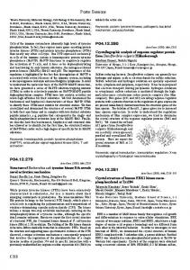
RECQ-like helicases are ATP- and Mg2+-dependent enzymes that are involved in maintaining genome integrity. The RECQ helicase family has five representatives in the human genome. Here we describe the 2Å crystal structure of human RECQ1 in complex with Mg-ADP. Overall, the structural architecture closely resembles that of bacterial RECQ albeit with altered relative domain positioning. All domains are conserved, including two RecA-like modules, the RECQ-specific zinc-binding and a winged-helix (WH) domain. The orientation of the two RecA domains, believed to harbour the helicases’ ATPdependent translocation activity, exhibits considerable variability as adjudged from the overall conformation adopted by the protein in multiple crystal forms. The C-terminal WH domain is positioned in a novel orientation in the human enzyme resulting in a more elongated molecule. This domain also exhibits a prominent betahairpin structural element, not seen in the bacterial enzyme, that is reminiscent of the DNA strand separation pin of other DNA helicases. The role of this pin is to act as an unwinding element by displacing the individual strands of duplex DNA. Mutation of the Tyr residue (Y564) that caps the separation pin as well as shortening the beta-hairpin abolishes DNA-unwinding activity confirming that this structural element plays a key role in DNA strand separation. The probable DNA-binding mode of RECQ1 can be inferred by comparison with other DNA-helicase complexes. The structure will be presented in detail along with implications for recognition and binding of DNA. In addition, progress on the structural characterisation of the other members of RECQ family will also be summarised. Keywords: DNA replication, protein-DNA interactions, structural genomics
C306
P04.06.241 Acta Cryst. (2008). A64, C306
A new nicking enzyme is developed from a mutant of the modified type II restriction enzyme scPvuII Chrysi Meramveliotaki1,2, Maria Androulaki1,2, Elias Eliopoulos2, Michael Kokkinidis1
1 University of Crete, Greece, Biology, Vassilika Vouton, Heraklion, Crete, 71409, Greece, 2Agricultural University of Athens, Iera Street 75, Athens, 11855, Greece, E-mail:
[email protected]
PvuII is the first restriction endonuclease (nuclease component of one of the type II restriction-modification systems of Proteus vulgaris) which has been converted from its wild-type (wt) homodimeric form into a single chain (sc) protein by tandemly joining the two subunits through the peptide linker GlySerGlyGly [1]. The DNA cleavage activity of the enzyme is thereby largely retained. The determined crystal structures (from twinned and un-twinned crystal forms) [2] show that the apo scPvuII adopts a more compact conformation compared to the wild-type form. Four mutants of scPvuII, which address specific aspects of its interactions with DNA have been crystallized and studied with similar results. In contrast, in equilibrium in solution, scPvuII and the mutants adopt two conformations, as proved from gel-filtration [3] and SAXS measurements. As proved from the crystal structure, the peptide linker forms new H-bonds in that area of the protein, which are possibly responsible for the two conformations of the apo enzyme. Several attempts for the co-crystallization of the scPvuII or a mutant - DNA complex were not successful, probably because of the serious aggregation problem of the proteins, as studied by Dynamic Light Scattering techniques [4]. The complex formation was also studied by SAXS method and it is actually formed in the case of scPvuII in the expected 1:1 molar stoichiometry. Later studies based on FRET technique proved that the DNA molecule in the complex is not bended [4], as it is also the case for the wtPvuII, but not for other type II restriction enzymes. The proteins - DNA interactions were also studied by EMSA techniques and the most outstanding result was that the D34G/K70A mutant of scPvuII proved to be a nicking enzyme. [1] A. Simoncsits et al., J. Mol. Biol., 2001, 309, 89-97. [2] C. Meramveliotaki et al., Acta Cryst. Sect F, 2007, 63, 836-8. [3] A. Simoncsits, private communication. [4] W. Wende, private communication. Keywords: scPvuII, nicking enzyme, mutant
P04.06.242 Acta Cryst. (2008). A64, C306-307
Structural characterization of ANAC019, a member of the NAC family of plant transcription factors Ditte H Welner1, Heidi A Ernst1, Addie N Olsen1, J. G Grossmann2, Charlotte Helgstrand1, Karen Skriver1, Leila L Leggio1
University of Copenhagen, Dept. of Chemistry, Universitetsparken 5, Copenhagen OE, Denmark, 2100, Denmark, 2School of Biological Sciences, The University of Liverpool, Liverpool L69 7ZX, UK, E-mail:
[email protected] 1
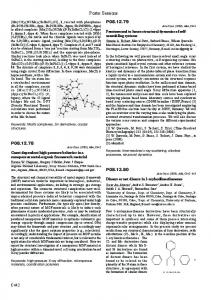
The NAC proteins constitute a large group of plant specific transcription factors which play important roles in biotic and abiotic stress responses and plant development (Olsen et al, 2005, Trends in Plant Science, 10:79-87). NAC proteins consist of two regions: a conserved N-terminal region (NAC domain) with DNA binding and oligomerization abilities, and a diverse C-terminal region which functions as a transcriptional activator. We have previously determined the first crystal structure of a conserved DNA-binding
NAC domain, the one of A. thaliana ANAC019 (Ernst et al, 2004, EMBO Rep, 5:297-303), revealing a novel dimeric transcription factor fold. The structure is now the basis for mutagenesis studies to identify the DNA-binding mode (Olsen et al, 2005, Plant Science 169: 785-797) Structure determination of a new crystal form of this NAC domain shows that the dimer is likely to have some flexibility, but the solution structure as determined by small angle X-ray scattering is in good agreement with the original crystal structure. Based on ongoing crystallographic, small angle scattering and mutagenesis studies, our current model for NAC proteins binding to DNA will be presented. Keywords: transcription factor, NAC protein, structure
P04.06.243 Acta Cryst. (2008). A64, C307
Structure of the topoisomerase IV from S. pneumoniae with a DNA target and quinolone drug Ivan Laponogov1,2, Maninder K Sohi1, Dennis A Veselkov1, Xiao-Su Pan2, Ritica Sawhney2, Andrew W Thompson3, L Mark Fisher2, Mark R Sanderson1
King’s College London, Randall Division of Cell and Molecular Biophysics, School of Biomedical and Health Sciences, Randall Division of Cell and Molecular Biophysics, New Hunt’s House, Guy’s Campus, King’s College London, London, N/A, SE1 1UL, UK, 2Molecular Genetics Group, Molecular and Metabolic Signalling Centre, Division of Basic Medical Sciences, St. George’s, University of London, UK, 3 Synchrotron SOLEIL, L’Orme de Merisiers BP, 48 St. Aubin 91191, Gif sur Yvette, France, E-mail:
[email protected] 1
Eighteen percent of all clinical bacterial infections are now treated with quinolone based antibiotics [1], which target the decatenating enzyme topoisomerase IV (a Class II topo) in gram-positive bacteria such as S. pneumoniae. Topoisomerase IV consists of both ParE and ParC domains. We have crystallised the complex of ParC55 (55 kDa) and ParE (30kDa) with a 32 base-pair DNA target and quinolone drug. Crystals were grown in space group P32 with cell dimensions a=b=118.30 Å, c=177.90 Å α=90° ,β=90° ,γ=120°both by conventional hanging drop vapour diffusion in 24-well limbro plates and by sitting drop in 96 well MRC crystallisation plates. The structure has been solved by molecular replacement (CNS) using as search models our ParC55 structure [2] and a ParE domain homology modelled on the basis of the structure of the TOPRIM domain of the yeast type IIa [3]. The DNA has been positioned from difference Fourier maps following refinement using CNS. The veracity of the model was confirmed by the ability of this phase set to determine the correct Pt sites for a K2PtCl4 heavy atom derivative. Subsequently multiwavelength anomalous diffraction data were collected at the SOLEIL synchrotron about the Pt absorption edge in order to calculate a MAD map. We should like to thank the beamline personnel headed by K. McAuley at the DIAMOND synchrotron, Chilton, Oxford for their help in collecting native and fixed Pt edge synchrotron data on station IO3. References: [1] Drilca,K., Malik,M., Kerns,R.J. and Zhaol X.L. (2008) Antimicrobial agents and Chemotherapy vol. 52 385-392. [2] Laponogov, I., Veselkov, D., Sohi, M.K., Pan, X.-S., Achari, A., Laponogov, I., Fisher, L.M. and Sanderson, M.R. PLoS ONE (2007) e301. [3] Dong, K.C. and Berger, J.M. (2008) Nature vol. 450 1201-1206. Keywords: protein-DNA, quinolone, S. pneumoniae
P04.07.244 Acta Cryst. (2008). A64, C307
RNA splicing related proteins; Crystal structure of RNA 3’-terminal phosphate cyclase Satoru Shimizu1, Masanori Ohki1, Nami Ohkubo1, Kaoru Suzuki2, Masaru Tsunoda3, Takeshi Sekiguchi2, Akio Takenaka1,3
Graduate School of Bioscience and Biotechnology, Tokyo Institute of Technology, 4259, Yokohama, Kanagawa, 226-8501, Japan, 2College of Science and Engineering, Iwaki Meisei University, 5-5-1, iino, chu-ohdai, Iwaki, Fukushima, 970-8551, Japan, 3Faculty of Pharmacy, Iwaki Meisei University, 5-5-1, iino, chu-ohdai, Iwaki, Fukushima, 970-8551, Japan, E-mail:
[email protected] 1
RNA 3’-terminal phosphate cyclase (Rtc) is an enzyme related to RNA splicing to convert the 3’-terminal hydroxyl group of truncated RNA to 2’,3’-cyclic phosphate which is required just before its ligation. This reaction may occur in the following two steps: (i) Rtc + ATP → Rtc-AMP + PPi and (ii) RNA-N3’p + Rtc-AMP → RNA-N>p + Rtc + AMP. To reveal the reaction mechanism, Rtc overexpressed in E.coli was crystallized in the following states, Rtc, Rtc-AMP, Rtc+Mn and Rtc+ATP, and their structures have been determined at 2.25, 2.25, 3.2 and 2.4 Å resolutions, respectively. Rtc is a single protein folded into two domains, the large domain being composed of three βαβαββ motifs arranged by psudo threefold symmetry, and the small domain being formed with a βαβββα motif inserted into the third motif of the large domain. The overall structures of other derivatives are almost the same as that of Rtc. At the catalytic site of Rtc-AMP, the α-phosphate group of AMP is covalently bound to the Nεatom of His307, and the adenine moiety of AMP is stacked between the side chains of Pro126 and His283. The two hydroxyl groups at 2’ and 3’ positions of the ribose are bound to the side chain of Asp286 through hydrogen bonds. These structure features suggest the following reaction scheme. A Mg2+ ion bound to Glu10 induces conformational changes of the α and β phosphate groups of ATP which is trapped by Arg17 and Arg39, so that the Nε atom of His307 easily attacks to the α-phosphate group to form a P-N bond in the first reaction. In the second reaction, when a truncated RNA is bound, its 3’-phosphate group might be forced to react with the phosphate group of AMP, and the activated 3’-phosphate group is attacked by the 2’-hydroxyl group to generate the 2’,3’-cyclic phosphodiester.
4 4
Keywords: RNA 3’-terminal phosphate cyclase, sulfolobus tokodaii, X-ray crystal structure analysis
P04.07.245 Acta Cryst. (2008). A64, C307-308
Crystal structure of human DGCR8 core Won Jin Bae, Sun Young Sohn, Jeong Joo Kim, Yunje Cho
National Creative Research Center for Structural Biology, Life Science, Room204, Dept of Life Science, POSTECH, san31 Hyojadong Namgu, Pohangsi, Kyungbuk, 790-784, Korea (S), E-mail:
[email protected]
A complex of Drosha with DGCR8 (or its homolog Pasha) cleaves primary microRNA (pri-miRNA) substrates into precursor miRNA and initiates the microRNA maturation process. Drosha provides the catalytic site for this cleavage, whereas DGCR8 or Pasha provides a frame for anchoring substrate pri-miRNAs. To clarify the molecular basis underlying recognition of pri-miRNA by DGCR8 and Pasha, we determined the crystal structure of the human DGCR8 core (DGCR8S, residues 493−720). In the structure, the two doublestranded RNA−binding domains (dsRBDs) are arranged with pseudo two-fold symmetry and are tightly packed against the
C307
Poster Sessions