Journal of Cardiovascular and Thoracic Research, 2013, 5(3), 127-128 doi: 10.5681/jcvtr.2013.027 http://journals.tbzmed.ac.ir/JCVTR
Simultaneous Operation of Hydatid Cyst of the Heart and Liver: A Case Report Rezayat Parvizi1*, Hossein Namdar1, Eissa Bilehjani1, Amrollah Bayat2, Mohammad ali sheikhalizadeh1 Cardiovascular Research Center, Tabriz University of Medical Sciences, Tabriz, Iran Department of General Surgery, Tabriz University of Medical Sciences, Tabriz, Iran
1 2
ARTICLE INFO Article Type: Case Report Article History: Received: 1 August 2013 Accepted: 4 September 2013 Keywords: Hydatid Cyst Liver Heart
ABSTRACT
Primary echinococcosis of the heart is exceptionally uncommon and is reported 0.5% to 2% of all hydatid cyst sites in comparison with liver (70%) or lung (20%) involvement. Hydatid disease of the heart is caused by the cestode tapeworm echinococcosis granulosis or alveolaris. We present a 29-year-old female with hydatid disease of the liver and heart. She only complained of abdominal pain and palpitation. Echocardiography and multi-slice computed tomography (MSCT) showed a 120×101 mm cyst in the liver and 64 mm in the right ventricular free wall. Both cysts were excised within one procedure successfully.
Case Report A 29-year-old woman was admitted to the cardiology unit with a progressive abdominal pain and occasional brief episodes of palpitations. Multi-Slice computed tomography (MSCT) showed a 120×101 mm hydatid cyst in the left lobe of the liver and a 64 mm cyst in the free wall of the right ventricle; later, TEE study confirmed diagnosis. Further surgical consultation suggested a simultaneous operation on both cysts. Initially, general surgeon team made a sub costal incision to approach the liver. A huge cyst was found at the left lobe that was punctured and its content aspirated. Then warm hypertonic saline (5%) was instilled into the cyst and after 3-4 minutes it was opened and containers (daughter cysts) were evacuated. Subsequently, open heart surgery was performed via median sternotomy. The cyst area was distinguished from surrounding to prevent rupture. Cold hypertonic saline (5%) was instilled into the cyst and after 3-4 minutes, infected cyst was opened and evacuated which produced a large cavity (60 ml) at site which was sutured afterward. Discussion Cardiac involvement of hydatid cyst is rare and usually with minimal symptoms always overlooked by the patient as well as physicians. Human echinococcosis is caused most commonly by echinococcus granulosus.1 The reported prevalence is about 0.5-2%. Human beings are only incidental hosts by contamination from contact with animals.2 Chitinous eggs are lysed in the proximal digestive tract releasing six *Corresponding author: Rezayat Parvizi, E-mail:
[email protected] Copyright © 2013 by Tabriz University of Medical Sciences
larvae which would ultimately reach the liver via portal venous system and rarely the heart via thoracic lymphatic duct.3 In the liver, larvae can pass through sinusoids, suprahepatic veins and right chambers of the heart and eventually reside the lungs.4 In the parenchyma of the liver and the lungs, the larvae could growth and produce hydatid cysts.5 After infection, the embryo usually reaches the myocardium via coronary circulation from the left side of the heart.6 The cyst is then formed within a period of 1 to 5 years.7 Myocardial reaction consists of a fibrous adventitial pericyst layer surrounding the laminated membrane.8 The left ventricular and interventricular septum free walls are the most common locations of the cysts in the myocardial region right-sided cysts have a tendency to expand intracavitarily and subendocardially, whereas the left-sided cysts tend to grow sub-epicardially.9 This may be due to the thicker and denser myocardium of the left than the right heart.10 It has been reported that most of the cysts locate in the left ventricular wall because of the rich vascular supply of the left ventricle.10 Cardiac hydatid cysts produce a large variety of symptoms via any of four mechanisms.11 They can obstruct blood flow or cause valve dysfunction. They can lead to arrhythmias or pericardial effusion with tamponade by local invasion. Rupture of the cyst causes embolization leading to systemic reaction when the cysts are on the left side of the heart, or pulmonary embolism when the cysts are on the right side.12 Moreover, some cardiac hydatid cysts may stay asymptomatic for many years so that the diagnosis of cardiac echinococcosis can be difficult and must be suspected in any patient from endemic or sheep
Parvizi et al.
farming areas especially with a cystic tumor of other infected organs such as liver.13 2D echocardiography, computed tomography scan or MRI may be helpful but trans-esophageal echocardiography is the gold standard in detecting intracardiac echinococcosis.10 In conclusion, the treatment of hydatid cyst is surgical and should not be delayed. Extirpation of the lesion is recommended under cardiopulmonary bypass. Serologic and echocardiographic controls are recommended for 5 years after extirpation to detect recurrences after surgical manipulation or cysts that were not discovered at operation (Figures 1-3). Ethical issues: This study was reviewed and confirmed by the ethics committee of Tabriz University of Medical Sciences.
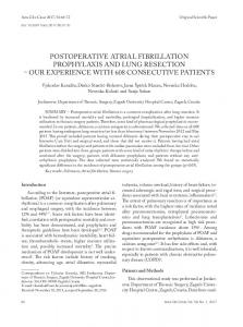
Figure 1. Multi-slice computed tomography
Conflict of interests: The authors declare no conflicts of interest.
References
Figure 2. Removed cyst from left lobe of liver
Figure 3. Hydatid cyst at free wall of right ventricle
128
Journal of Cardiovascular and Thoracic Research, 2013, 5(3), 127-128
1. Sokouti M, Golzari S, Aghdam BA. Surgery of uncomplicated pulmonary hydatid cysts: capitonnage or uncapitonnage? Int J Surg 2011;9:221-4. 2. Sokouti M, Pezeshkian M, Ghabili K, Golzari SE. Surgical Procedures and Postoperative Complications in Patients with Giant and Non-giant Pulmonary Hydatid Cysts. Life Sci J 2013;10: 138-42. 3. Golzari SE, Sokouti M, Ghaffari A, Bazzazi AM, Ghabili K. Ultrasonography in diagnosis of pulmonary hydatid cysts. Lancet Infect Dis 2013;13:294. 4. Sokouti M, Golzari SE, Tizro P, Khanli HM, Ghabili K. Genitourinary hydatid disease. Int Urol Nephrol 2013;45:759. 5. Golzari SE, Sokouti M, Bazzazi AM, Khanli HM, Ghabili K. Serodiagnostic tests in musculoskeletal hydatid disease. Spine (Phila Pa 1976) 2013;15;38:1797. 6. Nazyrov FG, Abdumadzhidov KhA, Buranov KhD, Akbarov MM, Aliev ShM, Mukaddirov MM. Surgical treatment of the combined heart, lung and liver hydatid disease. Khirurgiia (Mosk) 2009;5:23-6 7. Onursal E, Elmaci TT, Tireli E, Dindar A, Atilgan D, Ozcan M. Surgical treatment of cardiac echinococcosis: report of eight cases. Surg Today 2001;31:325–30. 8. Calamai G, Perna AM, Venturini A. Hydatid disease of the heart report of five cases and review of the literature. Thorax 1974;29: 451-8. 9. Erentuğ V, Bozbuğa N, Kirali K, Mataraci I, Kaymaz C, Balkanay M, et al. Cardiac hydatid cysts: surgical treatment and results. J Card Surg 2004;19:358-60. 10. Kojundzic SL, Dolic K, Buca A, Jankovic S, Besenski N. Hydatid disease with multiple organ involvement: a case report. Macedonian Journal of Medical Sciences 2010;3:154-8. 11. Saad RA, Amer KM, Migliore M, Aziz T, Azzu A. Right intraventricular hydatid cyst of the heart. Asian Cardiovasc Thorac Ann 2003;11:160-62. 12. Shehatha J, Alward M, Saxena P, Konstantinov IE. Surgical management of cardiac hydatidosis. Tex Heart Inst J 2009; 36:72–3. 13. Parvizi R, Joudati AR, Montazeri V, Susan Hassanzadeh Salmasi S, Varshouchi M. Cardiac Echinococcosis: Surgical Treatment and Results in 10 Cases. Iranian Heart Journal 2006;7:67-71.
Copyright © 2013 by Tabriz University of Medical Sciences