May 16, 1988 - Baim, S. B., and F. Sherman. 1988. mRNA structures .... Booth. 1987. Cytochrome c protein-synthesis rates and mRNA contents during atrophy ...
Vol. 8, No. 11
MOLECULAR AND CELLULAR BIOLOGY, Nov. 1988, p. 4625-4633
0270-7306/88/114625-09$02.00/0 Copyright X) 1988, American Society for Microbiology
Posttranscriptional Regulation of Cytochrome c Expression during the Developmental Cycle of Trypanosoma brucei AL F. TORRI AND STEPHEN L. HAJDUK*
Department of Biochemistry, University of Alabama at Birmingham Schools of Medicine and Dentistry, Birmingham, Alabama 35294 Received 16 May 1988/Accepted 1 August 1988
We examined the expression of a nucleus-encoded mitochondrial protein, cytochrome c, during the life cycle of Trypanosoma brucei. The bloodstream forms of T. brucei, the long slender and short stumpy trypanosomes, have inactive mitochondria with no detectable cytochrome-mediated respiration. The insect form of T. brucei, the procyclic trypanosomes, has fully functional mitochondria. Cytochrome c is spectrally undetectable in the bloodstream forms of trypanosomes, but during differentiation to the procyclic form, spectrally detected holo-cytochrome c accumulates rapidly. We have purified T. brucei cytochrome c and raised antibodies that react to both holo- and apo-cytochrome c. In addition, we isolated a partial cDNA to trypanosome cytochrome c. An examination of protein expression and steady-state mRNA levels in T. brucei indicated that bloodstream trypanosomes did not express cytochrome c but maintained significant steady-state levels of cytochrome c mRNA. The results suggest that in T. brucei, cytochrome c is developmentally regulated by a posttranscriptional mechanism which prevents either translation or accumulation of cytochrome c in the bloodstream trypanosomes. c mRNA in the cells. Thus, in yeast cells, cytochrome c can be regulated transcriptionally. Transcriptional regulation of cytochrome c has also been reported for Neurospora crassa, Manduca sexta, and Drosophila melanogaster (22, 41). It has been reported that cytochrome c levels can also be subject to posttranscriptional regulation. In rat skeletal muscle, immobilization-induced muscle atrophy results in mild, transient suppression of cytochrome c expression during a period when intracellular levels of cytochrome c mRNA appear stable (30). However, transcriptional regulation of cytochrome c is observed during extended periods of muscular immobilization. Cytochrome c mRNA levels decrease in rat skeletal muscle as the cells are allowed to atrophy, resulting in dramatic reductions in cytochrome c. In this paper we present evidence that the developmental regulation of cytochrome c in T. brucei occurs by a unique mechanism. Although the apo- and holo-proteins are not detected in the bloodstream forms, the transcripts for the protein are present in all life stages. These results suggest that cytochrome c regulation in trypanosomes occurs at the translational or posttranslational level.
Trypanosoma brucei is a protozoan parasite with a complex life cycle which encompasses both a mammalian host and a specific insect vector, the tsetse fly (44). In the mammal, the early bloodstream developmental stage, the rapidly dividing long slender trypanosome, differentiates into the nondividing short stumpy trypanosome (13, 44). These short stumpy trypanosomes are preadapted to life in the insect vector, where they differentiate into the early insect developmental stage, the procyclic trypanosomes (29, 45). The bloodstream forms of T. brucei have an inactive mitochondrion which lacks cytochrome-mediated respiration (5, 6, 9, 23). The procyclic form of T. brucei has a mitochondrion which is larger and has a more expansive network of cristae than is found in the bloodstream trypanosomes. The mitochondria of procyclic trypanosomes are active in cytochrome-mediated respiration. The regulation of mitochondrial biogenesis in T. brucei and the adaptations in energy metabolism that accompany this regulation have a profound effect on the expression of both mitochondrionencoded and nucleus-encoded mitochondrial proteins. To gain insight into this process, an investigation into the regulation of a nucleus-encoded mitochondrial protein, cytochrome c, was initiated. Cytochrome c serves as a component in the electron transport chain. The protein is encoded in the nucleus, synthesized in the cytosol off free ribosomes, and transported into the mitochondria, where a heme prosthetic group is attached, forming mature holo-cytochrome c (24, 28). Regulation of cytochrome c has been best characterized in Saccharomyces cerevisiae. In S. cerevisiae, cytochrome c expression is dependent on the availability of heme. Intracellular heme stimulates production of the heme activation proteins, which bind to upstream activation sites of the cytochrome c transcription unit to allow transcription of the gene (18, 19, 35). Catabolite repression reduces heme levels within S. cerevisiae, resulting in a reduction of cytochrome *
MATERIALS AND METHODS
Growth and isolation of trypanosomes. Bloodstream forms of T. brucei (TREU 667) were grown in female Wistar rats (approx. 200 g) or female CD-1 mice (approx. 5 to 10 weeks old), which were gamma irradiated with 600 rads prior to infection. The animals were infected by interperitoneal injection of blood from a female CD-1 mouse infected for 3 to 4 days from frozen stocks stored at 196°C in 7.5% dimethyl sulfoxide. Long slender trypanosomes were recovered from a 2- to 3-day infection, and short stumpy trypanosomes were recovered from an 8- to 9-day infection. In both cases, trypanosomes were harvested by cardiac puncture and anion-exchange chromatography over a Whatman diethylaminoethyl cellulose column (DE52, pH 8.0) with a phosphatebuffered saline solution containing glucose and heparin -
Corresponding author. 4625
4626
TORRI AND HAJDUK
(PSGH: 0.057 M Na2HPO4, 3 mM NaH2PO4, 0.044 M NaCI, 10 g of glucose per liter, 10 U of heparin per ml, pH 8.0). Procyclic trypanosomes (TREU 667) were grown in culture in a semidefined medium (SM medium) containing gentamicin (25 ,ug/ml) and supplemented with 10% fetal calf serum (FCS) (Hyclone), heat inactivated at 56°C for 30 min (12, 29). The cultures were grown under sterile conditions with continuous shaking at 26°C and harvested at late log phase (approx. 2 x 10⁷ trypanosomes per ml) by sedimentation at 5,000 x g for 15 min at 4°C. Differentiation of trypanosomes from the short stumpy bloodstream form to the procyclic culture form was initiated by suspending the short stumpy trypanosomes in SM medium at 26°C at a concentration of 5 x 10⁶/ml. The short stumpy trypanosomes were isolated as above with two exceptions: sterile conditions were maintained throughout the procedure, and 10% FCS was added to the DE52 resin and to PSGH to improve organism viability. Holo-cytochrome c determinations. Trypanosome cell pellets were washed in isotonic buffer (0.1 M NaCI, 0.05 M Tris hydrochloride pH 8.0), resuspended 0.01 volume of 10 mM Tris hydrochloride (pH 7.0), and sonicated with a Heat Systems Ultrasonics Cell Disruptor with a model H-1 microtip set at maximum output. Total cellular disruption was achieved with repeated 1-min sonication cycles performed on ice. The crude cell lysate was then centrifuged at 10,000 x g to sediment debris. A partial purification of the crude lysate supernatants was achieved by cation-exchange chromatography over carboxymethyl-Sephadex (CM) columns. The proteins were eluted with 10 mM Tris hydrochloride (pH 7.0)-0.5 M NaCl. The absorption spectra of the partially purified lysate from 300 to 600 nm was determined with a Varian 2290 spectrophotometer. Relative holo-cytochrome c concentrations were calculated based on absorption at the Soret peak maximum of 419 nm and corrected for lysate cell number. Purification and characterization of cytochrome c. T. brucei cytochrome c was isolated from 20 liters of procyclic trypanosomes cultured to late log phase by the procedure outlined by Hennig (20) with several modifications. Cellular debris was sedimented by centrifugation at 10,000 x g for 1 h at 4°C prior to the first chromatographic step, and all chromatography was performed in Tris hydrochloride buffers containing the protease inhibitors leupeptin (0.5 ,ug/ml), pepstatin (1 p.g/ml), and phenylmethylsulfonyl fluoride (17.5 p.g/ml). The final purification step was high-pressure liquid chromatography (HPLC) size exclusion chromatography on a TSK 3000 column, eluting with 10 mM Tris hydrochloride (pH 7.0). The elution of cytochrome c during each purification step was monitored by absorption at 405 nm and assayed by absorption spectra and by electrophoresis (26). Purified cytochrome c was digested with trypsin to generate peptides for protein sequencing. Approx. 8 pmol of TLCK-treated trypsin was added to approx. 800 pmol of trypanosome cytochrome c in 100 mM Tris (pH 8.0)-S50 mM NaCl and incubated at 37°C for 3 h. The same amount of trypsin was again added to the solution, and incubation was continued overnight. Generated peptide fragments were isolated by HPLC with a reverse-phase C-18 column. Recovered peptides were sequenced with an Applied Biosystems 470A protein sequencer, with the individual amino acids being identified by reverse-phase HPLC. Apo-cytochrome c was prepared by chemically removing the heme prosthetic group from holo-cytochrome c as described by Ambler and Wynn (1). SDS-PAGE. Sodium dodecyl sulfate-polyacrylamide gel
MOL. CELL. BIOL.
electrophoresis (SDS-PAGE) was performed by the method of Laemmli (26) with a 5% acrylamide stacking gel and a 15% acrylamide resolving gel. Proteins were visualized by Coomasie blue staining or silver staining. Production of antibodies. Antibodies to T. brucei cytochrome c were prepared by immunization of both New Zealand White rabbits and BALB/c mice with monomeric cytochrome c isolated from procyclic trypanosomes as described by Reichlin et al. (36) with several modifications. Inoculations of the rabbits were performed at monthly intervals with 100 to 200 ,ug of trypanosome cytochrome c in an emulsion with Freund adjuvant, distributed along a series of subcutaneous sites. Inoculations of mice were also performed at monthly intervals with 20 to 30 ,ug of trypanosome cytochrome c in an emulsion with Freund adjuvant, injected interperitoneally. In both cases, the initial inoculations were prepared with Freund complete adjuvant, and all subsequent inoculations were prepared with Freund incomplete adjuvant. Rabbit antisera were affinity purified with yeast cytochrome c coupled to cyanogen bromide-activated Sepharose as described by Axen et al. (2) and Cuatrecasas et al. (11). Western blot (immunoblot) analysis. Proteins were electrophoretically transferred from SDS-PAGE gels onto nitrocellulose (Schleicher & Schuell BA85) in 20 mM Tris-150 mM glycine-20% methanol at 200 V for 4 h at 10°C (17, 43). The filters were washed, blocked with gelatin, probed with the T. brucei cytochrome c antibodies, and visualized by using a second antibody coupled to alkaline phosphatase (Bio-Rad Immun-Blot assay kit). Cytochrome c cDNA synthesis and isolation. RNA was isolated from procyclic trypanosomes by the guanidine isothiocynate procedure of Maniatis et al. (27). Polyadenylated [poly(A)+] mRNA was selected by affinity chromatography over an oligo(dT) column (27). A cDNA library was constructed in Xgtll from this RNA by using the Amersham cDNA synthesis kit and the Amersham cDNA cloning kit. The cDNA library was plated, and duplicate plaque lifts were performed on nitrocellulose (39, 46). The cDNA library was screened for the expression of T. brucei cytochrome c by using both the rabbit and mouse antibodies against trypanosome cytochrome c, and the positive plaques were visualized by using second antibodies conjugated to alkaline phosphatase. The DNA inserts from dual positive plaques were sequenced by primer extension (United States Biochemical Sequenase sequencing kit). The T. brucei cytochrome c cDNA was released by EcoRI digestion, ligated into Bluescript KS' (Stratagene), and designated pAT31. The pAT32 subclone was prepared by digestion of pAT31 with both EcoRI and XhoI; the 190-base-pair fragment was then ligated into Bluescript KS'. RNase T, analysis. RNA from long slender, short stumpy, and procyclic forms of T. brucei was prepared from total cellular nucleic acid by DNase I (RNase-free) treatment as described by Rohrer et al. (37). The 32P-labeled RNA probes and unlabeled RNA sense strand controls were prepared as described elsewhere (37). The hybridization of riboprobes and all cellular RNA or control RNAs and the RNase T1 digestions were performed by the method of Mueller et al. (32) with several modifications. The hybridization mixes were incubated overnight in a water bath that was initially 100°C and was allowed to cool slowly to 65°C. The digestions were performed with 2 U of RNase T1 per ,ul at 37°C for 1 h. The RNase T1 products were precipitated and analyzed by electrophoresis through a 6% denaturing polyacrylamide gel.
CYTOCHROME c EXPRESSION IN TRYPANOSOMES
VOL. 8, 1988 a
0 co
o 118
1i3 o
)
-
le
M
E-
0
C~~~~~~~~~~
J)
ct
UI
x
,_
cc CD
.-
o
en
0m ._
m
('
*y
11
CD
4627
-
e
CA '1 -I-
2
1
c
20
'0 Eo
1 00
E
66.000-
0
36&000-
C
0.c
1
20,100-
0
14,200-
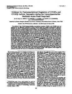
4 0 6 0 8 0 10 0 Hours at 260C cr FIG. 1. Holo-cytochrome c expression in differentiating trypanosomes. Short stumpy trypanosomes were placed in SM medium at 26°C to initiate differentiation to the procyclic form. At the indicated times, portions were removed and assayed for spectrally detectable holo-cytochrome c expression as described in Materials and Methods. The open squares represent the relative concentration of holo-cytochrome c in the differentiating trypanosomes based on absorbance at 419 nm and corrected for cell number. The solid squares represent cell growth in the culture, plotted as number of trypanosomes per milliliter. The time course followed the differentiation of trypanosomes starting from the short stumpy form (0 h) and continuing for 96 h into the differentiation process. co
0
20
RESULTS
Expression of spectrally detectable holo-cytochrome c. Cytochrome c expression during the developmental life cycle of T. brucei was examined by absorption spectroscopy. The absorbance spectrum from 350 to 600 nm was determined for the partially purified cell lysates from long slender, short stumpy, and procyclic trypanosomes. Lysates of the bloodstream forms of T. brucei do not absorb to any significant extent over this range. Lysates of the procyclic trypanosomes showed absorbance maxima at 419 nm, 523 nm, and 556 nm, which is characteristic of cytochrome c (results not shown). To establish the kinetics of cytochrome c accumulation, the absorption spectra of partially purified cell lysates prepared from trypanosomes in the process of differentiation were determined (Fig. 1). The short stumpy trypanosomes (0 h) had no spectrally detectable cytochrome c; however, holo-cytochrome c was detected in the trypanosomes 3 h after the short stumpy forms were shifted into SM medium at 26°C. The holo-cytochrome c concentration increased in the differentiating cells, approaching the level in fully differentiated procyclic trypanosomes within 30 h. Cell growth in the differentiating culture experienced a lag which coincided with the time required for cytochrome c levels to reach a plateau. These results indicate that cytochrome c is developmentally regulated in T. brucei. Purification and characterization of cytochrome c. Cytochrome c was purified from procyclic forms of T. brucei so that the protein could be characterized and used in the production of antibodies. Ion-exchange chromatography of the trypanosome lysate, utilizing TEAE cellulose, CM Sephadex, and Bio Rex 70, was followed by gel filtration chromatography over G-50 Sephadex and HPLC size exclusion on a TSK 3000 column (20). Cytochrome c was monitored throughout the purification by absorbance at 405 nm. At various steps during the purification, portions were removed and characterized by SDS-PAGE (Fig. 2). The chromatog-
6,200-
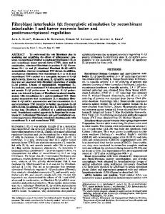
FIG. 2. SDS-PAGE of cytochrome c purification from procyclic trypanosomes. Portions from each step in the purification of cytochrome c were analyzed on 15% SDS-PAGE and visualized by silver staining of the gel. The samples included the procyclic trypanosome lysate supernatant (Lysate), the flowthrough off the TEAE-cellulose column (TEAE), the eluate from the CM-Sephadex column (CM), the eluate from the Bio-Rex 70 column (Bio-Rex), the pooled fractions collected off the Sephadex G-50 column (G-50), and the pooled fractions off the HPLC TSK-3000 column (HPLC). In addition, cytochrome c isolated from C. fasciculata (Crithidia), horse, and S. cerevisiae (yeast) were also analyzed. Approximately 10 ,ug of protein from the lysate and 5 ,ug of protein from the TEAE flowthrough were loaded onto the gel. All other samples were equally loaded onto the gel based on cytochrome c content, approximately 2 ,ug of cytochrome c from each.
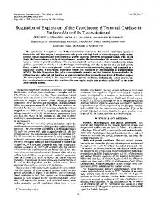
raphy resulted in the purification of a 12,500-dalton protein from the procyclic lysate. The T. brucei protein was similar in size to purified cytochrome c from Crithidia fasciculata, horse, and S. cerevisiae. Protein migration in the gel was affected by the different salt concentrations in the various samples. Spectral analysis of the isolated T. brucei protein revealed an absorbance profile characteristic of cytochrome c, with absorbance maxima at 419 nm, 523 nm, and 556 nm (results not shown). The T. brucei protein was digested with trypsin, and the peptide fragments were separated by reverse-phase HPLC. The sequence of seven tryptic fragments, containing 60 nonoverlapping amino acids, was determined and compared with the complete C. fasciculata and S. cerevisiae cytochrome c sequences (Fig. 3). The sequenced portion of the T. brucei protein showed more than 85% homology with cytochrome c from C. fasciculata and almost 45% homology with cytochrome c from S cerevisiae. Thus, the 12,500-dalton protein isolated from procyclic forms of T. brucei had the size, spectral properties, and amino acid sequence expected for cytochrome c. Expression of apo-cytochrome c during developmental differentiation. The expression of apo-cytochrome c during the developmental cycle of T. brucei was examined by Western blots probed with affinity-purified rabbit antibodies to T. brucei cytochrome c. The Western blot assays were performed on partially purified cell lysates of trypanosomes at various stages of differentiation from the short stumpy to the procyclic form. The Western blot results (Fig. 4) indicated that the antibodies detected both apo- and holo-cytochrome c from either T. brucei (compare procyclic with procyclic-H) or yeast cells (compare yeast holo-cyt c with yeast apo-cyt c). The results also demonstrated that the apo-protein from
4628
MOL . CELL . BIOL
TORRI AND HAJDUK
S.c. C.f. T.b.
CGU P. LWe la Gy a Al Lye Ly Arq Glu Pro Lou Pro Pro Gly Lsp Ala Ala Lys Thr
C.f. T.b. S.c. C.f. T.b.
5
1
-9
S.c. Pro
A Ala
Ala Ala Lou Pro Pro Gly Asp Ala Ala
20 6 Gly Ala Thr Lou Pbh Lys Thr Arg Cy* Lnu Cln Cys His Thr Va1 Gly Clu Lys Il Ph. Lys Giy Arg Ala Ala Gin Cy. His Thr Giy Ala Ala Gin 21
35
Clu Lou Gly Gly Pro His Lys Val Gly Pro Asn Lou His Gly Ala Lys Cly Gly Ala Amn Gly Va1 Gly Pro Amn Lou Pb. Gly Cly Gly Sor Asn Gly Val Gly Pro amn L.u Tyr Gly
Ile Ile
51-CL ATT
CDIR S.c. C.f. T.b.
cDNL
Ile 50
36 Ph Cly Arg His Sor
Gly Gln Ala Clu Gly Tyr Sor Tyr Thr Ap Val Asn Arg Gly Ser Gly Thr Va1 Clu Gly Ph. Ala Tyr Ser Lys hL Val Gly AM Lys Usr Gly Thr YA, Clu CT CCC CdT AAA TCC ca ACT CTT GAG CCT TTT ACd TC ACC
AAA
S.c. C.f. T.b.
65 51 Ala Asn Ile Lys Lys Asn Val Lou Trp Asp Glu An Asn Hat Sor Ala Acn Ala Asp Sor Gly Val Val Trp Thr Pro Clu Va1 Lou Asp ou Pro Gin Val rA ALa Asn Gin Asp Sor Gly Val Hat M
cDk
c:c AAT CRA CAT TOC
S.c.
T.b.
80 76 Clu Tyr Lou Thr Asn Pro Lys lys Tyr I11 Pro Gly Thr Lou Hat V.1 Tyr Lou Clu Asn Pro Lys Lys Pho Hat Pro Gly Thr Lys Hat Val Tyr Lou Clu Asn Pro Ph. :4L
cDNL
CTC TAT TMC CLG AAT CCA AAG ALA TTT ATC CCC CCC ACT ALL ATG
S.c.
81 Ala Pho Gly Gly Lou Lys Lye Clu Lys ap Arg ASn Asp Lou Sor Ph. Ala Cly Il Lys Lye Pro Cln Clu Arg Ala Asp Lou Ser Phe Ala Glv Lou Lye LyS Pro Cln Clu Ar Ala Asp Lou
C.f.
C.f.
T.b.
cDM& S.c. C.f. T.b.
cDNa
=T
ATG TGC ACT aCC CAC CEL CTT CGC
CGT
TTT CCA CCT TEL AAC AAA CCL CAC CAA
95
Il Ie
Il
CCC CCC GAC CTC ATC
103 96 Thr Tyr ILu Lys Lys Ala Cy& Clu Ala Tyr Lou Clu Asn Lou Lys Cly Ala Tyr Lou Clu
= Lou Lys AM
CCA TAC CTC CAG ACA TTA AG CAC TAL AAT CAG ATAL ATA MCA AAa
TTT ATA ATA TTE A
ATA TAL CEL CAT TAT TGL AAL
TmT
ATG
TTL
TCA TAC ATG TAT TAT TA AaC AGA CCL TOCG AGC GA GAL AGG AGC cc CGtC CCT TAT GTT ATC ATC GCA ACG ATT CA TEL TTA TTA TE
TMA TM T
TAC T =C ACT ATC AT AMT ATE ATE TAT ATT T
TAT TMI GCC CTM TaC GAG CMA CAL LAC GA
CAC AL CAT AAC
C"A TAG TEA AMC TEL TM TE TE TE M ATE CTE T TGA TAA GAG GAL ALA GALGa C
TE
ACG CCA CtA ACC ALL AA AMA
GjA GAA CLT aaA TGA ATT ACA ALA AA ARA ALL A-3' D X c cDNA Amino Acids
Cyt
.
35
-
I
D
3 103
Coding 3T-untranslated (372 bases) (207 bases) FIG. 3. Comparison of cytochrome c sequences. The partial amino acid sequence of T. briucei cytochrome c (T.b.) is aligned with the amino acid sequence of cytochrome c from both C. fascicilata (C.f.) (23) and S. cerevisiae (S.c.) (38). The sequence of the partial T. brucei cytochrome c cDNA is aligned by the protein sequence it encodes. T. brucei cytochrome c amino acid sequence based only on the cDNA sequence data is underlined. The bottom diagram is of the partial T. bricei cytochrome c cDNA. The size, orientation, and approximate positions of certain restriction sites are indicated (D, DraI sites; X, XhoI site). EcoRI sites flank the cDNA.
CYTOCHROME c EXPRESSION IN TRYPANOSOMES
VOL. 8, 1988
o 6
I
eL E
cLoo
Hours
at 0 o
I
260 0
0 X o ° °X0
66.000-
36.000-
20.10014.2006.200-
FIG. 4. Analysis of cytochrome c expression by Western blot. Trypanosome cell lysates were partially purified by chromatography over CM-Sephadex as described in Materials and Methods. The eluted proteins were fractionated on 15% SDS-PAGE, blotted onto nitrocellulose, and probed with affinity-purified rabbit antibodies against T. brucei cytochrome c. The antigen-antibody complexes were visualized by using a goat anti-rabbit immunoglobulin antibody conjugated to alkaline phosphatase. The blot includes a cell lysate of 2 x 10⁹ short stumpy trypanosomes, cell lysates of 1 x 10⁹ trypanosomes at specific times after initiating differentiation from the short stumpy to the procyclic form in SM medium at 26°C (5, 10, 20, and 40 h), and a cell lysate of 2 x 10⁹ trypanosomes from an established procyclic culture. The blot also includes a CM-Sephadex-purified lysate of 2 x 10⁹ procyclic trypanosomes which had been chemically treated to remove heme (Procyclic-H) and a similiarly treated lysate of 1 x 10⁹ procyclic trypanosomes which was then rechromatographed on CM-Sephadex (Procyclic-H + CM). The final two lanes contain 1 jig of yeast cytochrome c (Yeast Holo-Cyt c) and 1 ,ug of yeast cytochrome c chemically treated to remove the heme prosthetic group (Yeast Apo-Cyt c). Sizes are shown to the left (in daltons). a CM-Sephadex col(compare procyclic-H with procyclic-H + CM and note difference in cell equivalents used, 2 x 10⁹ and 1 x 10⁹, respectively). No cytochrome c was detected in the short stumpy cell lysate; however, in the differentiating trypanosomes (trypanosomes at 26°C for 5, 10, 20, and 40 h), cytochrome c was detectable within 20 h of placing the short stumpy forms in SM medium at 26°C. The expression of cytochrome c increased during the differentiation process. Cytochrome c was detected in Western blots from lysates of 2 x 10⁷ procyclic trypanosomes (data not shown). Thus, the level of cytochrome c in the procyclic form is at least 100 times greater than that in the bloodstream forms. The Western blots indicated that neither apo- nor holo-cytochrome c was present at significant levels in the bloodstream trypanosomes and that the expression of cytochrome c occurred during differentiation of the short stumpy form to the procyclic form. Similar results were obtained when total-cell lysates were used directly without partial purification on CM columns (data not shown). Cytochrome c cDNA cloning and analysis of transcript levels. To determine whether cytochrome c expression in T. brucei was regulated at the transcriptional level, quantitative RNase T1 protection studies were performed with the trypanosome cytochrome c cDNA (32). A T. brucei cDNA expression library was constructed in Xgtll from poly(A)+ procyclic RNA. Dual screening of the cDNA library with
trypanosomes could be isolated with umn
4629
both rabbit and mouse antibodies against T. brucei cytochrome c identified a partial cDNA to T. brucei cytochrome c (Fig. 3). The cDNA contained 207 nucleotides of the 3' region of T. brucei cytochrome c encoding amino acids 35 to 103. In addition, the cDNA contained the entire 3' untranslated flanking region of the procyclic trypanosome cytochrome c mRNA, consisting of 372 nucleotides. The T. brucei cytochrome c amino acid sequence derived from the cDNA agreed with the amino acid sequence of the purified protein. The full-length cDNA was excised from Xgtll by EcoRI digestion and ligated into the plasmid vector Bluescript to yield the subclone pAT31. The protein-coding region of the cDNA was isolated from pAT31 by digestion with both EcoRI and XhoI, and the 190-nucleotide fragment was ligated into Bluescript to yield the subclone pAT32. The pAT32 clone was linearized by digestion with BamHI, and the antisense strand to the cytochrome c coding region was transcribed by using T7 polymerase in the presence of [a-32P]GTP. The 250-nucleotide RNA was the T7 transcription product from the BamHI-truncated pAT32 template. The labeled high-molecular-weight RNA smear resulted from run-on transcription of undigested pAT32. The radiolabeled transcripts were then incubated in the presence of unlabeled RNA, and the hybrids were analyzed by digestion with RNase T1 and gel electrophoresis under denaturing conditions (Fig. 5). Hybridizations with equal amounts (3 jig) of total cellular RNA isolated from the three developmental stages of T. brucei demonstrated that the long slender, short stumpy, and procyclic form RNAs contained transcripts which protected the probe from RNase T1 digestion (Fig. 5A). This surprising result implies that the bloodstream forms of T. brucei, which do not express cytochrome c, nevertheless possess mRNA for the protein. Hybridizations to serial dilutions of both the procyclic RNA (0.3, 3, and 30 ,ug of total cellular RNA) and the unlabeled sense strand of the cytochrome c cDNA (500, 50, and 5 pg of pAT31 T7 polymerase transcripts) suggested that there was approximately 10 pg of cytochrome c mRNA in 1 ,ug of procyclic RNA. The RNase T1 analysis indicated that there were differences in the steady-state level of cytochrome c mRNA between the long slender, short stumpy, and procyclic forms of T. brucei. When hybridizations were conducted to compare equal amounts of total cellular RNA between the life stages (3 ,ug of total cellular RNA), a threefold difference in cytochrome c mRNA levels was observed between the bloodstream and procyclic trypanosomes (Fig. 5A). Hybridizations comparing equal cell equivalents of RNA (5 x 10⁶ cell equivalents of total RNA) indicated a fivefold difference in cytochrome c mRNA levels (Fig. 5B). The variations in the results probably reflected different cellular RNA concentrations between the bloodstream and procyclic forms of T. brucei. These differences in cytochrome c mRNA levels do not account for the observed 100-fold regulation in cytochrome c expression. As a control for mRNA levels within the isolated RNA, RNase T1 protection studies were also performed with the T. brucei ,B-tubulin gene subcloned into the transcription vector GEM-I (pgTub). Radioactively labeled antisense transcripts and unlabeled sense strand transcripts to T. brucei ,B-tubulin were synthesized off pgTub linearized by digestion with HindIII or PvuII, respectively. The radioactively labeled antisense strand of T. brucei ,3-tubulin was hybridized to its sense strand transcript and to equal amounts of total cellular RNA (2 j,g) isolated from the long slender, short stumpy, and procyclic forms of T. brucei. The hybrids were analyzed
4630
TORRI AND HAJDUK
MOL. CELL. BIOL.
la
< ZZ