1Department of Molecular Biology, Tokyo University of. Pharmacy and Life Science, 1432-1 Horinouchi,. Hachioji, Tokyo 192-0392, Japan. 2Japan Collection of ...
bs_bs_banner
Environmental Microbiology (2015) 17(5), 1817–1835
doi:10.1111/1462-2920.12648
Potential for biogeochemical cycling of sulfur, iron and carbon within massive sulfide deposits below the seafloor
Shingo Kato,1,2 Kei Ikehata,3 Takazo Shibuya,4 Tetsuro Urabe,5 Moriya Ohkuma2 and Akihiko Yamagishi1* 1 Department of Molecular Biology, Tokyo University of Pharmacy and Life Science, 1432-1 Horinouchi, Hachioji, Tokyo 192-0392, Japan. 2 Japan Collection of Microorganisms, RIKEN BioResource Center, 3-1-1 Koyadai, Tsukuba, Ibaraki 305-0074, Japan. 3 Faculty of Life and Environmental Sciences, University of Tsukuba, 1-1-1 Tennodai, Tsukuba, Ibaraki 305-8572, Japan. 4 Submarine Resources Research Project (SRRP) & Precambrian Ecosystem Laboratory (PEL), Japan Agency for Marine-Earth Science and Technology (JAMSTEC), 2-15 Natsushima, Yokosuka, Kanagawa 237-0061, Japan. 5 Department of Earth and Planetary Science, University of Tokyo, Hongo, Bunkyo-ku, Tokyo 113-0033, Japan. Summary Seafloor massive sulfides are a potential energy source for the support of chemosynthetic ecosystems in dark, deep-sea environments; however, little is known about microbial communities in these ecosystems, especially below the seafloor. In the present study, we performed culture-independent molecular analyses of sub-seafloor sulfide samples collected in the Southern Mariana Trough by drilling. The depth for the samples ranged from 0.52 m to 2.67 m below the seafloor. A combination of 16S rRNA and functional gene analyses suggested the presence of chemoautotrophs, sulfur-oxidizers, sulfate-reducers, iron-oxidizers and iron-reducers. In addition, mineralogical and thermodynamic analyses are consistent with chemosynthetic microbial communities sustained by sulfide minerals below the seafloor. Although distinct bacterial community compositions
were found among the sub-seafloor sulfide samples and hydrothermally inactive sulfide chimneys on the seafloor collected from various areas, we also found common bacterial members at species level including the sulfur-oxidizers and sulfate-reducers, suggesting that the common members are widely distributed within massive sulfide deposits on and below the seafloor and play a key role in the ecosystem function. Introduction Seafloor massive sulfide (SMS) deposits are often found at marine hydrothermal vent fields that mainly occur at and around plate boundaries that have a total strike length of 89 000 km in the oceans, including mid-ocean ridges (64 000 km), submarine volcanic arcs and back-arc basins (25 000 km) (Hannington et al., 2011). Heavy metal ions (e.g., Fe2+, Zn2+ and Cu2+) and hydrogen sulfides contained in hydrothermal fluids are precipitated as metal sulfides such as pyrite/marcasite (FeS2), sphalerite (ZnS) and chalcopyrite (CuFeS2) by cooling with cold seawater (Herzig and Hannington, 1995). The global mass of SMS deposits is estimated to be in the order of 108 tons (Hannington et al., 2011). In some cases, the SMS deposits are distributed at depths of tens to hundreds of metres below the seafloor (mbsf) (Humphris et al., 1995; Zierenberg et al., 1998) and persist for several thousand years or more (Lalou et al., 1995; You and Bickle, 1998; Takamasa et al., 2013; Ishibashi et al., in press). The SMS deposits usually contain pyrite, which is the most common of all metal sulfide minerals in nature. The oxidation of pyrite produces ferrous iron in oxygenated environments as follows:
FeS2 + 3.5 O2 + H2O → Fe2 + + 2 SO4 2 − + 2 H+
Sequentially, goethite is rapidly precipitated by oxidation of the dissolved Fe2+ with O2 at circumneutral pH as follows:
Fe2 + + 0.25 O2 + 1.5 H2O → FeO (OH) + 2 H+ Received 3 March, 2014; revised 13 August, 2014; accepted 25 September, 2014. *For correspondence. E-mail yamagish@ toyaku.ac.jp; Tel. +81 426 76 7139; Fax +81 426 76 7145.
© 2014 Society for Applied Microbiology and John Wiley & Sons Ltd
(1)
(2)
Reaction 2 is an exergonic reaction, and the supplied energy can support the growth of iron-oxidizers (Emerson
1818
S. Kato et al.
et al., 2010). In addition, intermediate reduced sulfur species, such as SO32− and S2O32−, are produced during electron transfers from sulfide in pyrite to sulfate in reaction 1 (Moses et al., 1987). The oxidation of these reduced sulfur species can also supply energy for the growth of sulfur-oxidizers. The oxidation of pyrite is the dominant process causing the mining-related acidification on land as shown in reactions 1 and 2. However, when the pyrite oxidation occurs in marine environments, which are buffered to weak alkaline pH by carbonic acids contained in seawater, excess acidity is buffered effectively. The overall reaction of pyrite oxidation in an alkaline carbonate solution can be represented by:
Multi-Coring System (BMS), and collected two sulfide core samples, BMS9 at the Pika site and BMS3 at another hydrothermal site (Archaean) in the SMT (Fig. S1). The age of the sulfide samples at the two sites ranges from 300 to 9000 years old depending on the depth from the seafloor (Ishibashi et al., in press). Here, we report the abundance, composition and potential biogeochemical roles of microbial communities in the sub-seafloor sulfide samples as characterized by cultureindependent molecular analyses targeting functional genes involved in sulfur cycling and carbon fixation, in addition to 16S rRNA genes, and geochemical and thermodynamic analyses.
FeS2 + 3.75 O2 + 2.5 H2O + 4 HCO3 − → FeO (OH) + 2 SO4 2 − + 4 H2CO3
Results
(3)
A variety of chemolithoautotrophs thrive in active hydrothermal fields using the reduced chemical species (e.g., H2, H2S, Fe2+ and CH4) contained in hydrothermal fluids as energy sources (Nakagawa and Takai, 2008). Even if the supply of hydrothermal fluids ceases, abundant reduced sulfur species and ferrous iron in the SMS deposits themselves can be utilized as energy sources for chemolithoautotrophs, as described previously (Wirsen et al., 1993). Culture-independent analyses have shown that there are abundant and diverse microbes including Gammaproteobacteria, Deltaproteobacteria, Nitrospirae and Bacteroidetes present in inactive chimneys (Suzuki et al., 2004; Kato et al., 2010; Sylvan et al., 2012; 2013). Microbial community compositions in the inactive chimneys are clearly different from those of active sulfide chimneys, which include thermophilic archaea and epsilonproteobacteria (Kato et al., 2010; Sylvan et al., 2012). However, the metabolic functions of the microbes detected in the inactive chimneys are still poorly understood. Furthermore, there is no information about the vertical distribution and composition of microbial communities within massive sulfides below the seafloor. Considering the wide distribution and vast amount of SMS deposits containing heavy metals (e.g., iron, zinc and copper), sulfur and bioavailable energy, study of the microbial ecology of the SMS deposits is important for better understanding of biogeochemical cycling and energy flow in global oceans. SMS deposits have been also found in the hydrothermal fields of the Southern Mariana Trough (SMT) (Kakegawa et al., 2008; Kato et al., 2010; Yoshikawa et al., 2012), which are located in the southern extension of the Izu-Bonin arc, eastern Philippine Sea Plate. Shallow sub-seafloor drilling surveys have indicated that SMS deposits are distributed at least 5 mbsf at an offridge hydrothermal site (Pika) of the SMT (Kakegawa et al., 2008). In the present study, we carried out shallow sub-seafloor drilling investigations using the Benthic
Mineralogy The sulfide core samples collected in the two hydrothermal sites using BMS were used for mineralogical and microbiological analyses. The subsamples used are listed in Table 1. The subsamples were collected from the portions at 0.52–2.67 mbsf. The visual core descriptions and photographs of the core samples are shown in Figs S2 and S3. More detailed descriptions have been reported by Nakamura and colleagues (in press). The mineral contents of the subsamples were determined by reflected-light microscopy, field-emission electron probe micro-analyser (FE-EPMA) and Raman spectroscopy (Table 1). Representative reflected-light photomicrographs of the subsamples are shown in Fig. S4. BMS9A consisted mainly of iron hydroxide minerals (such as goethite) and hydrated sulfate minerals (such as jarosite) with a lesser amount of pyrite/marcasite (hereafter, simply called pyrite) and trace amounts of barite, sphalerite and iron oxide minerals. These iron (hydr)oxide and hydrated sulfate minerals must have resulted from pyrite weathering. The other samples consisted mainly of pyrite. The samples from the Archaean site also contained sphalerite. Trace amounts of barite were also detected in all of the samples. Furthermore, iron (hydr)oxide minerals were detected in the fractured surfaces of the subsamples of BMS3A and BMS3B, as well as BMS9A. Anhydrite was not observed in any of the subsamples, as well as other parts of the core samples (Ishibashi et al., in press).
Thermodynamic calculation We simulated the chemical reaction of pyrite, the most abundant sulfide mineral in the samples, in seawater using thermodynamic calculations (Fig. S5). The calculations indicated that pyrite dissolves until its reacted amount reaches 6.35 mg in 1 kg NaCl-O2-CO2 solution as
© 2014 Society for Applied Microbiology and John Wiley & Sons Ltd, Environmental Microbiology, 17, 1817–1835
− − + + + +
− −
+ + +
−
− − − + − +
− −
− − −
−
+ + + −
Microbial abundance and phylogenetic diversity
+ + + + 97.5 95.1 6.1.E + 07 6.4.E + 07 1.1.E + 07 2.1.E + 07 + , amplified; −, not amplified; NA, not analysed.
28.4 53.2 BMS3C Archaean 2.48 BMS3D Archaean 2.67
BMS3B Archaean 1.96
BMS9C Pika 2.33 BMS3A Archaean 1.86
1.36 BMS9B Pika
(4)
After the goethite is completely depleted, the concentrations of Fe2+ and H2S reach to the equilibrium values with pyrite. The pH of reacted fluid finally reached to 7.3.
2.5 4.9
+ + 170
94.0 4.2.E + 08 6.8.E + 07
6.0
+ + + + 98.9 97.5 9.3.E + 07 3.4.E + 08 NA 187
NA 7.5.E + 07
1.1 2.5
+ + 99.1 NA
NA
1.9.E + 07
0.9
+ + 5.6 94.4 8.2.E + 08 4.0.E + 08 990
Iron hydroxides, Pyrite, barite, hydrated sulfates sphalerite, iron oxides Pyrite Barite, amorphous silica Pyrite Barite Pyrite, sphalerite Barite, iron hydroxides Pyrite, sphalerite Barite, iron hydroxides Pyrite, sphalerite Barite Pyrite, sphalerite Barite 0.52 BMS9A Pika
Minor
Number of Number of Bacterial Archaeal Amount of extracted DNA total cells total cells propotion propotion Primer Primer Archaeal (cells g−1) (cells g−1) (%) (%) set A set B 16S rRNA AprA cbbM cbbL (ng g−1) Depth from the seafloor Major (m)
1819
the assumed seawater (reaction 1). Simultaneously, oxygen is consumed by the oxidation of Fe2+ released from pyrite, resulting in the precipitation of goethite (FeO(OH)) (reaction 2). After the O2 depletion at the reacted pyrite amount of 6.8 mg, goethite is reduced and redissolved by hydrogen sulfides. Simultaneously, the concentration of Fe2+ (and associated Fe2+-complexes) increases, and a proton is consumed (i.e., the pH increases) as follows:
2FeO (OH) + 2 H2S + 2H+ → Fe2 + + FeS2 + 4 H2O
Sample Sampling ID site
Mineralogy
DNA extraction
Table 1. Summary of the samples used, mineralogy and results of Q-PCR and PCR amplification.
Q-PCR results
Bacterial 16S rRNA
PCR amplification
Biogeochemical cycling in sub-seafloor sulfides
Numbers of total cells were estimated from the amount of the extracted DNA (Table 1), assuming an average value of 2.5 fg of DNA per cell (Button and Robertson, 2001). The estimated numbers of total cells ranged from 1.1 × 10⁷ to 4.0 × 10⁸ cells g−1. In addition, the numbers of bacterial or archaeal cells were estimated from Quantitative polymerase chain reaction (Q-PCR) data (Table 1), assuming an average 16S rRNA gene copy number of 4.2 or 1.7 per cell as per the database rrnDB version 3.1.221 (Lee et al., 2009). The total numbers of cells (the sum of the estimated numbers of bacterial and archaeal cells) ranged from 1.9 × 10⁷ to 8.2 × 10⁸ cells g−1. Bacteria were heavily dominant (94.0–99.1%) over archaea (0.9–6.0%) in all the subsamples. These results are consistent with previous reports on inactive chimneys (Suzuki et al., 2004; Sylvan et al., 2013). Polymerase chain reaction (PCR) amplification of 16S rRNA genes was successful for all subsamples with the bacterial primer sets, and for four out of seven subsamples with the archaeal primer set (Table 1). After denoising and chimera removal, 2862–3641 high-quality bacterial reads and 73–96 bacterial clones were obtained by pyrosequencing and Sanger sequencing for each subsample (Table S1). Remarkably, some archaeal 16S rRNA gene sequences were found in the sequence data that were obtained with the bacterial primer set A. The high coverage values (over 80%) of the bacterial and archaeal libraries for all subsamples were attained by Sanger sequencing and/or pyrosequencing (Table S1), indicating that the majority of bacterial members in the subsamples could be recovered. The rarefaction curves for the bacterial and archaeal libraries did not reach the plateau stage (Fig. S6) and Chao1 richness estimates were greater (1.2- to 5.8-folds) than the number of observed operational taxonomic units (OTUs) at a 97% similarity level, indicating the presence of more rare species of bacteria and archaea in the subsamples. The
© 2014 Society for Applied Microbiology and John Wiley & Sons Ltd, Environmental Microbiology, 17, 1817–1835
1820 S. Kato et al.
Detection frequency (%)
100 90
Zetaproteobacteria Gammaproteobacteria Epsilonproteobacteria
80
Nitrospinae
Deltaproteobacteria Betaproteobacteria Alphaproteobacteria Unclassified Proteobacteria Planctomycetes
70 60 50
Nitrospirae Chloroflexi
40
Ignavibacteriae Bacteroidetes Actinobacteria
30 20
Other phyla
Primer set A
BMS3DB
BMS3BB
BMS3CB
BMS3AB
BMS9CB
BMS9AB
Uncultured clone groups Unclassified Bacteria BMS9BB
BMS3DBp
BMS3BBp
BMS3CBp
BMS3ABp
BMS9BBp
BMS9CBp
BMS9ABp
10 0
Fig. 1. Detection frequency of each taxonomic group in bacterial 16S rRNA gene libraries. ‘Other phyla’ include Acidobacteria, Caldithrix, Chlamydiae, Cyanobacteria, Elusimicrobia, Firmicutes, Gemmatimonadetes, Lentisphaerae, Poribacteria, Spirochaetes and Verrucomicrobia. ‘Uncultured clone groups’ include AC1, BRC1, CD12, FCPU426, GN01, GN02, GN04, GOUTA4, LD1, MVS-104, NKB19, NPL-UPA2, OD1, OP1, OP11, OP3, OP9, SAR406, SBR1093, SR1, TM6, TM7, WS1, WS2, WS3, ZB3 and KSB1. The detection frequency of each group for ‘Other phyla’ and ‘Uncultured clone groups’ is shown in Fig. S7.
Primer set B
Chao1 and Shannon values were higher in most of bacterial libraries than in the corresponding archaeal libraries. PCR amplification of functional genes, i.e., aprA, cbbM and cbbL, was successful for several subsamples (Table 1). Although the number of analysed clones ranged from 20 to 36, the coverage values (over 77.8%) indicated that the majority of the genotypes that are detectable by the PCR assays used here would represent in the libraries. Bacterial 16S rRNA genes Bacterial 16S rRNA genes detected in the subsamples were affiliated with several phyla and uncultured clone groups. Detection frequencies of major phyla and proteobacterial classes in the libraries are shown in Fig. 1 (See also Fig. S7 for other phyla and uncultured clone groups). Bacterial community structures detected with two primer sets were similar to each other. Gammaproteobacteria were dominant in the libraries constructed from the BMS9A subsample, and Zetaproteobacteria were detected only in this sample. In contrast, Nitrospirae were dominant in the other libraries. Each phylum and the proteobacterial class, i.e., Actinobacteria, Bacteroidetes, Ignavibacteriae, Chloroflexi, Planctomycetes, Nitrospinae and Deltaproteobacteria, accounted for over 10% each of the number of total clones/reads in some libraries. Phylogenetic trees for highly abundant representative OTUs in the major phyla (i.e., Actinobacteria, Bacteroidetes, Ignavibacteriae, Nitrospinae and Nitrospirae) and proteobacterial classes (i.e., Gammabacteria,
Deltabacteria and Zetaproteobacteria) are shown in Fig. 2 along with the relative abundance of OTUs in the libraries. The relative abundance of each OTU was similar between the libraries constructed with two primer sets. The representative OTUs in Nitrospirae were highly abundant in the bacterial libraries constructed from all subsamples except BMS9A. Some OTUs (e.g., BMS3CB50 and BMS3AB27) were clustered (defined as ‘Nitrospirae Cluster A’) with magnetotactic bacteria (MTB) such as Candidatus Magnetobacterium bavaricum and Candidatus Magnetoovum mohavensis with low similarities (∼87%). The other OTUs in Nitrospirae (such as BMS3AB26 and BMS3DB06) were clustered (defined as ‘Nitrospirae Cluster B’) with environmental clones. The OTUs in Nitrospirae Cluster A were detected abundantly in the samples BMS9 and BMS3, whereas the OTUs in Nitrospirae Cluster B were detected only in the sample BMS3. A deltaproteobacterial OTU (BMS3AB02) accounted for over 10% of OTUs in the libraries from the subsamples BMS3A and BMS3B. This OTU was related to sulfatereducing bacteria (SRB) Desulfobulbus spp. with 92% or lower similarities. The OTUs in a recently proposed phylum Nitrospinae (Lücker et al., 2013), which was formerly assigned to Deltaproteobacteria, accounted for over 10% of OTUs in the libraries constructed from subsample BMS3CB. In addition, the OTUs in Nitrospinae were also detected in the libraries from subsamples BMS9B, 9C, 3A and 3D. The OTUs have low similarities (∼85%) to the nitrite-oxidizing species Nitrospina gracilis. Four representative OTUs in Bacteroidetes were relatively abundant in the libraries constructed from
© 2014 Society for Applied Microbiology and John Wiley & Sons Ltd, Environmental Microbiology, 17, 1817–1835
Biogeochemical cycling in sub-seafloor sulfides Detection frequency 5−10%
p p p p p p p AB BB CB AB BB CB DB AB BB CB AB BB CB DB S9 S9 S9 S3 S3 S3 S3 S9 S9 S9 S3 S3 S3 S3 M M M M M M M B B B B B B B BM BM BM BM BM BM BM
>20%
>10−20%
BMS9AB11 ◆ 285−27, FN553610 A5−006, JN977231 Acidiferrobacter thiooxydans, AF387301 INOC12, AB600473 ◇ BMS9AB76 ◆ P9X2b3D12, EU491126 INOC19, AB600479 ◇ 3M23_080, JQ287106 ◇ Granulosicoccus antarcticus, EF495228 Thioalkalispira microaerophila, AF481118 64 Thiohalophilus thiocyanatoxydans, DQ469584 INOC60, AB600497 ◇ Belgica2005/10−1, DQ351764 100 BMS9AB51 ◆ ■ 3M33_017, JQ287233 ◇ Lucina nassula gill symbiont, X95229 Thioprofundum lithotrophica, AB468957 85 BMS9AB37 ◆ ■ 285−12, FN553596 85 9M32_077, JQ287030 ◇ 63 P9X2b8C01, EU491300 100 EPR3968−O8a−Bc12, EU491682 PltcGP02, AB424866 ◇ 73 INOC10, AB600472 ◇ 100 285−2, FN553586 BMS9AB33 ◆ AMSMV−15−B14, HQ588532 88 3M33_053, JQ287262 ◇ 52 Nitrococcus mobilis, L35510 Thioalkalivibrio denitrificans, AF126545 65 Halothiobacillus hydrothermalis, M90662 78 Thiofaba tepidiphila, AB304258 54 Nitrosococcus oceani, AY690336 94 51 YS16Uc23, AB329857 3M34_059, JQ287464 ◇ 7M24_018, JQ287300 ◇ Mariprofundus ferrooxydans, EF493243 65 Mariprofundus sp. GSB2, HQ206653 100 INOC38, AB600488 ◇ Poh_34, JF320724 92 16, FJ205310 94 BMS9AB48 ◆ YS18Us07, AB329954 80 T13J−B63, JN860378 87 9M4O_067, JQ287152 ◇ 96 IndB4−24, AB100012 ◇ BMS3AB02 ◆ ■ ELSC−TVG13−B89, GU220767 77 3M23_029, JQ287065 ◇ IndB4−15, AB100011 ◇ Desulfobulbus elongatus, X95180 65 Desulfobulbus mediterraneus, AF354663 97 Desulfocapsa thiozymogenes, X95181 Desulfurivibrio alkaliphilus, EF422413 Desulfobacterium anilini, AJ237601 96 BMS3AB08 ◆ 78 IndB3−24, AB100007 ◇ VHS−B3−88, DQ394969 98 SCS_HX28_23, HM598190 Desulfarculus baarsii, CP002085 100 P0X3b1C06, EU491343 55 SPG12_213_223_B3, FJ746243 51 IndB1−23, AB099997 ◇ BMS9AB90 ◆ 96 285−61, FN553636 BMS9AB29 ◆ INOC51, AB600494 ◇ 76 96 9NBGBact_19, DQ070796 PltcGP09, AB424871 ◇ 66 YdcGP69, AB425016 ◇ 100 100 BMS9AB65 ◆ HCM3MC78_9H_FL, EU374106 BMS3DB18 ◆ 100 IheB3−34, AB099989 ◇ 100 OXIC−007, JF344277 80 Acidimicrobium ferrooxidans, CP001631 64 100 Ferrimicrobium acidiphilum, AF251436 Ferrithrix thermotolerans, AY140237 75 Aciditerrimonas ferrireducens, AB517669 89 Iamia majanohamensis, AB360448 98 Ilumatobacter fluminis, AB360343 65 Streptomyces indigoferus, AB184214 71 Nitriliruptor alkaliphilus, EF422408 77 Gaiella occulta, JF423906 93 Rubrobacter radiotolerans, X98372 BMS3CB19 ◆ ■ 9M4O_017, JQ287126 ◇ IndB4−27, AB100014 ◇ 93 IheB3−8, AB099986 ◇ 91 YS16Us60, AB329906 87 89 BMS9CB55 ◆ 56 IheB3−31, AB099988 ◇ SPL_28, JF320754 96 100 Candidatus Magnetobacterium bavaricum, FP929063 Candidatus Magnetoovum mohavensis, GU979422 BMS3AB27 ◆ 76 EDW07B005_73, HM066572 52 Thermodesulfovibrio aggregans, AB021302 91 Thermodesulfovibrio yellowstonii, CP001147 Thermodesulfovibrio thiophilus, AB231857 93 99 Thermodesulfovibrio hydrogeniphilus, EF081294 99 P9X2b8D07, EU491240 55 BMS3AB26 ◆ BMS3BB52 ◆ 93100 FeOrig_B_108, GQ357017 YS16Uc19, AB329854 56 58 YS16Bc22, AB329994 P0X3b1C12, EU491370 66 63 YS16Us74, AB329911 BMS3DB75 ◆ 60 BMS3DB06 ◆ 100 73 48, FJ024316 P9X2b7G12, EU491234 96 94 9M4I_008, JQ287168 ◇ P7X3b4F06, EU491045 99 Nitrospira marina, X82559 100 Candidatus Nitrospira defluvii, FP929003 99 Nitrospira moscoviensis, X82558 95 80 Leptospirillum ferriphilum, AF356829 Leptospirillum ferrooxidans, EF025338 100 Leptospirillum ferrodiazotrophum, JN007036 79 65 YdcBP02, AB424965 ◇ INOC50, AB600493 ◇ 52 IndB4-4, AB100009 ◇ 7M24_067, JQ287339 ◇ 9M4S_048, JQ287396 ◇ 9M4O_019, JQ287128 ◇ BMS3BB04 ◆ ■ B500b_A11, JF738101 73BMS9BB47 ◆ IndB4−9, AB100010 ◇ BMS9BB44 ◆ 9M32_007, JQ286981 ◇ 73 3M23_030, JQ287066 ◇ 98 91 BMS3AB03 ◆ 100 livecontrolB6, FJ264762 Bacteroides ovatus, X83952 93 Geofilum rubicundum, AB362265 96 Prolixibacter bellariivorans, AB541983 99 INOC81, AB600503 ◇ 61 S11−9, EU287192 80 UncDeep9, AM997511 99 BMS3DB12 ◆ 53 LC3−28, DQ289911 60 ANOX−024, JF344586 78 BMS3DB04 ◆ 75 YdcGP01, AB424999 ◇ 100 BMS3DB11 ◆ ■ 57 3M33_035, JQ287249 ◇ Ignavibacterium album, AB478415 BMS9BB01 ◆ 96 99 TS−31, FJ535328 99 IheB3-7, AB099985 ◇ BMS9BB51 ◆ Mn3b−B46, FJ264565 Melioribacter roseus, JQ951932 100 100 PltcBP12, AB424818 ◇ 80 098B71, EU735004 73 Nitrospina gracilis, L35504 60 BPS_L334, HQ857719 100 BMS3CB18 ◆ 100 AV08−BC−35, JQ712430 BMS9CB60 ◆ 3M33_004, JQ287221 ◇ 90 1WB_25, EU574654 0.1 99
Gammaproteobacteria (Chromatiales)
56
1821
Fig. 2. Phylogenetic tree and relative abundance of representative OTUs of bacterial 16S rRNA gene clones. Size of filled circles indicates the detection frequency of OTUs in each library. The ML tree was constructed using 662 homologous positions in the alignment data set with the nucleotide substitution model GTR+I + G. Bootstrap values (> 50 of 100 replicates) are shown at the branch points. The scale bar represents 0.1 nucleotide substitutions per sequence position. Filled or unfilled diamonds following the sequence names indicate the OTUs detected in the present study or in inactive sulfide chimneys reported previously. Filled squares following the names indicate the common OTUs (see text in details).
Zetaproteobacteria Deltaproteobacteria Actinobacteria
Actinobacteria Cluster Nitrospirae Cluster A
Nitrospirae
Nitrospirae Cluster B
Bacteroidetes
Bacteroidetes Cluster
Ignavibacteria Nitrospinae
© 2014 Society for Applied Microbiology and John Wiley & Sons Ltd, Environmental Microbiology, 17, 1817–1835
1822 S. Kato et al. Symbols: Active sulfide chimeny Inactive sulfide chimney Iron-rich mat
Basaltic rock Mn crust BMS sulfide core
0.3 7M24
EPO8a
0.2
KiMba IPltc
EPB2
ABEba EPO2 Inoc 953Mn P7X3b P9X2b YS18 P0X4b P0X3b YS16 EPMO EPI2
PC2 (7.7%)
0.1 0 −0.1 −0.2 −0.3
BMS9A
TuiMs ABEs
9M32
3M23 AAcs APbsc AFhm
9M4I
3M33 9M4S
IheB
APcsc 9M4O IndB
IYdc
BMS3A BMS3B BMS3D BMS9B BMS3C
BMS9C
−0.4 −0.4 −0.3 −0.2 −0.1 0 0.1 PC1 (8.9%)
0.2
0.3
0.4
Fig. 3. Comparison of microbial community compositions by PCoA. Each axis indicates the first and second principal coordinates (PC1 and 2). The percentages in the axis labels represent the percentages of variation explained by the principal coordinates. Colours of the symbols indicate sample types as shown in the box. The used samples in the PCoA are listed in Table S2.
subsamples BMS9B, 3A, 3B and 3C, although additional OTUs in Bacteroidetes were detected in all libraries expect BMS3DB. The OTUs were classified in the VC21_Bac22 group at a family level and form a cluster (defined as the ‘Bacteroidetes Cluster’) as supported by a high bootstrap value. This cluster corresponds well to the ‘Sulfiphilic Bacteroidetes’ group defined by Sylvan and colleagues (2013). Five representative OTUs affiliated with the phylum Ignavibacteria were relatively abundant in the libraries BMS9ABp, 9BB and 3DB, although additional OTUs in Ignavibacteria were detected in all libraries at lower frequencies. The five OTUs have 92% or lower similarities to the two anaerobic species Ignavibacterium album and Melioribacter roseus. OTUs in Actinobacteria were detected in all bacterial libraries. In particular, four representative OTUs accounted for over 5% each of the total OTUs in the libraries BMS9ABp, 9AB, 9CBp and 3ABp. The OTUs were classified as belonging to the order Acidimicrobiales and form a cluster (defined as the ‘Actinobacteria Cluster’) as supported by a high bootstrap value. This cluster corresponds well to the ‘Actinobacteria Ocean Crustal Clade V’ (Mason et al., 2007), but differs from the ‘Cluster C’ (Kato et al., 2010). The OTUs showed 88% or low simi-
larities to cultured species in the Acidimicrobiales, such as the iron-oxidizer Acidimicrobium ferrooxidans and the iron-reducer Aciditerrimonas ferrireducens. Five representative OTUs affiliated with Chromatiales in the Gammaproteobacteria were relatively abundant in the libraries constructed from BMS9A, 3A, 3B and 3C. Many sulfur-oxidizers including symbionts are affiliated with Chromatiales. The OTUs were not affiliated with known genera in Chromatiales by QIIME analysis. Indeed, the OTUs show 92% or lower similarities to the cultured species in Chromatiales. A zetaproteobacterial OTU (BMS9AB48) was only recovered from the subsample BMS9A. The OTU has 91% similarity to the microaerophilic iron-oxidizing species Mariprofundus ferrooxydans, and shows high similarities (∼99%) to environmental clones recovered from iron-rich marine environments (Kato et al., 2009a; McAllister et al., 2011). Comparative analysis of bacterial community The number of OTUs shared between our sub-seafloor sulfide samples and inactive chimneys collected in the Okinawa Trough (OT), Central Indian Ridge (CIR), East Pacific Rise (EPR), Lau Basin (LB) and SMT (Suzuki et al., 2004; Kato et al., 2010; in press; Sylvan et al., 2012; 2013) are shown in Venn diagrams (Fig. S8). Seven OTUs were shared among three or more communities. All of the shared OTUs except BMS3CB29 were relatively abundant in the libraries from the subsamples (Fig. 2). In contrast, 78.3% of the number of total OTUs detected in the subsamples was not shared with those detected in the inactive chimneys. We performed principal coordinate analysis (PCoA) to compare microbial community compositions between the sub-seafloor sulfide samples and several seafloor samples, i.e., active and inactive chimneys (Suzuki et al., 2004; Kato et al., 2010; Sylvan et al., 2012; 2013), ironrich mats (Kato et al., 2009a), a manganese crust (Nitahara et al., 2011) and basaltic rocks (Santelli et al., 2008; Sylvan et al., 2013). The PCoA showed that the communities of the sub-seafloor sulfide samples were not closely related to those of the other samples (Fig. 3), which was consistent with the presence of many unique OTUs. Moreover, the community of BMS9A differed from those of the other sub-seafloor samples. This difference could reflect the influence of oxidizing conditions on the microbial community and on the mineralogy; BMS9A was the only one showing massive rust-coloured deposits (Fig. S3B). In addition, the communities sampled from the active and inactive chimneys differed from those from basaltic rocks with some exceptions. This difference could reflect the mineralogical differences between sulfides and basalts as suggested previously (Toner et al., 2013).
© 2014 Society for Applied Microbiology and John Wiley & Sons Ltd, Environmental Microbiology, 17, 1817–1835
Biogeochemical cycling in sub-seafloor sulfides Archaeal 16S rRNA genes Archaeal 16S rRNA genes were detected in the subsamples BMS3A to D with the archaeal primer set, whereas we failed to detect them in the subsamples BMS9A to C (Table 1). The detected OTUs were classified in the Marine Benthic Groups B and Marine Benthic Groups E (MBGE), Deep Sea Euryarchaeotic Group and Thermoplasmata (Fig. S9). The OTUs in MBGE were dominant in the archaeal libraries. Remarkably, archaeal 16S rRNA gene OTUs were contained in the sequence data detected with the bacterial primer set A (Table S1). In fact, the primer set can cover a small fraction of the archaeal 16S rRNA genes (411 out of 135 658 sequences, 0.30%) deposited in public databases as shown by using the Probe Match tool in RIBOSOMAL DATABASE PROJECT (Cole et al., 2014). Most of the detected archaeal OTUs were classified in the Parvarchaea and MBGE, and others were in the Micrarchaea and Thermoplasmata (Fig. S10). The Parvarchaea and Micrarchaea are yet-uncultured euryarchaeotic groups, which were discovered in samples from an acid mine using culture-independent metagenomic analysis (Baker et al., 2010).
Functional genes To assess the metabolic functions of microbial communities, we performed functional gene analyses targeting aprA, which is involved in sulfur oxidation and sulfate reduction, and cbbL and cbbM, which are involved in carbon fixation. The aprA gene encodes adenosine-5′phosphosulfate reductase, which is a key enzyme of the dissimilatory sulfate reduction and sulfur oxidation pathways. The cbbL and cbbM genes encode ribulose-1,5bisphosphate carboxylase/oxygenase (RubisCO), which is a key enzyme in the Calvin–Benson–Basham cycle, one of the six carbon fixation pathways. RubisCO is categorized into four forms from I to IV based on differences in their sequences and structures (Tabita et al., 2008). The genes of RubisCO form I and II are called cbbL and cbbM respectively.
1823
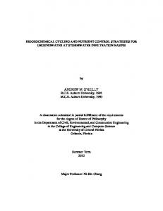
Cluster were detected in BMS3A, C and D, and accounted for 96.7% in the library BMS3Dapr. The OTUs in Nitrospirae-related Cluster and unassigned lineage have high similarity (76–77%) to the amino acid sequence of the aprA in Thermacetogenium phaeum within the Nitrospirae. cbbL and cbbM. cbbL genes were detected in the subsamples BMS3B and BMS9A, but cbbM genes were detected only in BMS9A (Table 1 and Fig. 5). OTUs of the cbbL/M genes were classified in gammaproteobacterial sulfur-oxidizing bacteria (Gamma-SOB), alphaproteobacterial sulfur-oxidizing bacteria (Alpha-SOB) and zetaproteobacterial iron-oxidizing bacteria (Zeta-FeOB) based on the phylogenetic tree (Fig. 5A and B). One cbbL OTU (BMS9AcbbL29) was not classified in the above clusters but related to the cbbL sequence of the deltaproteobacterial SAR324 cluster. The OTUs in Gamma-SOB accounted for over 90% in the libraries (Fig. 5C). We used the newly designed primers for cbbL in the present study (Fig. S11) because the previously designed primers (Alfreider et al., 2003; Campbell and Cary, 2004) have mismatches to the cbbL of A. ferrooxidans in Actinobacteria and M. ferrooxydans in Zetaproteobacteria; however, no cbbL related to these species was detected in the subsamples. Discussion SMS deposits in marine hydrothermal fields potentially support chemosynthetic ecosystems even after hydrothermal activity has ceased. Using 16S rRNA and functional gene analyses of sub-seafloor sulfide subsamples, we have provided the demonstration of the presence of diverse microbial species including chemoautotrophs, sulfur-oxidizers and sulfate-reducers. Based on the results of molecular biological, mineralogical and thermodynamic analyses, we discuss what reactions occur and which microbes are associated with them, and propose a model of biogeochemical cycling within the massive sulfide deposits below the seafloor (Fig. 6). Pyrite oxidation within SMS deposits
AprA. aprA genes were detected in the subsamples BMS3A to D and BMS9A (Table 1 and Fig. 4A). The aprA OTUs were classified in the sulfur-oxidizing bacteria (SOB) clusters I and II (Meyer and Kuever, 2007), SRB cluster, ‘Nitrospirae-related Cluster’ and ‘Unassigned lineage’ that are defined in the present study based on the phylogeny (Fig. 4B). All OTUs in the library BMS9Aapr were classified in the SOB clusters. The SOB clusters were also detected in the subsamples BMS3A to C. The OTUs in the SRB cluster were detected in BMS3A to D, but not in BMS9A. The OTUs in Nitrospirae-related
The mineralogical analyses and thermodynamic calculations suggest partial constraints on what reactions can take place within the SMS deposits. The detection of iron hydroxides (such as goethite) on the fractured surface of the subsample BMS9A (0.52 mbsf) provide direct evidence indicating that pyrite was oxidized by O2 contained in the oxygenated seawater, which passed through fractures within the shallower region. While the oxygenated seawater penetrated into the SMS deposits, the iron oxidation (reaction 2) could provide energy for the growth of (micro)aerobic iron-oxidizers.
© 2014 Society for Applied Microbiology and John Wiley & Sons Ltd, Environmental Microbiology, 17, 1817–1835
1824 S. Kato et al. 72
T13M−aprA−4, AFC36508
BMS3Bapr10 ◆
A89, ADW77065
B
A
BMS3Bapr04 ◆
61
SOB cluster I
57 64
SOB cluster II Unassigned lineage
SRB
BMS9Aapr15 ◆ BMS3Capr24 ◆
Thiothrix nivea, ABV80025 76 endosymbiont of Riftia pachyptila, ABV80043 Thiorhodococcus minor, ABV80013 66 T13M−aprA−29, AFC36517
Desulfobacteraceae
BMS3Bapr12 ◆
100
MR-MnCr-4, ABV00927 Candidatus Riegeria galatelae, AEI70745
90
BMS3Aapr09 ◆ BMS3Capr13 ◆
78 70
80
B11, ACL35747 delta proteobacterium SCGC AAA240-D19, ADX05651 T3AP−A9, AEJ86636 98 Desulfobacca acetoxidans, ABR92549 Syntrophobacterales aspA70m10, ADD84946 99
70 60
85
BMS3Capr06 ◆
50
74 56
40
91
30
BMS3Bapr17 ◆
Desulfobulbaceae
Ds_D3, AFV48113 Desulfofustis glycolicus, AAL57397 Desulfofaba gelida, AAL57385
85 80
20
Desulfobulbus rhabdoformis, AAL57377 Desulfobulbus elongatus, AAL57413 P_F7, AFV48094
BMS3Capr27 ◆ Desulfobacteraceae Desulfococcus multivorans, EPR35913 Desulfobacterium indolicum, ABR92477 Desulfovibrio alkalitolerans, EPR31030 Desulfonatronovibrio hydrogenovorans, AAL57378 98 Chlorobaculum thiosulfatiphilum, ABV79991 100 Chlorobium limicola, ABV79995 Chlorobi Pelodictyon clathratiforme, ABV79999 T8−aprA−13, AFC36464
SRB
Detection frequency (%)
BMS9Aapr03 ◆ BMS9Aapr28 ◆
apsA43, AEO13571 A53, AFK76440 T8−aprA−9, AFC36461
Nitrospirae-related cluster Syntrophobacterales (+Firmicutes) Desulfobulbaceae
83 97
10 69
BMS3Dapr
BMS3Capr
BMS3Bapr
63 BMS3Dapr23 ◆ 78 MEDEE_apr_C5, AGC09896 64 T8−aprA−15, AFC36465 56 BMS3Dapr17 ◆ 79 APS7.3, AAF16948 90 BMS3Dapr04 ◆
CVA−aprA− 28, CCG27943 Thermacetogenium phaeum, ABR92597 Thermodesulfovibrio islandicus, AAL57380 Thermodesulfovibrio yellowstonii, ABR92418 C20, ACL35753 MW2−4_61, BAN18085
58 98 56 56 89
Nitrospirae-related cluster
BMS3Aapr30 ◆
82
Nitrospirae Unassigned lineage
BMS3Capr28 ◆
BMS3Bapr30 ◆
99 55
apsA40B9, ADD84888
BMS3Bapr15 ◆
100
BMS3Bapr21 ◆
Pelagibacter ubique, YP_266257 A51, AFK76439 T13J−aprA−4, AFC36481
BMS3Bapr18 ◆
EBAC2C11, AAV31646 T8−aprA−43, AFC36478 61 100
BMS3Bapr05 ◆
SOB cluster I
Desulfomonile tiedjei, ABR92551 apsam29, ACF15348 Archaeoglobus veneficus, ABR92410 Archaeoglobus infectus, BAF64850 Archaeoglobi Archaeoglobus profundus, AAL57401 Allochromatium minutissimum, ABV80107 Thioalkalivibrio sulfidophilus, YP_002512435 Sideroxydans lithotrophicus, YP_003524335 92 Bathymodiolus brevior, ABV80099 Vesicomyosocius okutanii, YP_001218952 56 LSmat.aprA07, CBW37658
SRA
85 99
SRB
Desulfurispora thermophila, WP_018085432 Desulfotomaculum thermocisternum, ABR92594 78 Thermodesulforhabdus norvegica, ABR92558 Syntrophobacter fumaroxidans, AAL57405 81 T2AP−B5, AEJ86640
Syntrophobacterales (+Firmicutes)
T13M−aprA−1, AFC36505 58 T8−aprA−37, AFC36477
83
SRB
77
SOB
BMS3Aapr
(28) (32) (36) (27) (31) BMS9Aapr
0
SOB cluster II
Sulfuricella denitrificans, BAI66427 Thiobacillus denitrificans, ABV80031 Lamprocystis purpurea, ABV80005 Thiocystis gelatinosa, ABV80009
aprA−O−23, AEY77773 aprA−C−1, AEY77776 99 T13M−aprA−18, AFC36515
BMS9Aapr06 ◆
0.1
Fig. 4. A. Detection frequency and B. phylogenetic tree of aprA. B. The ML tree was constructed using 105 homologous positions in the alignment data set of amino acid sequences with the substitution model LG+G. Bootstrap values (> 50 of 100 replicates) are shown at the branch points. The scale bar represents 0.1 amino acid substitutions per sequence position. Filled diamonds following the sequence names indicate the OTUs detected in the present study.
© 2014 Society for Applied Microbiology and John Wiley & Sons Ltd, Environmental Microbiology, 17, 1817–1835
Biogeochemical cycling in sub-seafloor sulfides 79
1825
Fosmid_W, ACZ28640
BMS9AcbbL08 ◆
Suiyo (I)-8, BAD14298 endosymbiont BA−Sym(RI)−2, AAX48774
A
Gamma-SOB
BMS9AcbbL13 ◆ BMS3BcbbL08 ◆ BMS3BcbbL13 ◆
endosymbiont BA−Sym(RI)−1, AAX48773 Q673V5
58 Solemya velum gill symbiont, 53 BMS9AcbbL43 ◆
BMS3BcbbL44 ◆
OT-cbbL1.31, BAE80638 L1, ACN58123 Thiothrix nivea, WP_002707749 81
B 2KL−f2−otu1, AEN02439
BMS3BcbbL22 ◆ BMS9AcbbL03 ◆
BMS9AcbbM09 ◆
BMS9AcbbL29 ◆
55 Allochromatium minutissimum, ACC95828 Thiocapsa marina, WP_007190957 76 Halorhodospira halophila, YP_001002624 50 Methylococcus capsulatus, YP_115143 Ectothiorhodospira mobilis, ABN10961 59 Acidithiobacillus thiooxidans, WP_010637867 Acidimicrobium ferrooxidans, YP_003108761 Halothiobacillus neapolitanus, YP_003262812 Nitrosomonas eutropha, YP_747036 Acidithiobacillus ferrooxidans, YP_002221061 Nitrosococcus halophilus, YP_003528851 Sulfuricella denitrificans, YP_008546874 Thiomicrospira halophila, WP_019895625 Synechococcus sp., YP_478774
0.1
100 Detection frequency (%)
90 Other
70
Zeta-FeOB
60
Alpha-SOB
50
gamma proteobacterium str. SS-5, ADW09070
Thiothrix disciformis, WP_020395078 Thiomicrospira pelophila, ABC55009 V7cbbM87, BAK22521 Thiomicrospira crunogena, ABC55008
Thiomicrospira halophila, WP_019895620 97
SdcbbM01, BAK22503 SdcbbM24, BAK22506
BMS9AcbbM11 ◆ Thiotaurens thiomutagens, AFC88140
Accumulibacter phosphatis, YP_003166098 RccbbM16, BAK22493 V7cbbM18, BAK22518
BMS9AcbbM
10
BMS9AcbbM17 ◆
Rhodobacter capsulatus, YP_003577981
SdcbbM14, BAK22505 100 92 87
Alpha-SOB
Thioalkalicoccus limnaeus, AEN02459 hfmcbbM20, BAM15969 Magnetospira thiophila, ACM90120 61 Magnetovibrio blakemorei, AAL76921 81 Fosmid_aI, ACZ28620 endosymbiont of scaly snail, BAN68648 SdcbbM36, BAK22509 96 Rhodopseudomonas palustris, AAL14577 Rhodopseudomonas palustris, AAL14583
20
BMS9AcbbL
Lamprocystis purpurea, WP_020503383 Thiocystis violascens, YP_006416176 Thiothrix nivea, WP_002706799
Sulfuricella denitrificans, YP_008547681
30
BMS3BcbbL
BMS9AcbbM25 ◆ BMS9AcbbM14 ◆ BMS9AcbbM15 ◆
EBAC750−10A1, AAR38475
Gamma-SOB
40
0
59
RccbbM24, BAK22494 SdcbbM69, BAK22513
57
Thiorhodococcus drewsii, WP_007041869 Rhodoferax ferrireducens, YP_522655 Acidithiobacillus thiooxidans, WP_010637025 Thiobacillus denitrificans, WP_018078338 Acidithiobacillus ferrooxidans, YP_002220242 Halothiobacillus neapolitanus, YP_003262978 Sideroxydans lithotrophicus, YP_003522651 Thiomonas intermedia, YP_003643355 Thiobacillus denitrificans, YP_316396 Leptothrix ochracea, WP_009453497 Dechloromonas aromatica, YP_286836 Thiorhodovibrio sp. str. 970, WP_009149320 51 Vesicomyosocius okutanii, YP_001219490 99 Ruthia magnifica, YP_903909
C
80
73
Gamma-SOB
DI−cbbl 5, AAZ76716 L14, ACN58125 Mariprofundus ferrooxydans, WP_009850044 SCGC AAA007-C22, ADX05596 SCGC AB-629-O05, WP_018051788 SAR324-related
Leptonema illini, WP_002774391 Desulfovibrio aespoeensis, YP_004122290 Desulfovibrio hydrothermalis, YP_007327315 Magnetospirillum magnetotacticum, Q8RTI2
Phaeospirillum molischianum, WP_002725205 Magnetospirillum gryphiswalden, CAM77388
BMS9AcbbM13 ◆
Zetaproteobacterium SCGC AB-133-G06, WP_018288599 Mariprofundus ferrooxydans, WP_009851287
98 52
Thiomicrospira kuenenii, ABC55010 Hydrogenovibrio marinus, BAD15326 beta proteobacterium str. OYT1, BAM15628
100 100
0.1
Zeta-FeOB
Rhodospirillum rubrum, YP_427487 sulfur-oxidizing symbionts, WP_005960001 Verrucomicrobium sp. str. 3C, WP_018289723
100
Gallionella capsiferriformans, YP_003845843
Fig. 5. Phylogenetic tree of (A) cbbL and (B) cbbM and (C) detection frequency of the detected clones in each library. (A and B) The ML trees were constructed using 214 and 225 homologous positions in the alignment data sets of the amino acid sequences for cbbM and cbbL with the substitution model LG+G. Bootstrap values (> 50 of 100 replicates) are shown at the branch points. The scale bar represents 0.1 amino acid substitutions per sequence position. Filled diamonds following the sequence names indicate the OTUs detected in the present study.
© 2014 Society for Applied Microbiology and John Wiley & Sons Ltd, Environmental Microbiology, 17, 1817–1835
1826 S. Kato et al. Seawater O2
Seafloor
CO2
(1) FeS2
FeS2
(2)
CO2 OrgC FeOOH H2S CO2
Fe2+ CO2
SO42-
O2
OrgC SO42-
(3) (4)
OrgC
(5)
Sulfide deposit (1) Zetaproteobacteria, Acidimicrobiales (2) Chromatiales (3) Chromatiales, Deltaproteobacteria (4) Ignavibacteria, Acidimicrobiales, Deltaproteobacteria, Nitrospirae (5) Deltaproteobacteria, Nitrospirae Fig. 6. Model of biogeochemical cycling of Fe, S and C within the sub-seafloor massive sulfide deposits of the SMT. The bacterial groups associated with each reaction (1–5) are shown in the box at the bottom of the figure. Reactions 1–3 also represent carbon fixation. Ferrous iron and reduced sulfur species are provided from pyrite (FeS2). Oxygen, carbon dioxide and sulfate are provided from seawater. (Micro)aerobic iron-oxidizers in the Zetaproteobacteria and Acidimicrobiales oxidize ferrous iron to goethite (FeOOH) for their energy acquisition as shown by reaction 1. (Micro)aerobic sulfur-oxidizers in the Chromatiales oxidize reduced sulfur species to sulfate as shown by reaction 2. Reaction 3 includes abiotic H2S oxidation to elemental sulfur by iron hydroxides, and biotic S0 disproportionation to sulfide and sulfate performed by species in the Deltaproteobacteria and Chromatiales. Each reaction is shown in Fig. S12, but not in this figure to avoid complication. Anaerobic iron-reducers and sulfate-reducers in the Acidimicrobiales, Nitrospirae, Ignavibacteriae and Deltaproteobacteria reduce goethite and sulfate with organic carbon produced by the above chemoautotrophs and release ferrous iron and reduced sulfur species as shown by reactions 4 and 5. The released ferrous iron and reduced sulfur species can be oxidized once more by the iron-oxidizers and sulfur-oxidizers. Aerobic chemoorganotrophs are not shown in the carbon cycling. OrgC, organic carbon.
Pyrite was the dominant component of the deeper subsamples (1.36–2.67 mbsf) of the SMS deposits at either the Pika or Archaean site, except BMS9A, which is consistent with previous reports (Kakegawa et al., 2008; Ikehata et al., in press; Ishibashi et al., in press). Concentration of O2 in penetrating seawater might decrease via microbial respiration before it reached the deeper regions. However, it is unclear whether the O2 was completely depleted in the deeper regions or not. In fact, iron (hydr)oxide minerals were observed on the fractured surfaces of some subsamples collected from the deeper region (Table 1, Fig. S4). Microbial respiration on the fractured surfaces of the sulfide deposits would ensure that the interior of the deposits remained anoxic. Notably, pyrite oxidation by iron hydroxides does not occur in anoxic marine sediments as shown by laboratory incuba-
tion experiments, although this reaction is thermodynamically possible (Schippers and Jørgensen, 2002). Therefore, it is unlikely that the iron hydroxide is an oxidant for pyrite in the sulfide deposits. Overall, the energy provided via pyrite oxidation with O2 can be used for carbon fixation by primary producers. This organic carbon can then support the growth of secondary consumers including sulfate-reducers and iron-reducers. Thus, a chemosynthetic ecosystem could develop within the SMS deposits. The supply of hydrothermal fluids has probably stopped at the drilling points of both the Pika and Archaean sites: the absence of anhydrite in the sulfide samples supports this notion. Anhydrite, which is a typical component of active sulfide chimneys, is dissolved from sulfide chimneys after hydrothermal activity ceases (Haymon and Kastner, 1981). In fact, anhydrite has been detected in the active chimneys, but rarely in the inactive chimneys of the SMT (Ikehata et al., in press). Furthermore, anhydrite has not been detected in the other parts of the sulfide core samples of BMS03 and BMS09 (Ishibashi et al., in press) or in our subsamples. Therefore, the energy supporting the chemosynthetic ecosystem is likely provided by the pyrite oxidation, and not by reduced chemical species in hydrothermal fluids. This is consistent with the absence of Epsilonproteobacteria, which is a common member in the active chimney ecosystems of the SMT (Kato et al., 2010), in the sulfide samples as well as in the inactive chimneys. Similar observations and conclusions regarding the presence/absence of anhydrite and Epsilonproteobacteria between active and inactive chimneys have been reported previously (Sylvan et al., 2013). Potential for biogeochemical cycling Carbon fixation. The detection of cbbM/L indicates the presence of chemolithoautotrophic bacteria using the Calvin cycle within the SMS deposits. Based on the phylogeny, the cbbM/L are likely to be derived from sulfur-oxidizers in the Alphaproteobacteria and Gammaproteobacteria and iron-oxidizers in the Zetaproteobacteria. The cbbM/L were detected only in two subsamples (BMS9A and BMS3B) (Table 1). This result is consistent with the 16S rRNA gene analysis: the 16S rRNA genes in Chromatiales of Gammaproteobacteria and those in Zetaproteobacteria were detected in both subsamples. The 16S rRNA genes in Chromatiales were also detected in other subsamples, implying that not all of the detected members in Chromatiales possess cbbM/L genes detectable by the PCR assays used here. In addition, we designed a new primer set for cbbL covering A. ferrooxidans and M. ferrooxydans (Fig. S11) to detect more diverse chemolithoautotrophs including
© 2014 Society for Applied Microbiology and John Wiley & Sons Ltd, Environmental Microbiology, 17, 1817–1835
Biogeochemical cycling in sub-seafloor sulfides Zetaproteobacteria and Actinobacteria. However, cbbL related to A. ferrooxidans and M. ferrooxydans were not detected, implying that members possessing the 16S rRNA genes in Zetaproteobacteria and the Actinobacteria Cluster are not autotrophs, although there is a possibility that the designed primers still have mismatches to the cbbL of these members. Chemolithoautotrophs in the Epsilonproteobacteria and Aquificae, which are common in active sulfide chimneys, fix inorganic carbon via the reverse tricarboxylic acid (rTCA) cycle (Nakagawa and Takai, 2008). In the present study, the aclB gene encoding the beta subunit of Adenosine triphosphate (ATP) citrate lyase, which is a key enzyme of rTCA cycle, was not detected by PCR. This is consistent with the 16S rRNA gene analysis showing that Epsilonproteobacteria and Aquificae were not or were rarely detected in our samples (Fig. 1 and Fig. S7). Sulfur cycling. Phylogenetic analysis of the detected aprA indicates the presence of both of sulfur-oxidizers and sulfate-reducers in the SMS deposits. This result implies that local sulfur cycling driven by these bacteria takes place. Such local sulfur cycling by bacteria has been reported in other marine sediments and oxygen minimum zones (Jørgensen, 1977; Canfield et al., 2010). SRB belonging to the Deltaproteobacteria are likely present in the SMS deposits. In particular, the aprA OTUs (e.g., BMS3Bapr17) are likely derived from the Desulfobulbus-related species detected by the 16S rRNA gene analysis (e.g., BMS3AB02; Fig. 2). It is unlikely that the SRB belong to the Firmicutes because no 16S rRNA gene in that taxon was detected in the subsamples, except BMS9ABp (0.17% of the number of total reads). Although the taxonomic affiliation of the detected aprA of the SOB clusters is unclear, the detection of the 16S rRNA genes related to sulfur-oxidizers in the Chromatiales in Gammaproteobacteria implies that some or all of the aprA are derived from this group of sulfur-oxidizers. In addition, the detected deltaproteobacterial OTUs may contain species capable of elemental sulfur (S0) disproportionation, although we can only infer from 16S rRNA gene phylogeny (Fig. 2). Some deltaproteobacterial species in the genera of Desulfobulbus and Desulfocapsa can grow autotrophically on S0 via the disproportionation reaction under anoxic conditions (Finster, 2008) as follows.
4 S0 + 4 H2O → 3 H2S + SO4 2 − + 2 H+
(5)
The S0 can be produced as an intermediate by the abiotic reaction between iron hydroxides and H2S (Fig. S12) (Yao and Millero, 1996) as follows:
2 FeO (OH) + 3 H2S → S0 + 2 FeS + 2 H+
(6)
1827
The precipitated iron hydroxides are provided in reaction 3 under oxic conditions. The H2S can be produced by sulfate-reducers and also by sulfur-disproportionaters themselves (Fig. S12). It should be noted that the bacterial S0 disproportionation should ultimately depend on the chemosynthesis by pyrite oxidation with O2. Furthermore, we detected the OTUs (i.e., BMS9AB11 and BMS9AB76) related to Acidiferrobacter thiooxydans in the Chromatiales (Fig. 2). This bacterium can grow on S0 with iron hydroxides (Hallberg et al., 2011). This reaction can be represented as follows:
3 S0 + 2 FeO (OH) → 2FeS + SO4 2 − + 2 H+
(7)
It should be noted that reaction 7 results from a combination of reactions 5 and 6. These reactions of microbial S0 disproportionation and iron reduction have already reported by Thamdrup and colleagues (1993). Although details of the metabolic pathway of A. thiooxydans remain unclear, the iron hydroxides could be abiotically reduced by H2S that is being produced during sulfur disproportionation, as in the case of Acidithiobacillus ferrooxidans in the order Acidithiobacillales of the Gammaproteobacteria (Osorio et al., 2013). It is possible that some of these OTUs related to A. thiooxydans represent such sulfur-disproportionating bacteria and play a role in not only sulfur cycling, but also iron cycling within the SMS deposits. However, the similarities to A. thiooxydans were low (∼90%), and the bootstrap value supporting the cluster of the OTUs and A. thiooxydans was statistically insignificant (56%; Fig. 2). The OTUs also showed ∼91% similarity to (micro)aerobic or nitratereducing sulfur-oxidizers such as Thioalkalispira spp. and Thiohalophilus spp., and to chemoorganotrophs such as Granulosicoccus spp. Thus, in the present study, we cannot exclude another possibility that the OTUs represent these (micro)aerobic or nitrate-reducing sulfideoxidizers, or chemoorganotrophs. Based on the phylogeny, we infer that the aprA in the Nitrospirae-related cluster and unassigned lineage (Fig. 4) are derived from members possessing the 16S rRNA genes in Nitrospirae cluster A and/or B (Fig. 2). One possibility is that the aprA are derived from sulfuroxidizers. The Nitrospirae cluster A contains MTB, which seem to be microaerobic sulfur-oxidizers (Jogler et al., 2010; Lefèvre et al., 2011), although direct evidence has not been provided yet because no cultured isolates are available to date. The MTB produce crystals of submicron-sized magnetite (Fe3O4) and store them intracellularly as magnetosomes (Jogler et al., 2010; Lefèvre et al., 2011), a process that could take place in the SMS environment as long as O2 is present. Alternatively, the detected OTUs of 16S rRNA and aprA genes related to Nitrospirae may represent sulfate-reducers. The closest cultured species to both of these 16S rRNA and
© 2014 Society for Applied Microbiology and John Wiley & Sons Ltd, Environmental Microbiology, 17, 1817–1835
1828 S. Kato et al. aprA genes are the SRB, Thermodesulfovibrio spp. As described above, O2 could be depleted in the interior of the subsamples where these OTUs of 16S rRNA and aprA genes were detected. It is also possible that the detected OTUs contains both of MTB and sulfate-reducers. Although members of the Bacteroidetes are generally associated with the degradation of complex organic substrates, some strains with limited capacity to oxidize thiosulfate as an auxiliary electron donor have been reported (Teske et al., 2000). It is unclear whether the detected OTUs in the Bacteroidetes Cluster contain sulfur-oxidizers or not because they have low similarities to the reported sulfur-oxidizers (82–83%). However, environmental clones in this cluster have been commonly detected in the inactive chimneys (Suzuki et al., 2004; Kato et al., 2010; Sylvan et al., 2012; 2013), indicating that the members in the ‘Bacteroidetes Cluster’ (or the ‘Sulfiphilic Bacteroidetes’) preferentially colonize metal sulfides, as found previously (Sylvan et al., 2013). Iron cycling. Regarding potential ecosystem members associated with iron cycling, we can only infer from the phylogeny of the detected 16S rRNA genes because no functional gene approach for iron oxidation or reduction have been established. Fe2+ for iron-oxidizers can be released by pyrite oxidation (reaction 1) and also by reduction of iron oxides via abiotic reaction (reaction 4) or activity of iron-reducers. The zetaproteobacterial OTU, which was detected only in BMS9A mainly consisting of iron (hydr)oxides, potentially represents an iron-oxidizer. The Zetaproteobacteria include a microaerophilic ironoxidizer M. ferrooxydans (Emerson et al., 2007) and are dominant in iron-rich environments in marine hydrothermal fields (Kato et al., 2009a,b; Rassa et al., 2009). Our results suggest that putative microaerophilic ironoxidizers in the Zetaproteobacteria prefer habitats where seawater circulation introduces O2 rather than deeper anoxic SMS habitats. The detected OTUs in the ‘Actinobacteria Cluster’ in Acidimicrobiales of the Actinobacteria potentially represent iron-oxidizers and/or iron-reducers. To date, cultured species in the Acidimicrobiales are known to be associated with the leaching of sulfide minerals in terrestrial acid mines (Clark and Norris, 1996; Baker and Banfield, 2003); however, their biogeochemical role in SMS deposits had not been considered. In addition, the detected members related to Melioribacter in the Ignavibacteriae, to Thermodesulfovibrio in the Nitrospirae and to Desulfobulbus in the Deltaproteobacteria may contain iron-reducers. They potentially play a role in iron cycling in the SMS deposits, because some cultured species in these genera are known to reduce ferric iron as electron acceptors (Holmes et al., 2004; Sekiguchi et al., 2008; Podosokorskaya et al., 2013).
Commonality and difference in bacterial communities Our comparative analysis showed that there were several common members between our sub-seafloor sulfide samples and the inactive chimneys (Fig. S8), despite samples coming from different geographic locations (i.e., EPR, CIR, OT and LB). These common members are likely a core microbiome for microbial communities in the sulfide habitats. A core microbiome is defined as microbes that are common across microbial communities in certain habitats and are hypothesized to play a key role in ecosystem function (Shade and Handelsman, 2012). Common members among the sulfide habitats include the representative bacterial OTUs in the ‘Bacteroidetes Cluster’, Desulfobulbus-related group, Chromatiales, the ‘Nitrospirae Cluster A’ and Ignavibacteriae. They probably contribute to sulfur, iron and carbon cycling and play a key role in ecosystem function within the sulfide habitats. On the other hand, our comparative analysis showed that there were many unique bacterial OTUs in the subseafloor sulfide samples (Fig. S8). The presence of these unique OTUs resulted in the difference in bacterial communities between our samples and the others as shown by the PCoA (Fig. 3). It seems that the difference in the communities reflects environmental (e.g., oxygen concentration and mineralogy), temporal (i.e., elapsed time from the cessation of hydrothermal venting) and spatial (e.g., geological background and water depth) differences. However, in the present study, it remains unclear what factors contribute most to the observed differences between the bacterial communities. Previous studies have suggested that differences in mineralogy cause differences in bacterial communities in sulfide chimneys (Toner et al., 2013; Sylvan et al., 2013). Indeed, the observed difference in the bacterial communities between the BMS9A and the other sub-seafloor subsamples (Figs 1 and 3) likely reflect the difference in O2 availability that results in the observed difference in mineralogy of the main components (iron hydroxide vs. pyrite and/or sphalerite; Table 1). Although there are some differences in mineralogy among the sub-seafloor subsamples, they do not appear to be a major cause of the slight differences in their communities among the subsamples. Further analyses such as of pore-water chemistry of drilled cores will be needed to identify the major factors. Uncultured archaea We have previously reported that MGI was mainly detected in inactive sulfide chimneys of the SMT (Kato et al., 2010). However, MGI is a dominant member in oceans (DeLong et al., 1994; Karner et al., 2001) and has also been detected in bottom seawater in the SMT (Kato et al., 2009b). Hence, it is difficult to identify whether the
© 2014 Society for Applied Microbiology and John Wiley & Sons Ltd, Environmental Microbiology, 17, 1817–1835
Biogeochemical cycling in sub-seafloor sulfides
1829
MGI presence is because of contamination from surrounding seawater or originated from the chimneys. Indeed, in the present study, MGI was not detected. In contrast, in addition to MGI, MGBE was detected in the inactive sulfide chimneys of the CIR and OT (Suzuki et al., 2004). In the present study, MGBE predominated in the libraries (Fig. S9A). As far as we know, MBGE has not been detected in deep seawater. These facts indicate that there is a preferable habitat for MBGE within the sulfide deposits regardless of the geographical distance among SMT, CIR and OT. In addition, we detected archaeal OTUs in the Parvarchaea and Micrarchaea using the universal bacteria primers (Fig. S10). These archaea were not recovered using the universal archaea primers (Fig. S9). Furthermore, the archaeal diversity in some subsamples is likely to be comparable with or even higher than the bacterial diversity, as shown by the rarefaction curves and diversity values (Fig. S6, Table S1). These results suggest that diverse archaea in SMS deposits have been overlooked by PCR with the archaea-universal primers usually used. Metagenomic analysis independent of specific primers will reveal the presence of unusual archaea in the SMS deposits, as evidenced in the previous studies of acid mine environments (Baker et al., 2006; 2010).
suitable electron acceptors are available, or by elemental sulfur disproportionation, as long as sulfide is reoxidized to elemental sulfur by reactive metal oxides. Based on the combination of molecular biological, mineralogical and thermodynamic analyses, we propose a model of biogeochemical cycling in the ecosystem (Fig. 6). The detected bacteria (e.g., those in the Chromatiales, Deltaproteobacteria, Nitrospirae and Zetaproteobacteria) likely drive the cycling of sulfur, iron and carbon within the SMS deposits. Some of these bacteria are likely to be widely distributed in SMS deposits in various areas and play important roles in the ecosystem functioning as a core microbiome. The ecosystem within massive sulfides is clearly different from those in hydrothermally active sulfide chimneys. We showed that more diverse archaea are present in the SMS deposits than previously recognized, although their ecological roles remain unclear. It should be noted that the DNA-based analyses could not address whether the detected microbes are active in situ or not. In addition, the diversity and metabolic functions shown in the present study are limited by the PCR primers used. Further investigation, such as metagenomics and metatranscriptomics, will provide helpful information for the better understanding of the microbial ecology of the SMS deposits.
Contamination assessment
Experimental procedures
The sequence analyses indicate that microbial contamination from the bottom seawater on the seafloor could be minimized. Our previous study has suggested that the dominant bacteria and archaea in the bottom seawater in the SMT are SAR11 cluster in the Alphaproteobacteria, SUP05 group in the Gammaproteobacteria and MGI in the Archaea (Kato et al., 2009b). No sequence related to MGI and the SUP05 group was detected in the present study. SAR11 was detected only in the BMS9CBp library (1.4% of the total number of reads). In addition, the cell densities were far higher in the sulfide samples (107– 108 cells g−1, Table 1) than those in the seawater (104– 105 cells ml−1; (Kato et al., 2009b)). Therefore, even if the seawater microbes did contaminate the sulfide samples, they would be heavily outnumbered by the indigenous microbes. Thus, we conclude that the vast majority of the detected sequences were derived from the bacteria and archaea indigenous to the SMS deposits. Using the filtrated seawater to washout the shavings, together with careful handling of the cores, greatly minimized the contamination.
Study area and sampling
Conclusion A chemosynthetic ecosystem within the sub-SMSs of the SMT could be supported by pyrite oxidation, as long as
Samples of SMS deposits were collected at the Pika and Archaean sites in the SMT back-arc spreading basin during the TAIGA10M cruise of the R/V Hakurei-Maru No.2 (JOGMEG, Japan) (14–23 June 2010). The Pika site is located on an off-ridge knoll about 5 km away from the spreading axis, and the Archaean site is located at a giant sulfide mound on the eastern ridge flank of the spreading axis (Yoshikawa et al., 2012)(Fig. S1). Hydrothermally active and inactive chimneys have been found at both sites. We carried out shallow sub-seafloor drilling using BMS in the Pika and Archaean sites during the cruise. The BMS is a remotely operated seafloor-coring machine installed on the R/V Hakurei-Maru No.2 that is capable of coring continuously in hard rocks up to 20 mbsf using 10 core barrels (Marumo et al., 2008). In the present study, to avoid contamination from the seawater on the seafloor, during the drilling operation the shavings were washed out using filter-sterile bottom seawater that were produced by a filtration system with 10, 0.45 and 0.22 μm pore-size cartridge-type filters equipped with the BMS. The sulfide core samples were obtained from the boreholes BMS03 (12°56.3627’N, 143°37.9036’E) in the Archaean site at water depth of 3,024 m and BMS09 (12°55.1368’N, 143°38.9333’E) in the Pika site at a water depth of 2804 m respectively (Fig. S1C and D). Although some active vents were found at both sites, none was observed around the drilling point (or was at least 10 m away). No hydrothermal fluid emission was observed from the two boreholes after drilling.
© 2014 Society for Applied Microbiology and John Wiley & Sons Ltd, Environmental Microbiology, 17, 1817–1835
1830 S. Kato et al. Borehole BMS03 and BMS09 were 3.30 and 4.25 m in length respectively (Figs S2 and S3). Each core sample was collected using two or three core barrels of the BMS. For the core sample BMS3, subsamples were collected from the parts of the core sample corresponding to depths of 1.86, 1.96, 2.48 and 2.67 mbsf, defined as BMS3A, B, C and D respectively (Fig. S2, Table 1). For the core sample BMS9, subsamples were collected from the parts of the core sample corresponding depths of 0.52, 1.36 and 2.33 mbsf, defined as BMS9A, B and C respectively (Fig. S3, Table 1). Brownish, grayish or blackish materials were observed on the fractured surfaces of the subsamples (Figs S2 and S3). These materials could be scraped from the surfaces using sterile spatulas. For DNA analysis, these scraped materials were transferred into DNA/RNA-free plastic tubes in a clean bench on board the vessel. To avoid contamination all of the subsamples were carefully taken from those portions that were not attached to the inner wall of the core barrels. The scraped materials and the whole subsamples were stored at −80 °C until DNA extraction and mineralogical analyses were carried out.
Mineralogical analyses Mineralogy of the subsamples was determined using reflected-light microscopy, FE-EPMA (JEOL JXA-8530F, Tokyo, Japan) and Raman spectroscopy (MARS, PHOTON Design, Tokyo, Japan) at the University of Tsukuba, Japan.
Thermodynamic calculation We conducted thermodynamic modelling using the REACT module in the Geochemist’s Workbench computer code (Bethke, 2008), assuming a reaction temperature of 2 °C (bottom seawater temperature). In this calculation, pyrite (FeS2) was reacted with an assumed seawater composition (Na: 480 mmol kg−1, Cl: 550 mmol kg−1, O2(aq): 0.2 mmol kg−1, SO4: 28 mmol kg−1, total carbonic acid: 2.2 mmol kg−1, pH: 7.8). All calculations were carried out using a total pressure of 500 bar, which was higher than the actual pressure (∼300 bar), but pressure is a minor factor because the equilibrium constants are not sensitive to the changes in pressure at or around the seafloor condition. In this model, hematite was suppressed to precipitate, whereas ferric iron mineral was assumed to be goethite (FeO(OH)) to maintain compatibility with the observed ferric iron minerals in the SMS deposits. The thermodynamic database required for the calculations was generated by the SUPCRT92 computer program (Johnson et al., 1992), with thermodynamic data for mineral, aqueous species and complexes from previous reports (Helgeson et al., 1978; Shock and Helgeson, 1988; Shock et al., 1989; 1997; Shock and Koretsky, 1995; McCollom and Shock, 1997; Sverjensky et al., 1997; Majzlan et al., 2003a,b; Klein et al., 2009; McCollom and Bach, 2009). The B-dot activity model was used in the calculations (Helgeson, 1969; Helgeson and Kirkham, 1974). The temperature-dependent activity coefficient for aqueous CO2 was derived from the empirical relationship established by Drummond (1981), and the temperature-dependent activity of water in NaCl solution was derived from Bethke’s (2008) formulation. Cleverley and Bastrakov (2005) provide useful temperature-dependent
polynomial functions for both of these latter parameters. Na was used to compensate for imbalanced charges in the model.
DNA extraction, cloning and sequencing PCR clone libraries were constructed according to our previous report (Kato et al., 2009a) with some minor modifications. Genomic DNA was extracted from approximately 0.5 g of each subsample using a FastDNA kit for Soil (Qbiogene, Inc., Irvine, CA, USA). We unsuccessfully attempted to extract DNA from mineral-rich areas within the solid sulfide samples, but were able to obtain DNA from the relatively soft materials scraped from the fractured surface of the samples. Concentration of the extracted DNA was determined using Quant-iT PicoGreen dsDNA Assay Kit (Invitogen, CA, USA). Bacterial and archaeal 16S rRNA, aprA and cbbM genes were amplified by PCR using the following oligonucleotide primer sets: Arc9F (Kato et al., 2011) and Uni1406R (Kato et al., 2009a) for archaeal 16S rRNA genes; Bakt_341F and Bakt_805R (Herlemann et al., 2011) (defined as primer set A), and Bac27F (Lane, 1991) and Uni1406R (primer set B) for bacterial 16S rRNA genes; AprA-1-FW and AprA-5-RV for aprA (Meyer and Kuever, 2007); and cbbM343F and cbbM1226R for cbbM (Kato et al., 2012). Primer set A is the same as S-D-Bact-0341-b-S-17 and S-D-Bact-0785-a-A-21, and is the best for 454 pyrosequencing for bacteria as evaluated previously (Klindworth et al., 2013). In addition, we designed two new primers, cbbL571F (5′-GAYTTYACC AAGGAYGACGARA-3′) and cbbL1385R (5′-AAYTCGA ACTTGATYTCYTTCC-3′), based on 43 of cbbL gene sequences obtained from public genome sequences (see Fig. S11) and used them for the clone library construction. The thermal cycle programs used in PCR for each primer set are shown in Table S3. In addition, we tried unsuccessfully to amplify ATP citrate lyase genes as described previously (Campbell et al., 2003). PCR products were purified using Wizard SV Gel and PCR Clean-Up System (Promega, WI, USA) and cloned with a TOPO TA cloning kit (Invitrogen, CA, USA). The nucleotide sequences of randomly selected clones were determined with a BigDye Terminator v3.1 Cycle Sequencing kit (Applied Biosystems, CA, USA) using M13 forward and reverse primers (Invitrogen, CA, USA) on an ABI PRISM 3130xl Genetic analyzer (Applied Biosystems, CA, USA) at the Japan Collection of Microorganisms (JCM) in RIKEN BRC or on an ABI PRISM 3730xl Genetic analyzer (Applied Biosystems, CA, USA) at the Support Unit for Bio-Material Analysis in RIKEN BSI Research Resources Center (RRC).
Pyrosequencing In addition to the PCR clone analysis, we performed 454 pyrosequencing for the analysis of the bacterial communities. Bacterial 16S rRNA genes, including V3 and V4 regions, were amplified by PCR using the primer set A with the thermal cycle shown in Table S3. An additional PCR (5 or 10 cycles) was performed with the primers with 454 adaptor and multiplex identifier sequences for each sample (Table S4). The PCR products were purified as described above and quantified using a Quant-iT PicoGreen dsDNA Assay Kit
© 2014 Society for Applied Microbiology and John Wiley & Sons Ltd, Environmental Microbiology, 17, 1817–1835
Biogeochemical cycling in sub-seafloor sulfides (Invitrogen, CA, USA), and then mixed in equal concentrations. This mix was used for pyrosequencing with Titanium chemistry (GS Junior Titanium emPCR Lib-L and GS Junior Titanium Sequencing Kits) on a GS Junior platform (Roche 454 Life Sciences, Branford, CT, USA) in RIKEN BRC.
Sequence analysis For 16S rRNA genes in the PCR clone libraries, chimeric sequences were removed using MALLARD (Ashelford et al., 2006). Non-chimeric sequences were aligned using MUSCLE (Edgar, 2004) and clustered into OTUs at a 97% similarity level using MOTHUR (Schloss et al., 2009). Representative OTUs and their relatives in public databases were aligned once more using MUSCLE. Gap positions in the alignment data were removed using GBLOCKS (Castresana, 2000). Maximum-likelihood (ML) trees were constructed with the non-gapped alignment data using PHYML 3.0 (Guindon et al., 2010). We used MRAIC.PL (Nylander, 2004) to select the nucleotide substitution model that best fitted our alignment data. Taxonomic affiliation of bacterial and archaeal 16S rRNA genes was performed using QIIME 1.7.0 (Caporaso et al., 2010). Chao1 richness estimators, Shannon’s diversity index, Good’s coverage and the number of OTUs shared among communities were calculated using MOTHUR. Unweighted PCoA was performed using QIIME via UniFrac. For the aprA, cbbM and cbbL genes, the nucleotide sequences were translated into amino acid sequences based on the bacterial genetic code using EMBOSS TRANSEQ (Rice et al., 2000). The amino acid sequences were aligned and clustered into OTUs at a 100% similarity level using MOTHUR. Representative clones for each OTU and their close relatives in public databases were aligned, gap positions were removed from the alignment data and then ML trees were constructed as described above. We used PROTTEST 3 (Darriba et al., 2011) to select the amino acid substitution model that best fitted our alignment data set. For bacterial 16S rRNA genes obtained from pyrosequencing, a first filtering of the sequence data was performed using the amplicon signal-processing pipeline of GS RunProcessor (Roche 454 Life Sciences). Sequences shorter than 400 bases, with ambiguities or with noise characteristic of pyrosequencing, and also chimeric sequences, were removed from the sequence data using QIIME via denoiser and ChimeraSlayer and were not used for further sequence analyses. High-quality sequences were aligned and clustered into OTUs at a 97% similarity level, and then taxonomic affiliations of the OTUs were performed using QIIME. Chao1 richness estimators and Shannon’s diversity index were calculated using MOTHUR with the distance matrix data produced by the above QIIME analysis. We found some archaeal sequences in the data set obtained with the primer A, so archaeal and bacterial sequences were separated from each other within the data set based on the taxonomic affiliations by QIIME. Each of archaeal and bacterial sequence data were analysed separately.
Q-PCR The copy numbers of the bacterial and archaeal 16S rRNA genes in the extracted DNA were determined by quantitative
1831
PCR as previously described (Kato et al., 2009a). In brief, we performed quantitative PCR on an ABI PRISM 7000 SEQUENCE DETECTION SYSTEM (Applied Biosystems, CA, USA) using the following primers and TaqMan probes: Bac1369, Prok1492R and TM1389F for bacterial 16S rRNA genes (Suzuki et al., 2000), or Arc349F and Arc806R and Arc516F for archaeal 16S rRNA genes (Takai and Horikoshi, 2000). A dilution series of the purified PCR products of a bacterial clone (BMS3BB34) or an archaeal clone (BMS3AA50) obtained in the present study was used as the Q-PCR standard for bacterial (r2 value of the standard curve = 0.999) and archaeal analyses (r2 = 0.995). All assays were performed in triplicate.
Accession numbers The nucleotide sequences detected in the present study were submitted to the DNA Data Bank of Japan (DDBJ) database under accession numbers: AB722091 to AB722174 and AB858509 to AB858686 for bacterial 16S rRNA genes; AB858687 to AB858827 for archaeal 16S rRNA genes; AB858828 to AB858850 for aprA genes; AB858851 to AB858857 for cbbM genes; AB858858 to AB858866 for cbbL genes. The sequence data produced by pyrosequencing were submitted to the DDBJ Sequence Read Archive (DRA) under accession number DRA001169.
Acknowledgement We would like to thank the crew of R/V Hakurei-Maru No.2, the operation team of BMS and the scientific colleagues for their professional skills and careful consideration for collecting core samples during the TAIGA10 cruise. We are grateful to the Support Unit for Bio-Material Analysis in RIKEN BSI Research Resources Center (RRC) for DNA sequencing. We would like to thank two anonymous reviewers for their helpful comments and suggestions. This research was supported by the ‘TAIGA project’, which was funded by a Grant-in-Aid for Scientific Research on Innovative Areas (#20109006) from the Ministry of Education, Culture, Sports, Science and Technology (MEXT), Japan, by a Grant-in-Aid for Young Scientists (B) (#24770032), and partially by the RIKEN Special Postdoctoral Researchers Program. The authors have no conflict of interest to declare.
References Alfreider, A., Vogt, C., Hoffmann, D., and Babel, W. (2003) Diversity of ribulose-1,5-bisphosphate carboxylase/ oxygenase large-subunit genes from groundwater and aquifer microorganisms. Microb Ecol 45: 317–328. Ashelford, K.E., Chuzhanova, N.A., Fry, J.C., Jones, A.J., and Weightman, A.J. (2006) New screening software shows that most recent large 16S rRNA gene clone libraries contain chimeras. Appl Environ Microbiol 72: 5734– 5741. Baker, B.J., and Banfield, J.F. (2003) Microbial communities in acid mine drainage. FEMS Microbiol Ecol 44: 139–152. Baker, B.J., Tyson, G.W., Webb, R.I., Flanagan, J., Hugenholtz, P., Allen, E.E., et al. (2006) Lineages of acidophilic archaea revealed by community genomic analysis. Science 314: 1933–1935.
© 2014 Society for Applied Microbiology and John Wiley & Sons Ltd, Environmental Microbiology, 17, 1817–1835
1832 S. Kato et al. Baker, B.J., Comolli, L.R., Dick, G.J., Hauser, L.J., Hyatt, D., Dill, B.D., et al. (2010) Enigmatic, ultrasmall, uncultivated archaea. Proc Natl Acad Sci USA 107: 8806–8811. Bethke, C.M. (2008) Geochemical and Biogeochemical Reaction Modeling, 2nd edition. Cambridge, UK: Cambridge University Press. Button, D.K., and Robertson, B.R. (2001) Determination of DNA content of aquatic bacteria by flow cytometry. Appl Environ Microbiol 67: 1636–1645. Campbell, B.J., and Cary, S.C. (2004) Abundance of reverse tricarboxylic acid cycle genes in free-living microorganisms at deep-sea hydrothermal vents. Appl Environ Microbiol 70: 6282–6289. Campbell, B.J., Stein, J.L., and Cary, S.C. (2003) Evidence of chemolithoautotrophy in the bacterial community associated with Alvinella pompejana, a hydrothermal vent polychaete. Appl Environ Microbiol 69: 5070–5078. Canfield, D.E., Stewart, F.J., Thamdrup, B., De Brabandere, L., Dalsgaard, T., Delong, E.F., et al. (2010) A cryptic sulfur cycle in oxygen-minimum–zone waters off the Chilean Coast. Science 330: 1375–1378. Caporaso, J.G., Kuczynski, J., Stombaugh, J., Bittinger, K., Bushman, F.D., Costello, E.K., et al. (2010) QIIME allows analysis of high-throughput community sequencing data. Nat Methods 7: 335–336. Castresana, J. (2000) Selection of conserved blocks from multiple alignments for their use in phylogenetic analysis. Mol Biol Evol 17: 540–552. Clark, D.A., and Norris, P.R. (1996) Acidimicrobium ferrooxidans gen. nov., sp. nov.: mixed-culture ferrous iron oxidation with Sulfobacillus species. Microbiology 142: 785–790. Cleverley, J.S., and Bastrakov, E.N. (2005) K2GWB: utility for generating thermodynamic data files for the Geochemist’s Workbench at 0–1000 °C and 1–5000 bar from UT2K and the UNITHERM database. Comput Geosci 31: 756– 767. Cole, J.R., Wang, Q., Fish, J.A., Chai, B., McGarrell, D.M., Sun, Y., et al. (2014) Ribosomal database project: data and tools for high throughput rRNA analysis. Nucleic Acids Res 42: D633–D642. Darriba, D., Taboada, G.L., Doallo, R., and Posada, D. (2011) Prottest 3: fast selection of best-fit models of protein evolution. Bioinformatics 27: 1164–1165. DeLong, E.F., Wu, K.Y., Prezelin, B.B., and Jovine, R.V.M. (1994) High abundance of archaea in antarctic marine picoplankton. Nature 371: 695–697. Drummond, S.E. (1981) Boiling and Mixing of Hydrothermal Fluids: Chemical Effects on Mineral Precipitation. PhD Thesis. Pennsylvania State University. Edgar, R.C. (2004) MUSCLE: multiple sequence alignment with high accuracy and high throughput. Nucleic Acids Res 32: 1792–1797. Emerson, D., Rentz, J.A., Lilburn, T.G., Davis, R.E., Aldrich, H., Chan, C., et al. (2007) A novel lineage of proteobacteria involved in formation of marine Fe-oxidizing microbial mat communities. PLoS ONE 2: e667. doi:10.1371/journal .pone.0000667. Emerson, D., Fleming, E.J., and McBeth, J.M. (2010) Ironoxidizing bacteria: an environmental and genomic perspective. Annu Rev Microbiol 64: 561–583.
Finster, K. (2008) Microbiological disproportionation of inorganic sulfur compounds. J Sulfur Chem 29: 281–292. Guindon, S., Dufayard, J.-F., Lefort, V., Anisimova, M., Hordijk, W., and Gascuel, O. (2010) New algorithms and methods to estimate maximum-likelihood phylogenies: assessing the performance of PhyML 3.0. Syst Biol 59: 307–321. Hallberg, K., Hedrich, S., and Johnson, D.B. (2011) Acidiferrobacter thiooxydans, gen. nov. sp. nov.; an acidophilic, thermo-tolerant, facultatively anaerobic ironand sulfur-oxidizer of the family Ectothiorhodospiraceae. Extremophiles 15: 271–279. Hannington, M., Jamieson, J., Monecke, T., Petersen, S., and Beaulieu, S. (2011) The abundance of seafloor massive sulfide deposits. Geology 39: 1155–1158. Haymon, R.M., and Kastner, M. (1981) Hot spring deposits on the East Pacific Rise at 21°N: preliminary description of mineralogy and genesis. Earth Planet Sci Lett 53: 363– 381. Helgeson, H.C. (1969) Thermodynamics of hydrothermal systems at elevated temperatures and pressures. Am J Sci 267: 729–804. Helgeson, H.C., and Kirkham, D.H. (1974) Theoretical prediction of the thermodynamic behavior of aqueous electrolytes at high pressures and temperatures; II, Debye-Huckel parameters for activity coefficients and relative partial molal properties. Am J Sci 274: 1199–1261. Helgeson, H.C., Delany, J.M., Nesbitt, H.W., and Bird, D.K. (1978) Summary and critique of the thermodynamic properties of rock-forming minerals. Am J Sci 278–A: 1–229. Herlemann, D.P.R., Labrenz, M., Jurgens, K., Bertilsson, S., Waniek, J.J., and Andersson, A.F. (2011) Transitions in bacterial communities along the 2000 km salinity gradient of the Baltic Sea. ISME J 5: 1571–1579. Herzig, P.M., and Hannington, M.D. (1995) Polymetallic massive sulfides at the modern seafloor a review. Ore Geol Rev 10: 95–115. Holmes, D.E., Bond, D.R., and Lovley, D.R. (2004) Electron transfer by Desulfobulbus propionicus to Fe(III) and graphite electrodes. Appl Environ Microbiol 70: 1234–1237. Humphris, S.E., Herzig, P.M., Miller, D.J., Alt, J.C., Becker, K., Brown, D., et al. (1995) The internal structure of an active sea-floor massive sulphide deposit. Nature 377: 713–716. Ikehata, K., Suzuki, R., Shimada, K., Ishibashi, J., and Urabe, T. (in press) Mineralogical and geochemical characteristics of hydrothermal minerals collected from hydrothermal vent fields in the Southern Mariana spreading center. In Subseafloor Biosphere Linked to Global Hydrothermal Systems: Taiga Concept. Ishibashi, J., Okino, K., and Sunamura, M. (eds). Tokyo: Springer Japan. Ishibashi, J., Shimada, K., Sato, F., Uchida, A., Toyoda, S., Takamasa, A., et al. (in press) Dating hydrothermal mineralization in active hydrothermal fields in the Southern Mariana Trough. In Subseafloor Biosphere Linked to Global Hydrothermal Systems: Taiga Concept. Ishibashi, J., Okino, K., and Sunamura, M. (eds). Tokyo: Springer Japan. Jogler, C., Niebler, M., Lin, W., Kube, M., Wanner, G., Kolinko, S., et al. (2010) Cultivation-independent characterization of ‘Candidatus Magnetobacterium bavaricum’ via
© 2014 Society for Applied Microbiology and John Wiley & Sons Ltd, Environmental Microbiology, 17, 1817–1835
Biogeochemical cycling in sub-seafloor sulfides ultrastructural, geochemical, ecological and metagenomic methods. Environ Microbiol 12: 2466–2478. Johnson, J.W., Oelkers, E.H., and Helgeson, H.C. (1992) SUPCRT92: a software package for calculating the standard molal thermodynamic properties of minerals gases, aqueous species, and reactions from 1 to 5000 bar and 0 to 1000 °C. Comput Geosci 18: 899–947. Jørgensen, B.B. (1977) The sulfur cycle of a coastal marine sediment (Limfjorden, Denmark). Limnol Oceanogr 22: 814–832. Kakegawa, T., Utsumi, M., and Marumo, K. (2008) Geochemistry of sulfide chimneys and basement pillow lavas at the Southern Mariana Trough (12.55°N–12.58°N). Resour Geol 58: 249–266. Karner, M.B., DeLong, E.F., and Karl, D.M. (2001) Archaeal dominance in the mesopelagic zone of the Pacific Ocean. Nature 409: 507–510. Kato, S., Kobayashi, C., Kakegawa, T., and Yamagishi, A. (2009a) Microbial communities in iron-silica-rich microbial mats at deep-sea hydrothermal fields of the Southern Mariana Trough. Environ Microbiol 11: 2094– 2111. Kato, S., Yanagawa, K., Sunamura, M., Takano, Y., Ishibashi, J., Kakegawa, T., et al. (2009b) Abundance of Zetaproteobacteria within crustal fluids in back-arc hydrothermal fields of the Southern Mariana Trough. Environ Microbiol 11: 3210–3222. Kato, S., Takano, Y., Kakegawa, T., Oba, H., Inoue, K., Kobayashi, C., et al. (2010) Biogeography and biodiversity in sulfide structures of active and inactive vents at deepsea hydrothermal fields of the Southern Mariana Trough. Appl Environ Microbiol 76: 2968–2979. Kato, S., Itoh, T., and Yamagishi, A. (2011) Archaeal diversity in a terrestrial acidic spring field revealed by a novel PCR primer targeting archaeal 16S rRNA genes. FEMS Microbiol Lett 319: 34–43. Kato, S., Nakawake, M., Ohkuma, M., and Yamagishi, A. (2012) Distribution and phylogenetic diversity of cbbM genes encoding RubisCO form ii in a deep-sea hydrothermal field revealed by newly designed PCR primers. Extremophiles 16: 277–283. Kato, S., Suzuki, K., Shibuya, T., Ishibashi, J., Ohkuma, M., and Yamagishi, A. (in press) Experimental assessment of microbial effects on chemical interactions between seafloor massive sulfides and seawater at 4 °C. In Subseafloor Biosphere Linked to Global Hydrothermal Systems: Taiga Concept. Ishibashi, J., Okino, K., and Sunamura, M. (eds). Tokyo: Springer Japan. Klein, F., Bach, W., Jöns, N., McCollom, T., Moskowitz, B., and Berquó, T. (2009) Iron partitioning and hydrogen generation during serpentinization of abyssal peridotites from 15°n on the Mid-Atlantic Ridge. Geochim Cosmochim Acta 73: 6868–6893. Klindworth, A., Pruesse, E., Schweer, T., Peplies, J.R., Quast, C., Horn, M., et al. (2013) Evaluation of general 16S ribosomal RNA gene PCR primers for classical and nextgeneration sequencing-based diversity studies. Nucleic Acids Res 41: e1. Lalou, C., Reyss, J.-L., Brichet, E., Rona, P.A., and Thompson, G. (1995) Hydrothermal activity on a 105-year scale at a slow-spreading ridge, TAG hydrothermal field,
1833
Mid-Atlantic Ridge 26°N. J Geophys Res 100: 17855– 17862. Lane, D.J. (1991) 16S/23s rRNA Sequencing. New York, NY: John Wiley & Sons. Lee, Z.M.-P., Bussema, C., and Schmidt, T.M. (2009) rrnDB: documenting the number of rRNA and tRNA genes in bacteria and archaea. Nucleic Acids Res 37: D489–D493. Lefèvre, C.T., Frankel, R.B., Abreu, F., Lins, U., and Bazylinski, D.A. (2011) Culture-independent characterization of a novel, uncultivated magnetotactic member of the Nitrospirae phylum. Environ Microbiol 13: 538–549. Lücker, S., Nowka, B., Rattei, T., Spieck, E., and Daims, H. (2013) The genome of Nitrospina gracilis illuminates the metabolism and evolution of the major marine nitrite oxidizer. Front Microbiol 4: 27. doi: 10.3389/fmicb.2013 .00027. McAllister, S.M., Davis, R.E., McBeth, J.M., Tebo, B.M., Emerson, D., and Moyer, C.L. (2011) Biodiversity and emerging biogeography of the neutrophilic iron-oxidizing Zetaproteobacteria. Appl Environ Microbiol 77: 5445– 5457. McCollom, T.M., and Bach, W. (2009) Thermodynamic constraints on hydrogen generation during serpentinization of ultramafic rocks. Geochim Cosmochim Acta 73: 856–875. McCollom, T.M., and Shock, E.L. (1997) Geochemical constraints on chemolithoautotrophic metabolism by microorganisms in seafloor hydrothermal systems. Geochim Cosmochim Acta 61: 4375–4391. Majzlan, J., Grevel, K.-D., and Navrotsky, A. (2003a) Thermodynamics of Fe oxides: part II. Enthalpies of formation and relative stability of goethite (α-FeOOH), lepidocrocite (γ-FeOOH), and maghemite (γ-Fe2O3). Am Mineral 88: 855–859. Majzlan, J., Lang, B.E., Stevens, R., Navrotsky, A., Woodfield, B.F., and Boerio-Goates, J. (2003b) Thermodynamics of Fe oxides: part I. Entropy at standard temperature and pressure and heat capacity of goethite (α-FeOOH), lepidocrocite (γ-FeOOH), and maghemite (γ-Fe2O3). Am Mineral 88: 846–854. Marumo, K., Urabe, T., Goto, A., Takano, Y., and Nakaseama, M. (2008) Mineralogy and isotope geochemistry of active submarine hydrothermal field at Suiyo Seamount, Izu– Bonin Arc, west Pacific Ocean. Resour Geol 58: 220–248. Mason, O.U., Stingl, U., Wilhelm, L.J., Moeseneder, M.M., Di Meo-Savoie, C.A., Fisk, M.R., et al. (2007) The phylogeny of endolithic microbes associated with marine basalts. Environ Microbiol 9: 2539–2550. Meyer, B., and Kuever, J. (2007) Molecular analysis of the diversity of sulfate-reducing and sulfur-oxidizing prokaryotes in the environment, using aprA as functional marker gene. Appl Environ Microbiol 73: 7664–7679. Moses, C.O., Kirk Nordstrom, D., Herman, J.S., and Mills, A.L. (1987) Aqueous pyrite oxidation by dissolved oxygen and by ferric iron. Geochim Cosmochim Acta 51: 1561– 1571. Nakagawa, S., and Takai, K. (2008) Deep-sea vent chemoautotrophs: diversity, biochemistry and ecological significance. FEMS Microbiol Ecol 65: 1–14. Nakamura, K., Sato, H., Fryer, P., Urabe, T., and TAIGA10M Shipboard Scientific Party (in press) Petrography and geochemistry of basement rocks drilled from Snail, Yamanaka,
© 2014 Society for Applied Microbiology and John Wiley & Sons Ltd, Environmental Microbiology, 17, 1817–1835
1834 S. Kato et al. Archaean, and Pika hydrothermal vent sites at the Southern Mariana Trough by Benthic Multi-Coring System (BMS). In Subseafloor Biosphere Linked to Global Hydrothermal Systems: Taiga Concept. Ishibashi, J., Okino, K., and Sunamura, M. (eds). Tokyo: Springer Japan. Nitahara, S., Kato, S., Urabe, T., Usui, A., and Yamagishi, A. (2011) Molecular characterization of the microbial community in hydrogenetic ferromanganese crusts of the TakuyoDaigo Seamount, northwest Pacific. FEMS Microbiol Lett 321: 121–129. Nylander, J.A.A. (2004) MrAIC.pl. Program distributed by the author. Evolutionary Biology Centre, Uppsala University. Osorio, H., Mangold, S., Denis, Y., N˜ancucheo, I., Esparza, M., Johnson, D.B., et al. (2013) Anaerobic sulfur metabolism coupled to dissimilatory iron reduction in the extremophile Acidithiobacillus ferrooxidans. Appl Environ Microbiol 79: 2172–2181. Podosokorskaya, O.A., Kadnikov, V.V., Gavrilov, S.N., Mardanov, A.V., Merkel, A.Y., Karnachuk, O.V., et al. (2013) Characterization of Melioribacter roseus gen. nov., sp. nov., a novel facultatively anaerobic thermophilic cellulolytic bacterium from the class Ignavibacteria, and a proposal of a novel bacterial phylum Ignavibacteriae. Environ Microbiol 15: 1759–1771. Rassa, A.C., McAllister, S.M., Safran, S.A., and Moyer, C.L. (2009) Zeta-proteobacteria dominate the colonization and formation of microbial mats in low-temperature hydrothermal vents at Loihi Seamount, Hawaii. Geomicrobiol J 26: 623–638. Rice, P., Longden, I., and Bleasby, A. (2000) EMBOSS: the European molecular biology open software suite. Trends Genet 16: 276–277. Santelli, C.M., Orcutt, B.N., Banning, E., Bach, W., Moyer, C.L., Sogin, M.L., et al. (2008) Abundance and diversity of microbial life in ocean crust. Nature 453: 653–656. Schippers, A., and Jørgensen, B.B. (2002) Biogeochemistry of pyrite and iron sulfide oxidation in marine sediments. Geochim Cosmochim Acta 66: 85–92. Schloss, P.D., Westcott, S.L., Ryabin, T., Hall, J.R., Hartmann, M., Hollister, E.B., et al. (2009) Introducing mothur: open-source, platform-independent, communitysupported software for describing and comparing microbial communities. Appl Environ Microbiol 75: 7537–7541. Sekiguchi, Y., Muramatsu, M., Imachi, H., Narihiro, T., Ohashi, A., Harada, H., et al. (2008) Thermodesulfovibrio aggregans sp. nov. and Thermodesulfovibrio thiophilus sp. nov., anaerobic, thermophilic, sulfate-reducing bacteria isolated from thermophilic methanogenic sludge, and emended description of the genus Thermodesulfovibrio. Int J Syst Evol Microbiol 58: 2541–2548. Shade, A., and Handelsman, J. (2012) Beyond the Venn diagram: the hunt for a core microbiome. Environ Microbiol 14: 4–12. Shock, E.L., and Helgeson, H.C. (1988) Calculation of the thermodynamic and transport properties of aqueous species at high pressures and temperatures: correlation algorithms for ionic species and equation of state predictions to 5 kb and 1000 °C. Geochim Cosmochim Acta 52: 2009–2036. Shock, E.L., and Koretsky, C.M. (1995) Metal-organic complexes in geochemical processes: estimation of standard
partial molal thermodynamic properties of aqueous complexes between metal cations and monovalent organic acid ligands at high pressures and temperatures. Geochim Cosmochim Acta 59: 1497–1532. Shock, E.L., Helgeson, H.C., and Sverjensky, D.A. (1989) Calculation of the thermodynamic and transport properties of aqueous species at high pressures and temperatures: standard partial molal properties of inorganic neutral species. Geochim Cosmochim Acta 53: 2157– 2183. Shock, E.L., Sassani, D.C., Willis, M., and Sverjensky, D.A. (1997) Inorganic species in geologic fluids: correlations among standard molal thermodynamic properties of aqueous ions and hydroxide complexes. Geochim Cosmochim Acta 61: 907–950. Suzuki, M.T., Taylor, L.T., and DeLong, E.F. (2000) Quantitative analysis of small-subunit rRNA genes in mixed microbial populations via 5′-nuclease assays. Appl Environ Microbiol 66: 4605–4614. Suzuki, Y., Inagaki, F., Takai, K., Nealson, K.H., and Horikoshi, K. (2004) Microbial diversity in inactive chimney structures from deep-sea hydrothermal systems. Microb Ecol 47: 186–196. Sverjensky, D.A., Shock, E.L., and Helgeson, H.C. (1997) Prediction of the thermodynamic properties of aqueous metal complexes to 1000 °C and 5 kb. Geochim Cosmochim Acta 61: 1359–1412. Sylvan, J.B., Toner, B.M., and Edwards, K.J. (2012) Life and death of deep-sea vents: bacterial diversity and ecosystem succession on inactive hydrothermal sulfides. mBio 3: e00279-11. doi:10.1128/mBio.00279-11. Sylvan, J.B., Sia, T.Y., Haddad, A.G., Briscoe, L.J., Toner, B.M., Girguis, P.R., et al. (2013) Low temperature geomicrobiology follows host rock composition along a geochemical gradient in Lau Basin. Front Microbiol 4: 61. doi: 10.3389/fmicb.2013.00061. Tabita, F.R., Satagopan, S., Hanson, T.E., Kreel, N.E., and Scott, S.S. (2008) Distinct form I, II, III, and IV Rubisco proteins from the three kingdoms of life provide clues about Rubisco evolution and structure/function relationships. J Exp Bot 59: 1515–1524. Takai, K., and Horikoshi, K. (2000) Rapid detection and quantification of members of the archaeal community by quantitative PCR using fluorogenic probes. Appl Environ Microbiol 66: 5066–5072. Takamasa, A., Nakai, S.I., Sato, F., Toyoda, S., Banerjee, D., and Ishibashi, J. (2013) U-Th radioactive disequilibrium and ESR dating of a barite-containing sulfide crust from south Mariana trough. Quat Geochronol 15: 38–46. Teske, A., Brinkhoff, T., Muyzer, G., Moser, D.P., Rethmeier, J., and Jannasch, H.W. (2000) Diversity of thiosulfateoxidizing bacteria from marine sediments and hydrothermal vents. Appl Environ Microbiol 66: 3125–3133. Thamdrup, B., Finster, K., Hansen, J.W., and Bak, F. (1993) Bacterial disproportionation of elemental sulfur coupled to chemical reduction of iron or manganese. Appl Environ Microbiol 59: 101–108. Toner, B.M., Lesniewski, R.A., Marlow, J.J., Briscoe, L.J., Santelli, C.M., Bach, W., et al. (2013) Mineralogy drives bacterial biogeography of hydrothermally inactive seafloor sulfide deposits. Geomicrobiol J 30: 313–326.
© 2014 Society for Applied Microbiology and John Wiley & Sons Ltd, Environmental Microbiology, 17, 1817–1835
Biogeochemical cycling in sub-seafloor sulfides Wirsen, C.O., Jannasch, H.W., and Molyneaux, S.J. (1993) Chemosynthetic microbial activity at Mid-Atlantic Ridge hydrothermal vent sites. J Geophys Res 98: 9693–9703. Yao, W., and Millero, F.J. (1996) Oxidation of hydrogen sulfide by hydrous Fe(III) oxides in seawater. Mar Chem 52: 1–16. Yoshikawa, S., Okino, K., and Asada, M. (2012) Geomorphological variations at hydrothermal sites in the southern Mariana trough: relationship between hydrothermal activity and topographic characteristics. Mar Geol 303–306: 172–182. You, C.F., and Bickle, M.J. (1998) Evolution of an active sea-floor massive sulphide deposit. Nature 394: 668– 671. Zierenberg, R.A., Fouquet, Y., Miller, D.J., Bahr, J.M., Baker, P.A., Bjerkgard, T., et al. (1998) The deep structure of a sea-floor hydrothermal deposit. Nature 392: 485–488.
Supporting information Additional Supporting Information may be found in the online version of this article at the publisher’s web-site: Fig. S1. Bathymetry map of sampling sites. (A) General location and (B) close-up map of the Southern Mariana Trough, and the drilling points of (C) BMS3 at the Archaean site and (D) BMS9 at the Pika site are shown. Fig. S2. (A) Visual core description and (B and C) photos of the core sample BMS3. (A) Visual core description is slightly modified from Nakamura and colleagues (in press). Photos of the subsamples (B) BMS3A and BMS3B, (C) BMS3C and BMS3D are shown. (B and C) Brownish oxides are observed on the fractured surfaces of the core. Fig. S3. (A) Visual core description and (B and C) photos of the core sample BMS9. (A) Visual core description is slightly modified from Nakamura and colleagues (in press). Photos of the sub-samples (B) BMS9A, (C) BMS9B and (D) BMS9C are shown. (B) Red-brown oxides are observed on the fractured surface of BMS9A. Fig. S4. Reflected-light photomicrographs of the core samples. (A-C) BMS9A, (D) BMS9B, (E) BMS9C, (F, G)
1835
BMS3A, (H, I) BMS3B, (J) BMS3C, (K) BMS3D. Bars, 100 μm. Abbreviation of mineral types is shown in the box. Fig. S5. Results of thermodynamic modeling of the reaction between pyrite and NaCl-O2 -CO2 solution. (A) Concentrations of dissolved species, and (B) pH in reacted fluid. (C) Amount of minerals generated from the reaction. Fig. S6. Rarefaction curves for each library. (A) Curves for bacterial 16S rRNA libraries. (B) Close-up of a part of (A). (C) Curves for archaeal 16S rRNA libraries. Fig. S7. Detection frequencies of (A) other phyla and (B) uncultured clone groups in each library. Fig. S8. Venn diagrams showing the number of unique and shared OTUs among the sulfide samples. Fig. S9. (A) Detection frequency and (B) phylogenetic tree of archaeal 16S rRNA genes. (A) The total clone number in each library is shown in parentheses. (B) The ML tree was constructed using 741 homologous positions in the alignment data set with the nucleotide substitution model GTR+I + G. Bootstrap values (> 50 of 100 replicates) are shown at the branch points. The scale bar represents 0.1 nucleotide substitutions per sequence position. Filled or unfilled diamonds after the sequence names indicate the OTUs/clones detected in the present study or those in inactive sulfide chimneys as reported previously. Fig. S10. Detection frequency of archaeal 16S rRNA genes detected by using the bacterial primer set A. The number of total archaeal clones/reads is shown in parentheses. Fig. S11. Alignment of the cbbL gene sequences used for the design of the novel primers. The positions targeted by the designed primers are indicated in the boxes. Fig. S12. Details of the reactions between sulfur and iron under anoxic conditions. (1) H2S production by sulfatereducers. (2) Abiotic H2S oxidation by iron hydroxides. (3) S0 disproportionation by deltaproteobacterial species and/or by species in the Chromatiales. Table S1. Summary of results of diversity analysis for each library. Table S2. List of the samples used in PCoA. Table S3. Thermal cycle programs used in PCR. Table S4. List of adapter and MID sequences of the used primers for pyrosequencing.
© 2014 Society for Applied Microbiology and John Wiley & Sons Ltd, Environmental Microbiology, 17, 1817–1835