Accepted Manuscript Pre-treatment of soybean plants with calcium stimulates ROS responses and mitigates infection by Sclerotinia sclerotiorum Arbia Arfaoui, Abdelbasset El Hadrami, Fouad Daayf PII:
S0981-9428(17)30383-2
DOI:
10.1016/j.plaphy.2017.11.014
Reference:
PLAPHY 5054
To appear in:
Plant Physiology and Biochemistry
Received Date: 9 August 2017 Revised Date:
15 November 2017
Accepted Date: 24 November 2017
Please cite this article as: A. Arfaoui, A. El Hadrami, F. Daayf, Pre-treatment of soybean plants with calcium stimulates ROS responses and mitigates infection by Sclerotinia sclerotiorum, Plant Physiology et Biochemistry (2017), doi: 10.1016/j.plaphy.2017.11.014. This is a PDF file of an unedited manuscript that has been accepted for publication. As a service to our customers we are providing this early version of the manuscript. The manuscript will undergo copyediting, typesetting, and review of the resulting proof before it is published in its final form. Please note that during the production process errors may be discovered which could affect the content, and all legal disclaimers that apply to the journal pertain.
ACCEPTED MANUSCRIPT 1
Pre-treatment of soybean plants with calcium stimulates ROS responses
2
and mitigates infection by Sclerotinia sclerotiorum
3
Arbia Arfaoui1,2, Abdelbasset El Hadrami2, Fouad Daayf*1
5
1
6
MB, R3T 2N2, Canada
7
2
Department of Plant Science, 222, Agriculture Building, University of Manitoba, Winnipeg,
RI PT
4
OMEX Agriculture Inc. 290 Agri Park Road, Oak Bluff, Manitoba, R4G 0A5, Canada
8
10
Arbia Arfaoui, Ph.D. E-mail:
[email protected]
SC
9
11 Abdel El Hadrami, Ph.D.
13
R&D Director, OMEX Agriculture Inc.
14
290 Agri Park Road, Oak Bluff, Manitoba
15
R4G 0A5, Canada
16
Ph: 1-204-477-4052
17
Fax: 1-204-477-4057
18
E-mail:
[email protected]
TE D
19
M AN U
12
*Corresponding author
21
Fouad Daayf, Ph.D.
22
Professor & Department Head
23
University of Manitoba
24
Department of Plant Science
25
222, Agriculture Building,
26
Winnipeg, MB, R3T 2N2, Canada
27
Phone: 204-474-6096
28
Fax: 204-474-7528
29
E-mail:
[email protected]
AC C
EP
20
30
1
ACCEPTED MANUSCRIPT
Abstract
32
Considering the high incidence of white mold caused by Sclerotinia sclerotiorum in a variety
33
of field crops and vegetables, different control strategies are needed to keep the disease under
34
economical threshold. This study assessed the effect of foliar application of a calcium
35
formulation on disease symptoms, oxalic acid production, and on the oxidative stress
36
metabolism in soybean plants inoculated with each of two isolates of the pathogen that have
37
contrasting aggressiveness (HA, highly-aggressive versus WA, weakly-aggressive). Changes
38
in reactive oxygen species (ROS) levels in soybean plants inoculated with S. sclerotiorum
39
isolates were assessed at 6, 24, 48 and 72 hours post inoculation (hpi). Generation of ROS
40
including hydrogen peroxide (H2O2), anion superoxide (O2-) and hydroxyl radical (OH.) was
41
evaluated. Inoculation with the WA isolate resulted in more ROS accumulation compared to
42
the HA isolate. Pre-treatment with the calcium formulation restored ROS production in plants
43
inoculated with the HA isolate. We also noted a marked decrease in oxalic acid content in the
44
leaves inoculated with the HA isolate in presence of calcium, which coincided with an
45
increase in plant ROS production. The expression patterns of genes involved in ROS
46
detoxification in response to the calcium treatments and/or inoculation with S. Sclerotiorum
47
isolates were monitored by RT-qPCR. All of the tested genes showed a higher expression in
48
response to inoculation with the WA isolate. The expression of most genes tested peaked at 6
49
hpi, which preceded ROS accumulation in the soybean leaves. Overall, these data suggest that
50
foliar application of calcium contributes to a decrease in oxalic acid production and disease,
51
arguably via modulation of the ROS metabolism.
TE D
M AN U
SC
RI PT
31
EP
52
Key words: Sclerotinia sclerotiorum, soybean, calcium formulation, oxidative stress markers,
54
gene expression, ROS.
55
AC C
53
2
ACCEPTED MANUSCRIPT
Introduction
57
White mold caused by the fungal pathogen Sclerotinia sclerotiorum is a major disease in
58
many crops including soybeans. It causes significant yield losses and affects seed quality
59
(Hegedus and Rimmer, 2005). Commonly used disease management strategies include crop
60
rotation, sclerotia burial, fungicide treatments, along with the use of tolerant varieties. Taken
61
either separately or combined, none of these strategies provide a complete control of this
62
disease. Alternative control measures, such as those relying on the elicitation of the host’s
63
innate defense mechanisms are needed, more than ever, in order to keep the disease under
64
economical thresholds.
65
Oxalic acid (OA) is a critical and multifunctional pathogenicity factor that governs the
66
infection success of S. sclerotiorum in a variety of crops (Cessna et al. 2000). Several studies
67
using OA-deficient mutants suggested that the toxic acid secreted by S. sclerotiorum during
68
the first steps of infection weakens the plant cell walls by sequestering Ca2+ and by
69
compromising the functions and signaling that are calcium-dependent (Tian et al. 2002; Paula
70
Júnior et al. 2009). It also provides the right acidity level in the intercellular space for an
71
optimal activity of cell wall-degrading enzymes (CWDEs) such as chitinases, pectinases and
72
polygalacturonases (Cotton et al. 2003). OA can affect the programmed opening and closure
73
of stomata in plant leaves causing water loss and leaf wilting (Guimarães and Stotz, 2004). In
74
addition to its direct role in disease, OA can also indirectly impact pathogenesis by
75
manipulating some of the host signaling pathways (Kim et al. 2008, 2011). Recently OA has
76
been reported as an elicitor of plant programmed cell death (Da Silava et al. 2011) involving
77
increased levels of reactive oxygen species (ROS).
78
Reactive oxygen species (ROS), i.e., H2O2 (hydrogen peroxide), O2- (superoxide ion) and OH.
79
(hydroxyl radical), play a key role in both plant defense to pathogens (Sharma et al. 2012) as
80
well as in pathogenesis (Aguirre et al. 2005; Bailly et al. 2008). Colonization by necrotrophic
81
and hemi-biotrophic pathogens such as S. sclerotiorum is thought to be enhanced by ROS
82
(Walz et al. 2008) since these pathogens have developed effective mechanisms to cope with
83
ROS toxicity (Mayer et al. 2001).
AC C
EP
TE D
M AN U
SC
RI PT
56
84 85
Plants have also evolved antioxidant protective mechanisms to maintain ROS at their lowest
86
level in the cells (Wojtaszek, 1997; El Hadrami et al. 2005), hence lessening damage to the
87
cell walls and other organelles and cell constituents. These mechanisms encompass non-
88
enzymatic antioxidant protection, which involves ascorbates, glutathione, α-tocopherol,
3
ACCEPTED MANUSCRIPT carotenoids, flavonoids, phenolic compounds and alkaloids (Kesheri et al. 2011), as well as an
90
enzymatic anti-oxidative machinery (El Hadrami et al. 2005). The latter includes superoxide
91
dismutases (SOD), catalases (CAT), peroxidases such as ascorbate and glutathione
92
peroxidases (APX, GPX), and peroxiredoxins (Prx). The SOD catalyzes the dismutation of
93
the anion O2- into oxygen and hydrogen peroxide. APX plays a major role in detoxifying
94
hydrogen peroxide in the chloroplast and supports the activity of both SOD and CAT during
95
biotic and abiotic stresses (Shigeoka et al. 2002; El Hadrami et al. 2005). Peroxiredoxins (Prx)
96
convert hydrogen peroxide into water and oxygen (Pulido et al. 2010). These enzymes belong
97
to a family of proteins with a catalytic oxido-redox cysteine in their active site able to react
98
with a variety of peroxide substrates. The cysteine quenches the peroxide substrate and
99
reaches an oxidative state as sulfenic acid, which reverts back to a reduced thiol after
100
interaction with other proteins such as glutaredoxin or thioredoxin, allowing the Prx to
101
proceed to the next catalytic cycle (Trujillo et al. 2007).
102
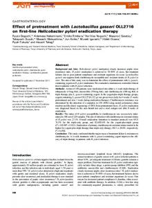
In a previous study, we had demonstrated that pre-treatment of soybeans with calcium
103
enhances defense responses with higher accumulation of isoflavone phytoalexins, thereby
104
reducing Sclerotinia infection (Arfaoui et al. 2016), and suggesting an indirect effect on the
105
pathogen.
106
In this study, we examined the effect of foliar calcium application on disease symptoms, on
107
oxalic acid production, and on ROS generation and detoxification in soybeans infected with
108
SS18 and SSPetri, a weakly- and highly-aggressive isolates of S. sclerotiorum, respectively.
109
The response to the oxidative stress caused by inoculation with either isolate was also
110
examined at the cellular level through overtime monitoring of changes in transcript levels of
111
four genes involved in ROS detoxification (Sodb2, Apx1, Prx2b and Thioredoxin).
114
M AN U
TE D
EP
AC C
112 113
SC
RI PT
89
Materials and Methods
115
Fungal isolates
116
We tested S. sclerotiorum SSPetri and SS18, a highly- (HA) and weakly-aggressive (WA)
117
isolates, respectively, as previously described (Arfaoui et al. 2016). Both isolates were grown
118
on PDA (Potato Dextrose Agar) at 20°C.
119 120
Plant material
4
ACCEPTED MANUSCRIPT We used seeds of soybean cv. Thunder 27005RR2 from Thunder Seed, Canada to grow
122
seedlings for 21 days in a growth chamber (20oC, 60-80% RH, and a 16 h photoperiod). For
123
each treatment, three pots, filled with soil and sand (1:1), were used, and six seeds were
124
planted per pot. Two days before inoculation, the seedlings were divided into two sets. One
125
set was sprayed with a 0.1% calcium formulation (P3; OMEX Agriculture Inc., Canada) until
126
the leaves were wet. 5 millimiter plugs of 5 day old culture were used to inoculate plants at
127
the V2 leaf stage. Plugs with Sclerotinia inoculum were placed in the middle of each leaf.
128
After inoculation, all of the plants were kept at 20oC and 90 % RH to provide adequate
129
conditions for infection. Leaf samples were collected at 6, 24, 48 and 72 hours post
130
inoculation (hpi) and immediately used for ROS production analysis or frozen in liquid
131
nitrogen and stored in -80oC, until used for RNA isolation. Leaves were also photographed
132
and the percentage of diseased area was calculated using the lesion assay software ASSESS
133
2.2.
134
Six treatments were compared: (i) water control (Control); (ii) control pre-treated with
135
calcium (Control + Ca); (iii) inoculated with the HA isolate (SSPetri); (iv) pre-treated with
136
calcium and inoculated with the HA isolate (SSPetri + Ca); (v) inoculated with the WA
137
isolate (SS18), and (vi) pre-treated with calcium and inoculated with the WA isolate (SS18 +
138
Ca). The experimental design consisted of three biological replicates for each treatment that
139
were completely randomized. Leaves from 6 different plants were pooled together to form
140
one of three biological replicates. The whole experiment was repeated twice.
SC
M AN U
TE D
141
RI PT
121
Determination of oxalic acid content in soybean leaves
143
Total oxalic acid concentration was determined in soybean leaf tissues using the protocol of
144
Xu and Zhang (2000) with a few modifications. Briefly, one g of fresh leaf material was
145
extracted in 50 ml of water at 80°C while shaking for 15 min at 120 r.p.m. The suspension
146
was centrifuged at 5000 g for 15 min and the supernatant was filtered through a Whatman
147
paper before being subjected to an oxalic acid assay. The assay consisted of 50 µl sample (or
148
standard OA solution), 27.5 µl of 1 mM bromophenol blue, 49.5 µl of 1M sulfuric acid, 44 µl
149
of 100 mM potassium dichromate and 1.2 ml water. The mixture was vortexed and incubated
150
in a water bath at 60°C for 10 min. After incubation, the reaction was quenched with 110 µl of
151
2 M sodium hydroxide. The absorbance was measured at 600 nm. Oxalic acid concentration
152
was expressed as µg.g-1 FW by comparison with an OA standard curve. Assays were
153
conducted in triplicate and the whole experiment was independently repeated three times.
AC C
EP
142
5
ACCEPTED MANUSCRIPT Oxalic acid released in PDB medium by S. sclerotiorum isolates was also quantified by the
155
same protocol of Xu and Zhang (2000). Briefly S. sclerotiorum isolates were grown in 100 ml
156
flask containing PDB media amended with 0.1% (v/v) of calcium-based formulation. Flasks
157
were statically incubated for 3 and 7 days at room temperature. Cultures filtrates were used
158
for oxalic acid determination and mycelial fractions dry weight were determined after drying
159
at 60°C for 3 days. Oxalic acid concentration was expressed as µg.mg-1 dry weight mycelium.
RI PT
154
160 161 In situ detection of ROS
163
Superoxide ion (O2-) was detected in soybean tissues 48 hpi Leaf tissues were placed for 2
164
hours in an aqueous solution in 0.5 mg.ml-1 Nitroblue Tetrazolium (NBT) (10 mM potassium
165
phosphate buffer, pH 7.5). After incubation, leaves were rinsed in 70% ethanol and mounted
166
in 50% glycerol and photographed (Hernandez et al. 2001).
167
Detection of hydrogen peroxide (H2O2) was conducted by staining the tissues in 3,3-
168
diaminobenzidine (DAB) as described by Thordal Christensen et al. (1997). H2O2 reacts with
169
DAB to form a reddish-brown stain. Leaves were incubated in DAB solution (1 mg.mL-1, pH
170
3.8) for 2 hours and vacuum infiltrate at 120 mbar for 1 min. After that, the leaves were
171
incubated in plastic boxes for 6-8 hours under high humidity conditions until brown
172
precipitate was observed. Stained leaves were fixed in a solution of ethanol: lactic acid:
173
glycerol (3:1:1, v/v/v) and photographed.
TE D
M AN U
SC
162
174
Quantification of ROS production
176
The production of hydrogen peroxide (H2O2), the anion superoxide (O2-) and hydroxyl radical
177
(OH.) were determined in soybeans leaves issued from Ca-treated and untreated plants
178
infected with SS18 or SSPetri isolates.
179
The production of O2- was quantified based on NBT reduction activity, as described by Doke
180
(1983) and El Hadrami et al. (2005). Briefly, leaves were washed using dH2O and placed for 1
181
hour in 3 ml of 10 mM potassium phosphate buffer, pH 7.8, containing 0.05% NBT and 10
182
mM NaN3. This mixture was heated for 15 min at 85°C and then cooled quickly. The
183
reduction activity of the soybean leaf extract was followed by measuring the absorbance at
184
580 nm.
185
The production of H2O2 was estimated as described by Tiedemann (1997) and El Hadrami et
186
al. (2005). Soybean leaves were incubated for 2 hours in the dark at room temperature in 2 ml
AC C
EP
175
6
ACCEPTED MANUSCRIPT of a reagent mixture containing 50 mM phosphate buffer pH 7.0, 0.05% guaicol and
188
peroxidase. The release of H2O2 was monitored by measuring absorbance at 450 nm.
189
The production of OH. was estimated as described by Tiedemann (1997) and El Hadrami et
190
al. (2005). Leaves were incubated in 1 ml of 1 mM 2-Deoxyglucose then incubated for 45 min
191
in the dark at room temperature. Five hundred microliters of this solution were added to 500
192
µl of 1% (w/v) thiobarbituric acid and 1 ml of 2.8% (w/v) trichloroacetic acid. The mixture
193
was boiled for 10 min and immediately cooled in ice for 10 min. The production of the
194
hydroxyl radical was followed by measuring absorbance at 540 nm.
RI PT
187
195 RNA extraction and RT-qPCR
197
RNA was extracted from the leaf tissues using the RNeasy plant mini kit (QIAGEN) and
198
treated
199
recommendations. The RNA was quantified at 260 nm using a NanoDrop ND-2000
200
spectrophotometer (Thermo Scientific, Wilmington, DE, USA), and its quality checked on 1%
201
agarose gel.
202
The first cDNA strand was synthesized using the total RNA with a Promega reverse
203
transcription kit (Promega, Madison, WI, USA) as recommended by the manufacturer. QRT-
204
PCR was carried out for both the target and reference (actin) genes using CFX96TM Real
205
Time System (C1000TM Thermal Cycler, Biorad) and SsoFast EvaGreen Supermix (Biorad,
206
Hercules, CA, USA) as recommended by the manufacturer. The amplification was carried out
207
using 95°C for 2 min to activate the hot-start recombinant Taq DNA polymerase, followed by
208
39 cycles of amplification at 95°C for 30 s, 60°C for 30 s, 72°C for 30 s, and 78°C for 11 s to
209
avoid primer-dimer formation. Following amplification, a melting curve program (55-95°C
210
with a heating rate of 0.5°C.s-1) at 95°C for 0.05 s, 65°C for 0.05 s, and 95°C for 0.5 s was
211
performed to ensure that only a single product was generated at the end of the assay. All
212
samples were run in parallel with the housekeeping gene actin to normalize cDNA loading.
213
Three biological replicates were used per run including the reference gene. Gene expression
214
results were analyzed using the 2−∆∆Ct method after verification that the primers amplified
215
with an efficiency of approximately 100% as described by Livak and Schmittgen (2001).
216
Specific primers were designed based on the soybeans mRNA sequences available and
217
GenScript
218
bin/tools/primer_genscript.cgi) to test the expression of Sodb2, Apx 1, Prx2b and Thio genes
219
(Table 1). The constitutively expressed gene actin was used for normalization of each target
220
gene
RNase-free
DNase
I
(Ambion)
according
to
the
manufacturer’s
AC C
EP
TE D
M AN U
with
SC
196
Real-time
PCR
Primer
Design
7
Software
(http://www.genscript.com/cgi-
ACCEPTED MANUSCRIPT Statistical analysis
222
All data sets from OA content, ROS accumulation and RT-qPCR were analyzed by ANOVA
223
using Statistica Software (Statsoft, Tulsa, OK, USA). The data sets were from three
224
independent replicates and were tested for variance homogeneity and significance of the
225
“experiment” effect. In the absence of significant “experiment” effect, data were pooled and
226
presented as averages. Differences among the means were assessed based on the Duncan
227
Multi-range test at P < 0.05.
RI PT
221
228
Results
230
Disease assessment
231
Five days post inoculation, soybean leaves inoculated with the HA isolate SSPetri displayed a
232
lesion of 40% of the leaf area. In contrast, inoculation with the WA isolate SS18 resulted in
233
limited infection of 8% (Fig.1). Application of calcium onto the leaves 48 h prior to
234
inoculation significantly restricted the lesion development, especially by the HA isolate
235
SSPetri with more than 50 % reduction in lesion size (Fig. 1)
M AN U
SC
229
236 Oxalic acid content
238
The HA isolate SSPetri produced more oxalic acid in the infected soybean leaves than the
239
WA isolate SS18 (Fig. 2). Pre-treatment with calcium onto the leaves 48 hours prior to
240
inoculation significantly reduced the production of oxalic acid in planta, especially by the HA
241
isolate SSPetri. At 3 and 7 days post inoculation, OA content was significantly lower (30%) in
242
the pre-treated than the non-treated leaves in response to inoculation with the HA isolate
243
SSPetri.
244
The amount of oxalic acid released by SSPetri (286.8µg) in the PDB medium was much
245
higher than that released by SS18 (163.6 µg) (Table 2). Adding calcium to the PDB medium
246
at a concentration of 0.1% decreased the level of the OA released by both isolates by 10%
247
(Table 2).
EP
AC C
248
TE D
237
249
ROS accumulation
250
H2O2 production was discernable as a reddish-brown stain by DAB. It was more noticeable in
251
SS18-infected plants than with SSPetri. A clear zone free of DAB staining was observed in
252
the cells directly adjacent to the inoculation site (Fig. 3). A significant change was perceived
8
ACCEPTED MANUSCRIPT in the Ca-treated soybean tissues inoculated with either isolate but the response was stronger
254
in response to the HA isolate.
255
Quantification of H2O2 showed an early response to inoculation with S. sclerotiorum. The
256
WA isolate SS18 induced an increase in H2O2 content as early as 6 hpi while the H2O2 content
257
in the tissues challenged with the HA isolate SSPetri was similar to that of the untreated, non-
258
challenged tissues (Fig. 4 a,b). After pre-treatment with calcium, the concentration of H2O2
259
was higher in response to the HA SSPetri (SSPetri+Ca) than to the WA isolate SS18 (SS18-
260
Ca). The maximum induction was observed at 72 hpi with 40% increase in response to
261
SSPetri+Ca as compared to SS18-Ca. No significant change was observed in the control
262
plants whether untreated or pre-treated with calcium.
263
An inhibition of O2- production was observed at the site of inoculation with the HA isolate,
264
with absence of a blue color around the lesions (Fig. 3). A noticeable production of O2- was,
265
on the other hand, observed in the tissues inoculated with SS18. The application of calcium
266
prior to inoculation restored the production of O2- in the leaves inoculated with the HA isolate
267
SSPetri. The content in O2- in the leaves inoculated with the WA isolate reached its maximum
268
at 72 hpi (Fig. 4 c,d).
269
The highest OH. production occurred 24 hpi in response to the WA isolate SS18. With the HA
270
isolate SSPetri, the level was similar to that of the untreated and non-challenged control. On
271
the other hand, pre-treatment with calcium restored the OH. production in the leaves
272
inoculated with the HA isolate SSPetri. The maximum production in response to SSPetri+Ca,
273
which was three times higher than in response to SS18-Ca, was observed 24 hpi (Fig. 4 e,f).
TE D
M AN U
SC
RI PT
253
EP
274
Expression patterns of genes controlling anti-oxidative enzymes
276
All of the tested genes that control anti-oxidative enzymes showed higher expression in
277
response to inoculation with the WA isolate SS18. Most of them peaked as early as 6 hpi,
278
which precedes ROS accumulation in the soybean leaves (Fig. 5).
279
Sodb2 transcripts showed more abundance in response to the WA isolate SS18 than with the
280
HA isolate SSPetri (Fig. 5 a,b). Pre-treatment with calcium caused a drop in sodB2 transcripts
281
in response to both isolates.
282
Both SS18 and SSPetri isolates induced the expression of Apx1. This expression was stronger
283
and peaking almost 10-fold higher at 24 hpi in response to the WA isolate SS18 as compared
284
to the HA isolate SSPetri. Pre-treatment of soybeans with calcium increased the level of
285
expression of the Apx1 gene in response to inoculation with the HA isolate SSPetri (Fig. 5
286
c,d).
AC C
275
9
ACCEPTED MANUSCRIPT Prx2b gene had a higher expression in the leaves inoculated with SS18 than with SSPetri.
288
Accumulation of transcripts in response to SSPetri was stable over time and similar to the
289
untreated control. Pre-treatment with calcium induced a slight increase in this gene’s
290
expression in response to inoculation with the HA isolate SSPetri (Fig. 5 e,f).
291
The thioredoxin-encoding gene showed a gradual increase in expression over time, with a
292
peak at 48 hpi. Its transcripts’ level was 3-fold higher in response to the WA isolate SS18 than
293
to the HA isolate SSPetri. Pre-treatment with calcium induced a 2-fold higher expression of
294
this gene in response to the HA isolate SSPetri as early as 6 hpi (Fig. 5 g,h).
RI PT
287
295
Discussion
297
Sclerotinia sclerotiorum is considered an atypical necrotroph fungus. It colonizes plant tissues
298
according to several distinct phases dictated by a precise reprogramming of the pathogen
299
transcriptional and physiological machinery (Kabbage et al. 2013, 2015). Our results indicate
300
that adding calcium to the growth medium reduce the amount of OA released by both isolates.
301
This is not surprising since OA is intrinsically linked to low pH and adding calcium to the
302
medium will reduce the acidity and increase the pH, which impact the OA production. Pre-
303
treatment of soybeans with calcium significantly reduced the amount of oxalic acid produced
304
in planta, leading to a substantial decrease in disease progress. The response was more
305
pronounced with the highly aggressive (HA) isolate SSPetri than with the weakly aggressive
306
(WA) isolate SS18. The HA isolate SSPetri produces more oxalic acid and causes more
307
disease severity (40%) than the WA isolate SS18 (8.0%). This corroborates other findings
308
showing a positive correlation between the rate of production of oxalic acid and the virulence
309
of S. sclerotiorum isolates (Durman et al. 2003; 2005). Therefore, chelating produced oxalic
310
acid by applying exogenous calcium could likely attenuate the pathogen ability to infect and
311
progress on/in the tissues, with consequences on pathogenesis and defense responses.
312
The reduction of OA is important in crosslinking calcium ions bound to pectins, which protect
313
host cell walls from fungi (Brisson et al. 1994). Gosh et al. (2016), suggested that calcium
314
may have a role in low oxalate-mediated cell wall rearrangement towards induced plant
315
defense.
316
ROS production is an integral part of the recognition process between S. sclerotiorum and its
317
host (Bolton et al. 2006; Perchepied et al. 2010). It is also a trigger for the signaling cascade
318
that allows plants to set defense responses remotely from the infection site. However,
319
pathogens such as S. sclerotiorum have evolved mechanisms to temporarily impede this
AC C
EP
TE D
M AN U
SC
296
10
ACCEPTED MANUSCRIPT oxidative burst, thereby allowing them to evade the effect of defense molecules such as
321
phytoalexins. The mechanisms underlying this early inhibition of the oxidative burst remain
322
unknown in S. sclerotiorum. In the present study, we assessed ROS content produced at the
323
site of inoculation and showed that soybean plants respond strongly to the infection by
324
releasing higher ROS levels in response to the WA isolate SS18. Inversely, the content of
325
ROS in the leaves inoculated with the HA isolate SSPetri was as low as that of untreated and
326
non-inoculated tissues. This reduction in ROS accumulation compromises the defence
327
responses and leads to a higher infection rate and disease severity. Similar results were
328
reported by Cessna et al. (2000) showing an inhibition of the production of H2O2 and a
329
suppression of the oxidative burst in tobacco and soybean cell cultures challenged by the
330
oxalic acid produced by a virulent isolate of S. sclerotiorum. Other studies found that the
331
reducing conditions that S. sclerotiorum engenders during the initial stages of colonization,
332
suppress host defense responses, including the oxidative burst and callose deposition
333
(Williams et al. 2011). Once infection is established, ROS production is restored leading to
334
oxidative burst and programmed cell death (Williams et al. 2011). This could be associated
335
with the trophic behavior of S. sclerotiorum as a necrotroph at start, then turning into a
336
hemibiotroph at a later stage.
337
Calcium pre-treatment induced an increase in ROS accumulation a few hours after inoculation
338
with the HA isolate SSPetri. This led to the restoration of the suppressed oxidative burst able
339
to elicit back the host defense responses. Similar results were reported in response to the pre-
340
treatment with thiamine or biological control agents in response to infection with the wild-
341
type of S. sclerotiorum (Jain et al. 2013; Zhou et al. 2013). This is not surprising since
342
calcium metabolism and ROS signaling are closely related. Ca2+ flux is known to operate both
343
upstream and downstream ROS production. In addition, Ca2+ influx is indispensable for the
344
initial ROS accumulation while ROS production is essential for later additional calcium
345
fluxes in the cell subsequent to pathogen elicitation (Levine et al. 1996; Blume et al. 2000).
346
In plants and fungi, oxalates play an important role in calcium regulation (Bush, 1995;
347
Franceschi et al. 2005) and their role in S. sclerotiorum pathogenesis is well established.
348
Calcium is a key factor acting as signal in many processes of plants, including growth,
349
response to both biotic and abiotic stress (Bush, 1995). In fungi, calcium gradients regulate
350
growth and the uptake of ions from their environment (Dutton and Evans, 1996; Lew, 2011).
351
However, free calcium may become a toxic cellular compound at high concentrations,
352
because it can build complexes with proteins, membranes, and organic acids. As a result of
AC C
EP
TE D
M AN U
SC
RI PT
320
11
ACCEPTED MANUSCRIPT fungal infection, calcium concentrations usually increase due to cell wall lytic activities by the
354
growing hyphae and host cells’ autolysis.
355
Several genes encoding for ROS-scavenging enzymes are often up-regulated hours after
356
infection to protect the host cells from the damage due to elevated ROS content (Levine et al.
357
1994). This was the case in the current study, where most tested genes were up-regulated 6 to
358
24 hpi. Their profile of expression was differential among the WA and HA isolates. Higher
359
transcripts levels were recorded early in response the WA isolate SS18 while those recorded
360
in response to HA isolate SSPetri were delayed and weaker. This suggests a negative
361
correlation between the ROS-scavenging capacity of the host and the success of the pathogen
362
in invading the issues. It also highlights the ability of HA isolate SSPetri to suppress ROS
363
accumulation at levels high enough to induce the synthesis/accumulation of ROS-scavenging
364
transcripts. Sod is one of the main constituents of the ROS scavenging machinery of the plant
365
defense system (Bowler et al. 1992; El Hadrami et al. 2005). Numerous reports revealed an
366
increase in SOD activity in plants under oxidative stress (El Hadrami et al. 2005; Nanda et al.
367
2010; Malencic et al. 2010; Morita et al. 2012). Our results showed a peak of accumulation of
368
Sodb2 transcripts as early as 6 hpi in response to infection with either SS18 or SSPetri,
369
followed by a decrease almost within 24 hpi after infection, which reflects the enzymes
370
involvement in the detoxification of O2-. This was confirmed with a positive correlation with
371
the O2- accumulation in the tissues. Inversely, lower levels of expression of Sodb2 along with
372
low amounts of O2- were detected in response to SSPetri, indicative of a suppression of the
373
oxidative burst and a surreptitiousness behavior of this HA isolate. Calcium pre-treatment did
374
not significantly induce any changes in sodb2 expression after infection with the HA isolate
375
SSPetri. These results suggest that the ROS scavenging mechanism of the HA isolate would
376
have been countered by one or more toxic metabolites produced by S. sclerotiorum during
377
pathogen invasion. Comparable observations were reported in B. napus exogenously supplied
378
with oxalic acid and showing a suppressed SOD activity (Liang et al. 2009).
379
The activation of Apx1 and Prx2b keeps the balance of cellular H2O2 in response to fungal
380
infection. Both genes were induced after infection with WA isolate SS18. However, the
381
expression of the Apx1 gene was stronger, suggesting that Apx1 was more efficient in
382
destroying H2O2 than Prx2b. Increase in Apx1 level was seen after the increase of Sodb2 (24
383
hpi) in response to both tested isolates whereas the increase in Prx2b transcripts was recorded
384
in response to WA isolate SS18 as early as 6 hpi and at the same time as Sod. These results
385
indicate that these two enzymes have different affinities for H2O2, confirming their belonging
AC C
EP
TE D
M AN U
SC
RI PT
353
12
ACCEPTED MANUSCRIPT to separate classes of H2O2-scavenging enzymes (Mittler et al. 2004; Dietz et al. 2006; Foyer
387
and shigeoka, 2010; Maruta et al. 2012, Arfaoui et al. 2013, 2014).
388
Pre-treatment with calcium increased the transcription of Apx1, Prx2b and Thioredoxin in
389
plants infected with the HA isolate SSPetri. This may be explained by the fact that the
390
calcium provided the plants with better elicitation of stress response. These results corroborate
391
reports showing the involvement of three biological control agents Pseudomonas aeruginosa,
392
Trichoderma harzianum and Bacillus subtilis in inducing changes in H2O2 and antioxidant
393
metabolism of peas challenged by S. sclerotiorum (Jain et al. 2013).
394
In light of the results of the present study, pre-treatment with calcium enhances plant
395
protection against oxalic acid toxicity and triggers oxidative stress and the antioxidant
396
systems in a balanced form to keep the alert system active without causing structural damage
397
to the tissues or the cell components. We demonstrated that intervening for the establishment
398
of an initial “reducing” status is critical in hampering the pathogenesis of S. sclerotiorum. At
399
the early stage of infection, calcium could effectively restore the suppressed oxidative burst,
400
essential to the signaling cascades to alert healthy tissues remotely from the infection site.
401
Further research on the mode of action of calcium and mechanisms of interaction with oxalic
402
acid would greatly assist the control of the white mold disease in many crops including
403
soybeans. Intensive efforts using fungal mutants that are compromised in Ca2+ efflux or
404
virulence factors and plant genotypes with various degrees of resistance to white mold will
405
help understanding the basis of calcium-plant-pathogen interactions. Given the importance of
406
oxalic acid and its interaction with calcium, these results suggest that foliar application of
407
calcium may constitute a new approach to control white/soft mold in field crops and
408
vegetables.
409
EP
TE D
M AN U
SC
RI PT
386
Acknowledgement
411
This research was supported by funding from Manitoba Rural Adaptation Council (MRAC)
412
and OMEX to Dr. Abdelbasset El Hadrami (MRAC-CAAP-MB0396) and from NSERC to
413
Fouad Daayf.
AC C
410
414 415
References
416
Arfaoui A, El Hadrami A, Adam L, Daayf F (2016) Pre-treatment with calcium enhanced
417
defense-related genes' expression in the soybean's isoflavones pathway in response to
418
Sclerotinia sclerotiorum. Physiol Mol Plant Pathol 93: 12–21
13
ACCEPTED MANUSCRIPT Arfaoui A, El Hadrami A, Daayf F (2013) Effect of foliar applications of a calcium-based
420
formulation on the antioxidant systems of soybeans affected by Sclerotinia sclerotiorum. Can
421
J Plant Pathol 36 (2): 280-281
422
Arfaoui A, El Hadrami A, Daayf F (2014) The use of calcium and manganese foliar nutrition
423
to induce resistance to Sclerotinia sclerotiorum. APS-CPS Joint Meeting, August 9-13, Hilton
424
Minneapolis, Minneapolis, Minnesota U.S.A
425
Bailly C, El-Maarouf-Bouteau H, Corbineau F (2008) From intracellular signaling networks
426
to cell death: the dual role of reactive oxygen species in seed physiology. Comptes Rendus
427
Biol 331: 806-814
428
Blume B, Nürnberger T, Nass N, Scheel D (2000) Receptor-mediated increase in cytoplasmic
429
free calcium required for activation of pathogen defense in parsley. Plant Cell 12: 1425-1440
430
Bolton MD, Thomma BPHJ, Nelson BD (2006) Pathogen profile Sclerotinia sclerotiorum
431
Lib.de Bary: Biology and molecular traits of a cosmopolitan pathogen. Mol Plant Pathol 7: 1-
432
16
433
Bowler C, Montagu MV, Inze D (1992) Superoxide dismutase and stress tolerance. Annu Rev
434
Plant Physiol Plant Mol Biolo 43: 83-116
435
Brisson LF, Tenhaken R, Lamb C (1994) Function of oxidative cross-linking of cell wall
436
structural proteins in plant disease resistance Plant Cell 6: 1703-1712
437
Bush DS (1995) Calcium regulation in plant cells and its role in signaling. Annu Rev Plant
438
Physiol Plant Mol Bio 46: 95-122
439
Cessna SG, Sears VE, Dickman MB, Low PS (2000) Oxalic acid, a pathogenicity factor for
440
Sclerotinia sclerotiorum, suppresses the oxidative burst of the host plant. Plant Cell 12: 2191-
441
2200
442
Cotton P, kasza Z, Bruel C, Rascle C, Fevre M (2003) Ambient pH controls the expression of
443
endopolygalaturonase genes in the necrotrophic fungus Sclerotinia sclerotiorum. FEMS
444
Microbiol Lett 227: 163-169.
445
Da Silva LF, Dias CV, Cidade LC, Mendes JS, Pirovani CP, Alvim FC, Pereira GAC, Aragao
446
FJL, Cascardo JCM, Costa MGC (2011) Expression of an oxalate decarboxylase impairs the
447
necrotic effect induced by nep1-like protein (NLP) of in transgenic tobacco. Mol Plant
448
Microbe Interact 24: 839-848.
449
Dietz KJ, Jacob S, Oelze ML, Laxa M, Tognetti V, Nunes de Miranda SM, Baier M,
450
Finkemeier I (2006) The function of peroxiredoxins in plant organelle redox metabolism. J
451
Exp Botany 57 (8): 1697-1709
AC C
EP
TE D
M AN U
SC
RI PT
419
14
ACCEPTED MANUSCRIPT Doke N (1983) Involvement of superoxide anion generation in the hypersensitive response of
453
potato tuber tissues to infection with an incompatible race of Phytophthora infestans and to
454
the hyphal wall components. Physiol Plant Pathol 23: 345-357
455
Durman SB, Menendez AB, Godeas AM (2003) Mycelia compatibility groups in Buenos
456
aires field populations of Sclerotinia sclerotiorum (Sclerotiniaceae). Soil Biol Biochem 51:
457
421-427
458
Durman SB, Menendez AB, Godeas AM (2005) Variation in oxalic acid production and
459
mycelia compatibility within field populations of Sclerotinia sclerotiorum. Soil Biol Biochem
460
37: 2180-2184
461
Dutton MV, Evans CS (1996) Oxalate production by fungi: its role in pathogenicity and
462
ecology in the soil environment. Can J Microbiol 42: 881-895
463
El Hadrami A, Kone D, Lepoivre P (2005) Effect of Juglone on Active Oxygen Species and
464
Antioxidant Enzymes in Susceptible and Partially Resistant Banana Cultivars to Black Leaf
465
Streak Disease. Eur J Plant Pathol 113: 241-254
466
Foyer CH, Shigeoka S (2010) Understanding Oxidative Stress and Antioxidant Functions to
467
Enhance Photosynthesis. Plant Physiol 155: 93-100
468
Franceschi VR, Nakata PA (2005) Calcium oxalate in plants: Formation and function. Ann
469
Rev Plant Biol 5: 641-671
470
Ghosh S, Narula K, Sinha A, Ghosh R, Jawa P, Chakraborty N, et al (2016)
471
Proteometabolomic analysis of transgenic tomato overexpressing oxalate decarboxylase
472
uncovers novel proteins potentially involved in defense mechanism against Sclerotinia. J
473
Proteomics 143: 242-253
474
Guimaraes RL, Stotz HU (2004) Oxalate production by Sclerotinia sclerotiorum deregulates
475
guard cells during infection. Plant Physiol., 136: 3703-3711
476
Hegedus DD, Rimmer SR (2005) Sclerotinia sclerotiorum: When “to be or not to be” a
477
pathogen? FEMS Microbiol Lett 251: 177-184
478
Hernández JA, Ferrer MA, Jiménez A, Ros Barceló A, Sevilla F (2001) Antioxidant systems
479
and O2•–/H2O2 production in the apoplast of pea leaves: its relation with salt induced necrotic
480
lesions in minor veins. Plant Physiol 127: 817–831
481
Jain A, Akanksha S, Surendra S, Harikesh S (2013) Microbial Consortium-Induced Changes
482
in Oxidative Stress Markers in Pea Plants Challenged with Sclerotinia sclerotiorum. J Plant
483
Grow Regul 32: 388-399
484
Kabbage M, Yaden O, Dickman MB (2015) Pathogenic attributes of Sclerotinia sclerotiorum:
485
switching from a biotrophic to necrotophic lifestyle. Plant Sci 233: 53-60
AC C
EP
TE D
M AN U
SC
RI PT
452
15
ACCEPTED MANUSCRIPT Kesheri M, Sinha R, Sinha, RP (2011) Antioxidants as natural arsenal against multiple
487
stresses in cyanobacteria. Int J Pharma Bio Sci 2: 168-187
488
Kim H, Chen C, Kabbage M, Dickman M B (2011) Identification and Characterization of
489
Sclerotinia sclerotiorum NADPH Oxidases. App Envi Microbiol 77 (21): 7721-7729
490
Kim KS, Min JY, Dickman MB (2008) Oxalic acid is an elicitor of plant programmed cell
491
death during Sclerotinia sclerotiorum disease development. Mol Plant Microbe Interact 21(5):
492
605-12
493
Lew RR (2011) How does a hypha grow? The biophysics of pressurized growth in fungi. Nat
494
Rev Microbiol 9: 509-518
495
Levine A, Tenhaken R, Dixon R, Lamb C (1994) H2O2 from the oxidative burst orchestrates
496
the plant hypersensitive disease response. Cell 79: 583-593
497
Levine A, Pennell RI, Alvarez ME, Palmer R, Lamb C (1996) Calcium-mediated apoptosis in
498
plant hypersensitive disease resistance response. Current Biol 6: 427-437
499
Liang Y, Strelkov SE, Kav NNV (2009) Oxalic acid-mediated responses in Brassica napus L.
500
Proteomics 9: 3156-3173
501
Livak KJ, Schmittgen, TD (2001) Analysis of relative gene expression data using real-time
502
quantitative PCR and the 2(-Delta Delta C (T)) Method. Methods 25(4): 402-408
503
Malencic D, Kiprovski B, Popovic M, Prvulovic D, Miladinovic J, Djordjevic V (2010)
504
Changes in antioxidant systems in soybean as affected by Sclerotinia sclerotiorum (Lib.) de
505
Bary. Plant Physiol Biochem 48: 903-908
506
Maruta T, Noshi M, Tanouchi A, Tamoi M, Yabuta Y, Yoshimura K, Ishikawa T, Shigeoka S,
507
(2012) H2O2-triggered retrograde signaling from chloroplasts to nucleus plays specific role in
508
response to stress. J Biol Chem 287: 11717-11729
509
Mayer AM, Staples RC, Gilad NL (2001) Mechanisms of survival of necrotrophic fungal
510
plant pathogens in hosts expressing the hypersensitive response. Phytochem 58(1): 33-41
511
Mittler R, Vanderauwera S, Gollery M, Van Breusegem F (2004) Reactive oxygen gene
512
network of plants. Trends Plant Sci 9: 490-498
513
Morita S, Tsukamoto S, Sakamoto A, Makino H, Nakauji E, Kaminaka H, Masumura T,
514
Ogihara Y, Satoh. S, Tanaka K (2012) Differences in intron-mediated enhancement of gene
515
expression by the first intron of cytosolic superoxide dismutase gene from rice in monocot
516
and dicot plants. Plant Biotechno 29: 115-119
517
Nanda AK, Andrio E, Marino D, Pauly N, Dunand C (2010) Reactive oxygen species during
518
plant-microorganism early interactions. J Integ Plant Biol 52: 195-204
AC C
EP
TE D
M AN U
SC
RI PT
486
16
ACCEPTED MANUSCRIPT Paula Júnior TJ, Vieira RF, Teixeira H, Carneiro JS (2009) Foliar application of calcium
520
chloride and calcium silicate decreases white mold intensity on dry beans. Trop Plant Pathol
521
34: 171-174
522
Perchepied L, Balagué C, Riou C, Claudel-Renard C, Rivière N, Grezes-Besset B, Roby D
523
(2010) Nitric oxide participates in the complex interplay of defense-related signaling
524
pathways controlling disease resistance to Sclerotinia sclerotiorum in Arabidopsis thaliana.
525
Mol Plant Microbe Interact 23: 846-860
526
Pulido P, Spinola MC, Kirchsteiger K, Guinea M, Pascual MB, Sahrawy M, Sandalio LM,
527
Dietz KJ, Gonzalez M, Cejudo FJ (2010) Functional analysis of the pathways for 2-Cys
528
peroxiredoxin reduction in Arabidopsis thaliana chloroplasts. J Exp Botany 61: 4043-4054
529
Sharma P, Jha AB, Dubey RS, Pessarakli M (2012) Reactive oxygen species, oxidative
530
damage, and antioxidative defense mechanism in plants under stressful conditions. J Botany
531
2012: 1-26
532
Shigeoka S, Ishikawa T, Tamoi M, Miyagawa Y, Takeda T, Yabuta Y, Yoshimura K (2002)
533
Regulation and function of ascorbate peroxidase isoenzymes. J Exp Botany 53: 1305-1319
534
Singh BN, Singh A, Singh SP, Singh HB (2011) Trichoderma harzianum-mediated
535
reprogramming of oxidative stress response in root apoplast of sunflower enhances defence
536
against Rhizoctonia solani. Eur J Plant Pathol 131: 121-134
537
Thordal CH, Zhang Z, Wei Y, Collinge DB (1997) Subcellular localization of H2O2 in plants.
538
H2O2 accumulation in papillae and hypersensitive response during the barley–powdery
539
mildew interaction. Plant J 11: 1187-1194
540
Tian SP, Fa, Q, Xu Y, Jiang AL (2002) Effects of calcium chloride on biocontrol activity of
541
yeast antagonists against the postharvest fungal pathogen Rizopus stolonifer. Plant Pathol 5:
542
352-358
543
Tiedemann A (1997) Evidence for a primary role of active oxygen species in induction of host
544
cell death during infection of bean leaves with Botrytis cinerea. Physiol Mol Plant Pathol 50:
545
151-66
546
Trujillo M, Ferrer SG, Thomson L, Flohé L, Radi R (2007) Kinetics of peroxiredoxins and
547
their role in the decomposition of peroxynitrite. Sub Biochem 44: 83-113
548
Walz A, Zingen-Sell I, Theisen S, Kortekamp A (2008) Reactive oxygen intermediates and
549
oxalic acid in the pathogenesis of the necrotrophic fungus Sclerotinia sclerotiorum. Eur
550
Journal Plant Pathol 120(4): 317-330
AC C
EP
TE D
M AN U
SC
RI PT
519
17
ACCEPTED MANUSCRIPT Williams B, Kabbage M, Kim HJ, Britt R, Dickman MB (2011) Tipping the balance:
552
Sclerotinia sclerotiorum secreted oxalic acid suppresses host defenses by manipulating the
553
host redox environment. PLOS Pathogens 7(6), e1002107
554
Wojtaszek P (1997) The oxidative burst: a plant’s early response against infection. Biochem J
555
322: 4158-4163
556
Xu XQ, Zhang ZQ (2000) Kinetic Spectrophotometric Determination of Oxalic Acid Based
557
on the Catalytic Oxidation of Bromophenol Blue by Dichromate, Microchim Acta 135: 169-
558
172
559
Zhou J, Sun AZ, Xing D (2013) Modulation of cellular redox status by thiamine-activated
560
NADPH oxidase confers Arabidopsis resistance to Sclerotinia scerotiorum. J Exp Botany 64:
561
3261-3272
SC
RI PT
551
562
M AN U
563 564 565 566 567
571 572 573 574 575 576 577 578
EP
570
AC C
569
TE D
568
579 580 581 582 583 584 18
ACCEPTED MANUSCRIPT 585 Table 1. Primers used in RT–qPCR for selectively amplifying ROS genes in soybean. Target gene and
Forward and reverse primer sequence (5′–
PCR
Primer
Genbank accession
primer name
3′)
product
Tm
No. for target gene
size (bp)
(ºC)†
66
62.18
Β-Actin
F: 5′-CAATCCCAAGGCCAACAGA-3′ R: 5′-ATGGCAGGCACATTGAAAGTC-3′
Sodb2
F: 5'-GCAACACAATTTGGTTCAGG-3'
62.23
82
R: 5'-AAGGAGGATTTGCTGCATTT-3' Apx1
F: 5'-TCACGGAGTTGTTGAGTGGT-3'
141
R: 5'-GTTCCTGAGCCAGGAGAAAG-3' Thioredoxin
F: 5'-CAAATTCATAGAGCCAGCGA-3'
587
o
†Tm: primers pair melting temperature ( C)
588 589 590
594 595 596 597 598 599 600 601 602
EP
593
AC C
592
TE D
591
603 604 605 606 607 19
NM_001250972.1
NM_001250856.1
58.67
99
59.08
AF145348.1
59.01
115
M AN U
R: 5'-CGCCTCCACATTAAACTCCT-3'
59.15
SC
F: 5'-TACAAGGAATCATGCCCTCA-3'
59.02
58.79
R: 5'-CCTCAGCGTAATCAGCAAAG-3’ Prx2b
AW350943
RI PT
586
59.35
59.05
NM_001250511.1
ACCEPTED MANUSCRIPT 608 609
Table 2. Oxalic acid concentration released by Seclerotinia isolates in PDB medium amended
610
or not with 0.1% (V/V) of calcium. Oxalic acid (µg oxalic acid mg-1 dry weight mycelium)
Treatments
7 days
SSPetri
247.69a±5.3
286.8a±15.7
SS18
126.80c±9.4
163.69c±8.0
SSPetri+Ca
213.24b±9.4
254.80b±10.3
SS18+Ca
110.36c±8.3
136.13c±7.0
RI PT
3 days
Each data point represents the average for three independent experiments with error bars
612
representing the standard error to the mean. Letters indicate significant differences among
613
treatments according to Duncan test at P < 0.05.
M AN U
614 615 616 617 618
623 624 625 626 627 628 629
EP
622
AC C
621
TE D
619 620
SC
611
630 631 632 633 634 635 20
ACCEPTED MANUSCRIPT 636 Figure captions
638
Fig. 1 Aggressiveness of the weakly (SS18) and the highly (SSPetri) aggressive isolates of S.
639
sclerotiorum in absence/presence of calcium.
640
(A) Lesion development on soybean leaves. Photographs were taken at 120 hours post-
641
inoculation.
642
(B) Relative lesion area on soybean leaves. Lesions area values were analyzed using the
643
lesion assay software ASSESS 2.2. Each data point represents the average for three
644
independent experiments with error bars representing the standard error to the mean. Letters
645
indicate significant differences among treatments according to Duncan test at P < 0.05.
SC
RI PT
637
646
Fig. 2 Oxalic acid content in soybean leaves. Soybean leaves were inoculated with a PDA
648
plug (5 mm in diameter) colonized with a weakly aggressive isolate (SS18) or a highly
649
aggressive isolate (SSPetri) of S. sclerotiorum in either absence or presence of calcium.
650
Each data point represents the average for three independent experiments with error bars
651
representing the standard error to the mean. Letters indicate significant differences among
652
treatments according to Duncan test at P < 0.05.
M AN U
647
653
Fig. 3 DAB and NBT staining to assess H2O2 and O2- production (arrows) in soybean leaves
655
in the different treatments: Control; SS18, SSPetri, Control+Ca, SS18+Ca and SSPetri+Ca.
656
TE D
654
Fig. 4 Time course of generation of H2O2, O-2 and OH. during infection of soybeans leaves
658
with the weakly (SS18) or the highly (SSPetri) aggressive S. sclerotiorum isolates in absence
659
or presence of calcium. Letters indicate significant differences among treatments according to
660
Duncan test at P < 0.05. Each data point represents the average for three independent
661
replicates with error bars representing the standard errors to the means.
AC C
662
EP
657
663
Fig. 5 Relative gene expression of target genes during the infection of soybean leaves with the
664
weakly (SS18) or the highly (SSPetri) aggressive S. sclerotiorum isolates in absence or
665
presence of calcium. Letters indicate significant differences among treatments according to
666
Duncan test at P < 0.05. Each data point represents the average for three independent
667
replicates with error bars representing the standard errors to the means.
668 669 21
ACCEPTED MANUSCRIPT
SC
RI PT
A
M AN U
670
671 672 673 674
Fig. 1
AC C
EP
TE D
B
22
ACCEPTED MANUSCRIPT
SC
RI PT
675
678
M AN U
676 677 Fig. 2
679
AC C
EP
TE D
680
23
M AN U
SC
RI PT
ACCEPTED MANUSCRIPT
681 682 683
Fig. 3
AC C
EP
TE D
684
24
685 686 687 688
Fig. 4
AC C
EP
TE D
M AN U
SC
RI PT
ACCEPTED MANUSCRIPT
25
AC C
EP
TE D
M AN U
SC
RI PT
ACCEPTED MANUSCRIPT
689 690
Fig. 5
691
26
ACCEPTED MANUSCRIPT
Highlights Foliar application of calcium contributes to disease reduction. In presence of calcium, OA decreased in leaves inoculated with S. sclerotiorum. Soybean inoculation with an aggressive isolate of S. sclerotiorum induced less ROS
RI PT
accumulation.
Pre-treatment with calcium restored ROS production in plants inoculated with S. sclerotiorum. Expression of genes involved in ROS detoxification increased in response to a weakly
AC C
EP
TE D
M AN U
SC
aggressive isolate of S. sclerotiorum.
ACCEPTED MANUSCRIPT
Contributions
AC C
EP
TE D
M AN U
SC
RI PT
All three authors contributed to the planning and experimental design of the studies. AA run the experiments, collected the data, and performed analysis and interpretation. AE and FD also contributed to the data analysis and interpretation. All three authors contributed to the writing of the manuscript. AE and FD co-supervised the work.