Review For reprint orders, please contact
[email protected]
Predicting recurrent stroke after minor stroke and transient ischemic attack Expert Rev. Cardiovasc. Ther. 7(10), 1273–1281 (2009)
Philippe Couillard, Alexandre Y Poppe and Shelagh B Coutts† Author for correspondence Department of Clinical Neurosciences and Radiology, University of Calgary, C1261, Foothills Medical Centre, 1403 29th St NW, Calgary, AB, T2N 2T9, Canada Tel.: 1 403 944 1594 Fax: 1 403 283 2270 shelagh.coutts@ albertahealthservices.ca †
The risk of a subsequent stroke following an acute transient ischemic attack or minor stroke is high, with 90-day risk at approximately 10%. Identification of those patients at the highest risk for recurrent stroke following a transient ischemic attack or minor stroke may allow risk-specific management strategies to be implemented, such as hospital admission with expedited work-up for those at high risk and emergency room discharge for those at low risk. Predictors of recurrent stroke, including the ABCD2 score, brain imaging and the stroke mechanism, are reviewed in this article, with a focus on recent literature. An emphasis is placed on the importance of early imaging of the brain parenchyma (diffusion-weighted imaging) and vascular imaging to identify patients at high risk for recurrence. The need for identification of the cause of the initial event, allowing therapies to be tailored to the individual patient, is discussed. Keywords : imaging • prevention • prognosis • recurrence • stroke • transient ischemic attack
Stroke is the second leading cause of death and is a major cause of adult disability in the world [1,2] . The incidence of stroke varies throughout the world from 240 per 100,000 people in Dijon, France, to approximately 600 per 100,000 people in Novosibirsk, Russia [3] . In the USA alone, there are approximately 795,000 new strokes each year [101] . In some stroke patients, the symptoms are mild (minor stroke) or even transient – a transient ischemic attack (TIA). Tragically, many of these patients with mild stroke experience either progression of their symptoms or a second, more severe stroke that leaves them disabled. One in five ischemic stroke patients report a TIA in the hours to days preceding the stroke [4–6] . In addition, 15–30% of disabling strokes are heralded by nondisabling stroke or TIAs, usually within 1 week [4] . After a TIA or minor stroke, there is an approximately 10% risk of subsequent stroke within 90 days, with 4–5% of this risk occurring in the first 48 h [7–15] . Patients with strokes considered too minor for tissue plasminogen activator represent an interesting group. Studies show that up to a third of patients die or become dependent at hospital discharge [16,17] . With the advent of thrombolysis for acute ischemic stroke, systems of care have improved and patients with mild or transient deficits often www.expert-reviews.com
10.1586/ERC.09.105
present very quickly after symptom onset. This provides both an opportunity and a challenge: an opportunity to identify high-risk patients early, especially when their deficits are transient; a challenge in managing and triaging a large contingent of patients in a system often already overloaded. This review will focus on determinants of stroke recurrence, prediction rules and imaging in minor stroke and TIA. Most of the evidence described in this review is leveltwo evidence, as it is derived from single‑center cohort studies. Definition of recurrent stroke
Ischemic stroke and TIA are on a spectrum of serious conditions involving brain ischemia. Both represent inadequate cerebral blood flow and cause patients to incur disability and death at higher rates. There are various definitions of stroke recurrence, but the one that we find of most practical use is a ‘functional deterioration in neurological status or a new sudden focal neurological deficit of vascular origin lasting more than 24 h’. This could represent either recurrent ischemia or hemorrhage; however, in most cases, the recurrent event after an ischemic stroke will be ischemic. The neurologic dysfunction is implicitly ischemic in nature and not epileptic or related to a migrainous phenomenon
© 2009 Expert Reviews Ltd
ISSN 1477-9072
1273
Review
Couillard, Poppe & Coutts
or other stroke mimics (e.g., infections, drug abuse and side effects, or intercurrent illnesses). In this article, for the most part, we consider TIA and minor stroke together as a spectrum of the same disease, as patients presenting very early in their event cannot be classified by the traditional method (symptoms resolved within 24 h of symptom onset) [14] . Rather than defining TIA and minor stroke as distinct syndromes, we prefer to look upon these as events causing minimal or no deficits. The Effect of Urgent Treatment of Transient Ischemic Attack and Minor Stroke on Early Recurrent Stroke (EXPRESS) study found no difference in recurrent stroke outcomes in TIA or minor stroke patients [18] . Other studies have also examined at patients in this manner rather than making an arbitrary distinction between the two conditions [19,20] . Short-term stroke risk after TIA?
Studies of prognosis after TIA from the early 1990s quoted annual stroke recurrence risk as between 3.7 and 7.8% per year [21] . Recently, more ominous results have been reported [22] . A Californian study of 1707 patients diagnosed with TIA in the emergency department found a 10.5% incidence of stroke at 90 days, with half of the strokes occurring in the first 2 days [14] . A Canadian study of 2285 TIA patients produced similar findings, with a 90-day rate of stroke of 9.5% and a 1-year stroke risk of 14.5% [13] . A re-analysis of the Oxford Community Stroke project showed an 8.6% risk of stroke within 7 days after TIA [6] . Moreover, a large proportion of ischemic events will recur in the first 24 h, with a risk of 5.1% at 24 h [23] . National stroke care guidelines echo this need for urgent assessment, in an effort to reduce disability associated with disease recurrence [24] . Clinical characteristics
Studies to identify patients who are at high risk for recurrent stroke after TIA have investigated the features of the patient (e.g., diabetes mellitus [14] and hypertension [14,25]), of the event (e.g., symptom duration > 10 min or weakness or speech disturbance [14,25]), of the mechanism [26] , of the vascular territory and of imaging to predict the risk of recurrence. Patient variables and event characteristics are used to predict the risk of recurrent stroke. Using a combination of many of these factors, clinical stratification tools (e.g., California, ABCD and ABCD2 scores [14,25,27]) have been developed to help identify patients at high risk of recurrent events, with the aim of urgent hospitalization and investigation. Conversely, the scoring systems could identify patients at low risk, who could be managed in the outpatient setting (or not enrolled in clinical trials). Recently, the California and ABCD scores were combined to produce the ABCD2 score. The total ABCD2 score, ranging from 0 to 7, relies on the summation of points associated with five clinical factors: • Aged 60 years or over (1 point) • Blood pressure higher than 140/90 mmHg (1 point) • Clinical features: unilateral weakness (2 points) or speech impairment without weakness (1 point) 1274
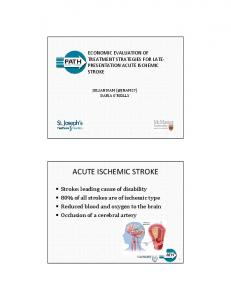
• Duration of symptoms lasting longer than 60 min (2 points) or 10–59 min (1 point) • Diabetes mellitus (1 point) The ability of the ABCD2 score to predict recurrent stroke risk has been validated in independent cohorts, with AUC values ranging from 0.62 to 0.83. It allows the stratification of patients into high risk (score: 6–7; 8.1% 2-day risk of stroke), moderate risk (score: 4–5; 4.1% 2-day risk of stroke) and low risk (score: 0–3; 1% 2-day risk of stroke) (Figure 1) . These scores are particularly useful for screening by nonexperts, but have some limitations: Validation studies assessing the ABCD and ABCD2 scores have generated mixed results; the applicability is potentially limited to TIA (although this is not clear), and the score is not informative on event mechanism [28,29] . Table 1 describes some of the factors that increase the stroke recurrence. Impact of mechanism on risk of recurrence
Strokes can be subdivided according to their mechanism for different purposes: clinical trials, epidemiological studies and for therapeutic decision-making. The Trial of Org 10172 in Acute Stroke Treatment (TOAST) classification, although imperfect, is often used in clinical practice and research protocols [30] . It uses five categories: large-artery atherosclerosis, small-vessel disease, cardioembolic, other determined and other undetermined. The risk of stroke recurrence is partly dependent on the mechanism underlying the index event [26] . Early identification of a mechanism may, therefore, improve our ability to prognosticate recurrence and deterioration. Events due to large-artery disease (primarily extracranial) have the highest risk of early recurrence, approaching eight times that of those due to small-vessel disease, which have the lowest risk. Cardioembolic events fall somewhere in between these two extremes [26,31] . The early risk of stroke after TIA due to large-artery disease has been well demonstrated in a study of patients with more than 50% carotid stenosis, among whom 20% had a stroke in the first 2 weeks prior to endarterectomy [9] . A post hoc analysis of the Warfarin versus Aspirin for Symptomatic Intracranial Disease study found a 6.7% risk of stroke within 90 days after TIA in patients with symptom-relevant 50–99% intracranial artery stenosis [32] . The vascular territory involved in a TIA also has prognostic significance. This is perhaps best described as the fact that retinal TIAs (amaurosis fugax) have a more favorable prognosis than events in other vascular territories [33,26] . Vertebrobasilar events may be associated with a higher risk of early recurrence [20,34,35], especially if stenosis is demonstrated in the posterior circulation [26,34] . Cerebral & vascular imaging as a predictor of recurrence
The limitations of previous predictive clinical scores may be due to the omission of hyperacute imaging data. MRI, specifically diffusion-weighted imaging (DWI), has revolutionized early ischemia detection in TIA and stroke. However, its lack of availability in a timely fashion has limited widespread use. By contrast, computerized tomography can be obtained expectantly in most Expert Rev. Cardiovasc. Ther. 7(10), (2009)
Predicting recurrent stroke after minor stroke & transient ischemic attack
www.expert-reviews.com
25 2 days 7 days 30 days
20
90 days Stroke risk (%)
settings. Both parenchymal and vascular imaging is important in working up a patient with TIA or stroke. Carotid imaging is particularly important, since symptom-relevant carotid stenosis of greater than 50% is associated with a higher risk of early stroke after TIA [36] , and such patients demonstrate robust benefit from early carotid revascularization. Evidence of an acute infarct on noncontrast computerized tomo graphy (NCCT) alone has been shown to be predictive of recurrent stroke in TIA patients [37] . In this study, the authors simply rated scans as having an acute infarct or not and found an adjusted odds ratio of 4.06 (95% CI: 1.16–14.1), for predicting recurrent stroke. However, the proportion of patients with evidence of acute infarcts was small (4%), therefore limiting its usefulness. A recent publication indicated that adding NCCT data increased the predictive value of the ABCD score [38] . There was a suggestion of improvement of the score with the addition of the NCCT information, but the effect was minimal, with the AUC being similar to that of the ABCD score in this population, with AUC values of 0.76 for ABCD and 0.79 for ABCD and computed tomography. This is unsurprising, as it seems unlikely that a NCCT alone would be the imaging answer to predicting outcome, as we know that MRI has greater sensitivity to smaller ischemic lesions compared with NCCT [39] . Computerized tomography angiography (CTA) uses the administration of intravenous, ionic contrast material to assess the intra cranial and extracranial vasculature with high spatial resolution. The addition of CTA adds less than 5 min to a standard CT brain examination and can be safely completed expediently in most patients with a small dose of contrast [40] . In most institutions, a NCCT is completed in the emergency room immediately after the initial clinical assessment of these patients, and adding a CTA to the protocol is more feasible than removing the patient from the scanner and proceeding to MRI. It has also been demonstrated that an intracranial arterial occlusion identified by CTA is an independent predictor of poor outcome in acute stroke patients [41] . CTA allows for the emergent identification of internal carotid artery stenosis [42] , can provide information regarding intracranial vasculature missed by carotid ultrasound, has a diagnostic accuracy at least as high as that of magnetic resonance angiography [43,44], and is comparable to digital subtraction angiography [45] . Prospective data suggest that the presence of intracranial occlusion or extracranial large-artery disease as diagnosed on CTA in TIA and minor stroke patients predicts disability at 90 days [46,47] . The negative prognostic value of intracranial vessel occlusion has also been shown using magnetic resonance angiography [47] and transcranial Doppler [20] . Magnetic resonance imaging is superior to CT for demonstrating focal ischemic change, especially in small-volume lesions [39,48] . DWI is particularly sensitive and specific for demonstrating ischemic damage and has revolutionized its detection when compared with standard MRI studies. A substantial proportion (40–60%) of patients with TIA have injuries that are observed on DWI [49,50] . Lesion pattern and location on DWI can change the suspected anatomic and vascular TIA localization in more than a third of patients [34,51] . Early DWI characteristics correlate well with etiological classification and allow for earlier determination of stroke mechanism [51,52] .
Review
15
10
5
0 0
1
2
3
4
5
6
7
ABCD2 score
Figure 1. Short-term risk of stroke by ABCD2 score in six groups combined (n = 4799). Reproduced with permission from [27] (2007) © Elsevier.
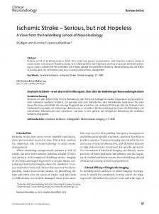
Hyperacute MRI in TIA and minor stroke patients can also stratify the risk of subsequent events and disability [53–57] . The presence of an infarct on brain imaging intuitively portends an increased risk of recurrence and has been reported to confer a two- to 15-fold increase in the short-term risk of stroke. Patients with a lesion identified with DWI are at higher risk of having a subsequent stroke as well as a disabled outcome (modified Rankin Scale score ≥ 2) than patients without a lesion [55] . The patients at highest risk are those with a lesion according to DWI and an intracranial occlusion. In one study of TIA and minor stroke, patients with no DWI lesion were at a much lower risk of recurrent events (the definition of recurrent events included patients with symptom progression) at 90 days (4.3%), compared with 10.8% with a DWI lesion and no vessel occlusion, and 32.6% with a DWI lesion and a large-vessel occlusion (Figure 2) [55] . The number of DWI lesions at baseline also predicts the likelihood of new lesions (symptomatic or asymptomatic) at 30-day follow-up MRI [58,59] . The presence of lesions of varying ages on the baseline MRI, which would suggest an active embolic process, is further associated with an increased risk of recurrent stroke [60] . MRI also has the capability of acting as a more sensitive measure of subclinical or ‘silent’ ischemic events than computed tomography. Up to 9.8% of TIA and minor-stroke patients may have new MRI lesions at 30-day follow-up, half of which are clinically silent [58] . This is relevant because the accumulation of silent infarcts does not appear to be benign in the longer term [61–63] . Recent studies have shown that a proportion of TIA patients have abnormal perfusion imaging despite resolution of symptoms. It is not clear at the current time whether this population has an increased risk of recurrent stroke [64,65] . Minor stroke and TIA patients with no DWI lesion have a low risk of recurrent clinical stroke and asymptomatic lesions on follow-up imaging [56–58] . It is not clear whether the initial 1275
Review
Couillard, Poppe & Coutts
Table 1. Clinical and imaging features that increase the risk of a recurrent stroke or symptom progression after transient ischemic attack or minor stroke. Feature
High risk
Low risk
Timing
Hours ago
Weeks ago
Age (years)
>60
140/90
60
50 None of 31.2%, and 22.9% of these patients were Lacunar perfusion abnormality Present Absent functionally impaired at 90 days. The value DWI: Diffusion-weighted imaging; MES: Microembolic signals; TCD: Transcranial Doppler. of MRI is best illustrated by the identification of a risk threshold that splits a group of clinievent in patients without DWI lesions is an ischemic event with cally high-risk patients in half. Among patients who were prea more benign prognosis or a nonischemic event, for example, dicted to have a 90-day stroke risk of 20% by ABCD2, ABCD2 migraine or seizure, which was misdiagnosed as stroke or TIA. plus MRI stratified 40% of the patients to a lower stroke risk of It is likely that both of these factors contribute to the better 7.1%, and the remaining 60% of patients to a higher stroke risk prognosis in patients without DWI lesions. Regardless of the of 28.6%. The addition of DWI positivity to the ABCD2 score underlying pathogenesis, the predictive value of a negative DWI has been incorporated into an automated calculator of stroke risk scan for a benign outcome is strong. Thus, the potential for after TIA [57] . Combined clinical and imaging assessment could benefit from enrolling these patients into clinical trials is low. increase the efficient use of resources, as patients could be triaged In a recent systematic review, the presence of a DWI lesion more appropriately into low- and high-risk groups. However, after a TIA correlated well with the known clinical predictors these results should be interpreted with caution until they have of stroke after TIA [66] . Not all studies have confirmed this, and been validated in independent populations. further work is needed into this area. Thus, whether a DWI Neuroimaging can also determine the cause of clinical worslesion in isolation from other clinical imaging [67] or etiologic ening more accurately. MRI might help distinguish if deteriofactors [68] predicts recurrent stroke is unclear. Recently, it has ration is due to a new infarct geographically separate from the been shown that almost 50% of TIA patients with DWI lesions initial infarct, growth of the initial infarct, or isolated clinical have evidence of an extracranial or intracranial large-artery worsening without a new infarct or infarct growth [78] . In addiocclusion or stenosis, suggesting that vascular imaging may be tion, MRI can identify clinically silent new infarcts or infarct the critical factor [56] . growth, both of which are known to occur within weeks after Gradient-echo sequences on MRI also provide information an initial TIA or minor stroke [58] . Therefore, similar to serial regarding the presence of cerebral microhemorrhages. These imaging in multiple sclerosis, attention should be paid to the microhemorrhages may be seen in up to 22% of patients with magnetic resonance lesion burden in TIA and stroke patients acute ischemic stroke, although they are less prevalent in patients and to how this burden changes over time. Use of imaging may with TIA [69–72] . Their presence appears to carry prognostic sig- be beneficial in studies of secondary prevention after TIA and nificance, being associated with an increased risk of recurrent minor stroke by allowing only patients at high risk for recurfatal and disabling strokes in stroke and TIA patients [70] . rence to be selected for enrolment, thereby reducing sample sizes Transcranial Doppler (TCD) with emboli detection is also without sacrificing statistical power. In addition, as in multiple useful in the assessment and risk stratification of TIA and sclerosis, using both clinical and MRI end points (i.e., new silent minor-stroke patients [73] . TCD can help predict early stroke lesion and/or microhemorrhage accumulation) might also allow 1276
Expert Rev. Cardiovasc. Ther. 7(10), (2009)
Predicting recurrent stroke after minor stroke & transient ischemic attack
Why is it important to identify patients at the highest risk of recurrent stroke?
With many recent negative trials in the stroke literature, the acute ischemic stroke drug market has been described as a ‘graveyard’ for pharmaceutical companies [79,80] . During the 20th Century, at least 178 randomized clinical trials were conducted in stroke, and of these, only three trials reported positive findings [81] . One reason for this is stroke pathophysiological heterogeneity [82] . The use of imaging to enrol patients is a potential way of targeting a more homogeneous population [83] . This approach could remove ‘noise’ from the trial, allowing a therapeutic effect to be seen with a relatively smaller sample size [84] . Accurate identification of those patients at the highest risk of worsening or having a recurrent event would allow the targeting of therapies to these TIA and minor-stroke patients. Patients with low-risk events who have nothing to gain could be excluded. There are a number of potential clinical and imaging factors that may be used to stratify risk in minor stroke and TIA. Clinical tools are important; however, those based on imaging and clinical factors are potentially better. In an ideal world, acute intervention trials in these patients would be based on MRI criteria; however, as this tool is not available in many (if not most) centers, it would limit trial enrolment and generalizability. Table 1 shows some of the important clinical and imaging factors that predict recurrent stroke after TIA or minor stroke. There are a number of effective treatments available to prevent stroke after a TIA. Available treatments include aspirin [85] or other antiplatelet medications [86] , antihypertensive agents [87] , statins [88] , anticoagulation for atrial fibrillation [89], and early carotid endarterectomy for symptomatic internal carotid artery stenosis of 50% or more [90] . Guidelines have been introduced to outline some of the treatment options for these patients [24,91] . Although there have been many interesting studies of antiplatelet agents in secondary stroke prevention, discussion of these studies is beyond the scope of this review article. Importantly, virtually all of these trials have avoided enrolling patients within 24 h of their event. With regards to emergent treatment (within 24 h), the Fast Assessment of Stroke and Transient Ischaemic Attack to Prevent Early Recurrence (FASTER) study was the first randomized trial to test the benefit of emergent preventive treatment of minor-stroke and TIA patients [92] . This study was the first to assess ‘emergent secondary stroke prevention’. This study was based in Calgary and was a pilot study testing the emergent use of aspirin versus aspirin plus clopidogrel. It demonstrated a promising, although not significant, 35% relative risk reduction in recurrent stroke in the combination treatment arm versus aspirin monotherapy. A larger, second FASTER trial is planned, to test the benefit of urgent combination treatment in a patient sample of several thousand. www.expert-reviews.com
No DWI lesion and no occlusion: 4.3%
1.0 Proportion free from stroke
for smaller sample sizes by increasing the number of relevant outcomes. However, it is important to note that accumulation of asymptomatic lesions has not been proven to predict long-term recurrent stroke, and these data are necessary before imaging can be used as a surrogate for clinical outcome.
Review
0.9
Yes DWI lesion and no occlusion: 10.8%
0.8 Yes DWI lesion and yes occlusion: 32.6%
0.7 0.6 0.5
Likelihood ratio test p-value = 0.02 0
30 60 Time after presenting with acute TIA or minor stroke (days)
90
Figure 2. Stroke-free survival curves for patients with and without diffusion-weighted imaging lesion and intracranial vessel occlusion. Curves are adjusted for NIH Stroke Scale score and baseline glucose level. The adjusted risks of stroke at 90 days are shown as percentages on the right side above the curves. DWI: Diffusion-weighted imaging; TIA: Transient ischemic attack. Reproduced with permission from [96] .
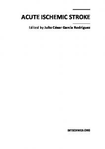
A recent publication suggested that if five, simple evidencebased interventions (i.e., dietary modification, exercise, aspirin, a statin and an antihypertensive agent) were used appropriately, then the risk of recurrent stroke might be reduced by up to 80% [93] . Thus, it would seem that urgent stroke prevention services for TIA promise to reduce recurrent stroke by facilitating more rapid and early access to the effective treatments, as discussed earlier. Additionally, two observational studies published in 2007 (EXPRESS [Figure 3] and Transient Ischaemic Attack Clinic with Round-The-Clock Access [SOS-TIA]) [18,94] indicate that hyperacute evaluation and treatment after TIA may significantly reduce subsequent stroke by 80% compared with expected rates. Expert commentary
The common thinking regarding TIA and minor stroke has been that early risk is assumed primarily by recurrent events. However, there is evidence that up to 90% of early clinical deterioration can actually be attributed to symptom progression or infarct growth rather than a true recurrent stroke [78] . This distinction may be important because specific therapeutic interventions might be tailored to attenuate infarct progression, while others would aim to minimize the risk of recurrence. Although EXPRESS [18] and SOS-TIA [94] have shown that existing evidence-based therapies for secondary stroke prevention appear to be of benefit in the acute phase, novel and potentially more effective therapies are still lacking. More randomized, controlled trials in the ilk of the FASTER study [92] are needed to investigate the value of different antiplatelet regimens, anticoagulation, blood pressure manage ment, high-dose statins, thrombolysis, endovascular stenting and 1277
Review
Couillard, Poppe & Coutts
Five-year view
Risk of recurrence (%)
14 Phase 1
12
Phase 2
10 8 6 4 2
p < 0.0001
0 0
10
20
30
40 50 60 Time (days)
70
80
90
Figure 3. Risk of recurrent stroke after first seeking medical attention in all patients with transient ischemic attack or stroke in the whole study population. Reproduced with permission from [18] .
neuroprotection in TIA patients and minor-stroke patients at high, early risk of deterioration. Therapies tailored to particular TIA and stroke mechanisms are also needed, especially for patients with intracranial atherosclerosis and small-vessel infarcts. For now, the therapeutic paradigm should consist of expedited evaluation, relevant bloodwork, parenchymal and vascular neuroimaging, cardiac investigations and a ‘cocktail’ therapeutic approach, with attention to mechanisms requiring specific treatment. Recurrent events, whether they are progression of the original event or a distinct new event, need emergent investigations and treatment to prevent recurrent stroke [78] .
Over the next 5 years, early brain and vascular imaging will be performed in all acute stroke patients – especially in TIA or minor-stroke patients. We expect that 5 years from now, most patients will have a MRI brain scan immediately, including detailed vascular imaging, allowing a more tailored approach to acute treatment immediately after the scan. Early identification of stroke subtype is important, as it will alter the longterm care of the patient, including secondary stroke prevention, as well as assigning prognosis, identification of patients with increased risk of neurological worsening, recurrent stroke and medical complications. Recurrent stroke prevention will involve tailoring therapy to an individual patient based upon imaging results. Clinical trials will investigate specific stroke etiologies and response to acute treatment. Currently, we feel that the most obvious treatment strategy to be tested is dual antiplatelet therapy (aspirin and clopidogrel) in large-artery disease [95] , but there will be other such targeted treatment strategies. Stroke care will move from an era of negative trials, due to lumping of different mechanisms of stroke together, to a more etiological approach to the disease. Repeat imaging at a few months poststroke, showing ongoing accumulation of disease, will also allow us to manage their vascular risk factors more aggressively. Financial & competing interests disclosure
The authors have no relevant affiliations or financial involvement with any organization or entity with a financial interest in or financial conflict with the subject matter or materials discussed in the manuscript. This includes employment, consultancies, honoraria, stock ownership or options, expert testimony, grants or patents received or pending, or royalties. No writing assistance was utilized in the production of this manuscript.
Key issues • Ischemic strokes are common and costly for the individual and society. • Early identification of patients at high risk for recurrent stroke is essential to reduce morbidity, mortality and allocate resources appropriately. • Identification of stroke mechanism is essential to tailoring treatment to the individual patient. • Diagnostic imaging allows triage and classification of patients into high- and low-risk groups. • Stroke is a disease of blood vessels – intracranial and extracranial vascular imaging is as important as brain parenchymal imaging. • Diffusion-weighted imaging can predict which patients are at high risk for recurrent stroke. • Many recurrent strokes occur in the hours to days after the initial event, making assessment and treatment of these patients an emergency. • Early initiation of treatment can prevent recurrent stroke in up to 80% of transient ischemic attacks and minor-stroke patients.
References Papers of special note have been highlighted as: • of interest •• of considerable interest 1
Donnan GA, Fisher M, Macleod M, Davis SM. Stroke. Lancet 371(9624), 1612–1623 (2008).
2
Murray CJ, Lopez AD. Mortality by cause for eight regions of the world: global burden of disease study. Lancet 349(9061), 1269–1276 (1997).
1278
3
Sudlow CL, Warlow CP. Comparing stroke incidence worldwide: what makes studies comparable? Stroke 27(3), 550–558 (1996).
4
Rothwell PM, Warlow CP. Timing of TIAs preceding stroke: time window for prevention is very short. Neurology 64(5), 817–820 (2005).
5
Hankey GJ, Warlow CP. Treatment and secondary prevention of stroke: evidence, costs, and effects on individuals and populations. Lancet 354(9188), 1457–1463 (1999).
6
Lovett JK, Dennis MS, Sandercock PA, Bamford J, Warlow CP, Rothwell PM. Very early risk of stroke after a first transient ischemic attack. Stroke 34(8), E138–E140 (2003).
7
Coull AJ, Lovett JK, Rothwell PM. Population based study of early risk of stroke after transient ischaemic attack or minor stroke: implications for public education and organisation of services. BMJ 328(7435), 326 (2004).
Expert Rev. Cardiovasc. Ther. 7(10), (2009)
Predicting recurrent stroke after minor stroke & transient ischemic attack
8
9
Dennis MS, Bamford JM, Sandercock PA, Warlow CP. The Oxfordshire Community Stroke Project. A comparison of risk factors and prognosis for transient ischemic attacks and minor ischemic strokes. Stroke 20(11), 1494–1499 (1989). Eliasziw M, Kennedy J, Hill MD, Buchan AM, Barnett HJ. Early risk of stroke after a transient ischemic attack in patients with internal carotid artery disease. CMAJ 170(7), 1105–1109 (2004).
10
Friedman GD, Wilson WS, Mosier JM, Colandrea MA, Nichaman MZ. Transient ischemic attacks in a community. JAMA 210(8), 1428–1434 (1969).
11
Giles MF, Rothwell PM. Risk of stroke early after transient ischaemic attack: a systematic review and meta-analysis. Lancet Neurol. 6(12), 1063–1072 (2007).
12
Gladstone DJ, Kapral MK, Fang J, Laupacis A, Tu JV. Management and outcomes of transient ischemic attacks in Ontario. CMAJ 170(7), 1099–1104 (2004).
13
Hill MD, Yiannakoulias N, Jeerakathil T, Tu JV, Svenson LW, Schopflocher DP. The high risk of stroke immediately after transient ischemic attack: a populationbased study. Neurology 62(11), 2015–2020 (2004).
14
Johnston SC, Gress DR, Browner WS, Sidney S. Short-term prognosis after emergency department diagnosis of TIA. JAMA 284(22), 2901–2906 (2000).
•• Landmark publication showing a high early risk of recurrent stroke after emergency room department diagnosis of transient ischemic attack. 15
Whisnant JP, Matsumoto N, Elveback LR. Transient cerebral ischemic attacks in a community. Rochester, Minnesota, 1955 through 1969. Mayo Clin. Proc. 48(3), 194–198 (1973).
16
Barber PA, Zhang J, Demchuk AM, Hill MD, Buchan A. Why are stroke patients excluded from TPA therapy? An analysis of patient eligibility. Neurology 24(56), 1015–1020 (2001).
17
Smith EE, Abdullah AR, Petkovska I, Rosenthal E, Koroshetz WJ, Schwamm L. Poor outcomes in patients who do not receive intravenous tissue plasminogen activator because of mild or improving ischemic stroke. Stroke 36, 2497–2499 (2005).
18
Rothwell PM, Giles MF, Chandratheva A et al. Effect of urgent treatment of transient ischaemic attack and minor stroke on early recurrent stroke (EXPRESS study):
www.expert-reviews.com
a prospective population-based sequential comparison. Lancet 370(9596), 1432–1442 (2007). •• Landmark paper that shows that implementation of rapid secondary stroke prevention therapies may reduce recurrent stroke risk of transient ischemic attack (TIA) or minor stroke by up to 80%.
Review
26
Lovett JK, Coull AJ, Rothwell PM. Early risk of recurrence by subtype of ischemic stroke in population-based incidence studies. Neurology 62(4), 569–573 (2004).
27
Johnston SC, Rothwell PM, NguyenHuynh MN et al. Validation and refinement of scores to predict very early stroke risk after transient ischaemic attack. Lancet 369(9558), 283–292 (2007).
19
Coutts SB, Eliasziw M, Hill MD et al. An improved scoring system for identifying patients at high early risk of stroke and functional impairment after an acute transient ischemic attack or minor stroke. Int. J. Stroke 3(1), 3–10 (2008).
20
Ois A, Gomis M, Rodriguez-Campello A et al. Factors associated with a high risk of recurrence in patients with transient ischemic attack or minor stroke. Stroke 39(6), 1717–1721 (2008).
28
Fothergill A, Christianson TJ, Brown RD Jr, Rabinstein AA. Validation and refinement of the ABCD2 score: a population-based analysis. Stroke 40(8), 2669–2673 (2009).
21
Wilterdink JL, Easton JD. Vascular event rates in patients with atherosclerotic cerebrovascular disease. Arch. Neurol. 49(8), 857–863 (1992).
29
22
Rothwell P. Incidence, risk factors and prognosis of stroke and TIA: the need for high-quality, large-scale epidemiological studies and meta-analyses. Cerebrovasc. Dis. 16(3 Suppl.), 2–10 (2003).
Shah KH, Metz HA, Edlow JA. Clinical prediction rules to stratify short-term risk of stroke among patients diagnosed in the emergency department with a transient ischemic attack. Ann. Emerg. Med. 53(5), 662–673 (2009).
30
Adams HP, Jr., Bendixen BH, Kappelle LJ et al. Classification of subtype of acute ischemic stroke. Definitions for use in a multicenter clinical trial. TOAST. Trial of Org 10172 in Acute Stroke Treatment. Stroke 24(1), 35–41 (1993).
31
Purroy F, Montaner J, Molina CA, Delgado P, Ribo M, Alvarez-Sabin J. Patterns and predictors of early risk of recurrence after transient ischemic attack with respect to etiologic subtypes. Stroke 38(12), 3225–3229 (2007).
32
Ovbiagele B, Cruz-Flores S, Lynn MJ, Chimowitz M. Early stroke risk after transient ischemic attack among individuals with symptomatic intracranial artery stenosis. Arch. Neurol. 65, 733–737 (2008).
33
North American Symptomatic Carotid Endarterectomy Trial Collaborators. Beneficial effect of carotid endarterectomy in symptomatic patients with high-grade carotid stenosis. N. Engl. J. Med. 325, 445–453 (1991).
34
Flossmann E, Redgrave JN, Briley D, Rothwell P. Reliability of clinical diagnosis of the symptomatic vascular territory in patients with recent transient ischemic attack or minor stroke. Stroke 39, 2457–2460 (2008).
35
Giles MF, Rothwell P. Prognosis and management in the first few days after a transient ischemic attack or minor ischaemic stroke. Int. J. Stroke 1(2), 65–73 (2006).
23
•
24
25
Chandratheva A, Mehta Z, Geraghty OC, Marquardt L, Rothwell PM. Populationbased study of risk and predictors of stroke in the first few hours after a TIA. Neurology 72(22), 1941–1947 (2009). Identifies that approximately half of all recurrent strokes during the 7 days after a TIA occur in the first 24 h, highlighting the need for emergency assessment. This paper emphasizes that this condition needs to be treated as an emergency. Easton JD, Saver JL, Albers GW et al. Definition and evaluation of transient ischemic attack: a scientific statement for healthcare professionals from the American Heart Association/American Stroke Association Stroke Council; Council on Cardiovascular Surgery and Anesthesia; Council on Cardiovascular Radiology and Intervention; Council on Cardiovascular Nursing; and the Interdisciplinary Council on Peripheral Vascular Disease. The American Academy of Neurology affirms the value of this statement as an educational tool for neurologists. Stroke 40(6), 2276–2293 (2009). Rothwell PM, Giles MF, Flossmann E et al. A simple score (ABCD) to identify individuals at high early risk of stroke after transient ischaemic attack. Lancet 366(9479), 29–36 (2005).
•• Describes the derivation of the ABCD2 score, which is now routinely used in many parts of the world to identify patients at high risk of recurrent stroke after TIA.
1279
Review 36
37
Couillard, Poppe & Coutts
Rothwell PM. Prediction and prevention of stroke in patients with symptomatic carotid stenosis: the high-risk period and the high-risk patient. Eur. J. Vasc. Endovasc. Surg. 35(3), 255–263 (2008). Douglas VC, Johnston CM, Elkins J, Sidney S, Gress DR, Johnston SC. Head computed tomography findings predict short-term stroke risk after transient ischemic attack. Stroke 34(12), 2894–2898 (2003).
38
Sciolla R, Melis F. Rapid identification of high-risk transient ischemic attacks: prospective validation of the ABCD score. Stroke 39(2), 297–302 (2008).
39
Chalela JA, Kidwell CS, Nentwich LM et al. Magnetic resonance imaging and computed tomography in emergency assessment of patients with suspected acute stroke: a prospective comparison. Lancet 369(9558), 293–298 (2007).
40
41
42
43
44
45
46
Krol AL, Dzialowski I, Roy J et al. Incidence of radiocontrast nephropathy in patients undergoing acute stroke computed tomography angiography. Stroke 38(8), 2364–2366 (2007). Smith WS, Tsao JW, Billings ME et al. Prognostic significance of angiographically confirmed large vessel intracranial occlusion in patients presenting with acute brain ischemia. Neurocrit. Care 4(1), 14–17 (2006). Josephson SA, Bryant SO, Mak HK, Johnston SC, Dillon WP, Smith WS. Evaluation of carotid stenosis using CT angiography in the initial evaluation of stroke and TIA. Neurology 63(3), 457–460 (2004). Bash S, Villablanca JP, Jahan R et al. Intracranial vascular stenosis and occlusive disease: evaluation with CT angiography, MR angiography, and digital subtraction angiography. AJNR Am. J. Neuroradiol. 26(5), 1012–1021 (2005). Schramm P, Schellinger PD, Fiebach JB et al. Comparison of CT and CT angiography source images with diffusion-weighted imaging in patients with acute stroke within 6 h after onset. Stroke 33(10), 2426–2432 (2002). Randoux B, Marro B, Koskas F et al. Carotid artery stenosis: prospective comparison of CT, three-dimensional gadolinium-enhanced MR, and conventional angiography. Radiology 220(1), 179–185 (2001). Kern R, Steinke W, Daffertshofer M, Prager R, Hennerici M. Stroke recurrences in patients with symptomatic
1280
vs asymptomatic middle cerebral artery disease. Neurology 65(6), 859–864 (2005). 47
48
Torres-Mozqueda F, He J, Yeh IB et al. An acute ischemic stroke classification instrument that includes CT or MR angiography: the Boston Acute Stroke Imaging Scale. AJNR Am. J. Neuroradiol. 29(6), 1111–1117 (2008). Awad I, Modic M, Little JR, Furlan AJ, Weinstein M. Focal parenchymal lesions in transient ischemic attacks: correlation of computed tomography and magnetic resonance imaging. Stroke 17(3), 399–403 (1986).
49
Inatomi Y, Kimura K, Yonehara T, Fujioka S, Uchino M. DWI abnormalities and clinical characteristics in TIA patients. Neurology 62(3), 376–380 (2004).
50
Kidwell CS, Alger JR, Di Salle F et al. Diffusion MRI in patients with transient ischemic attacks. Stroke 30(6), 1174–1180 (1999).
51
Lee LJ, Kidwell CS, Alger J, Starkman S, Saver JL. Impact on stroke subtype diagnosis of early diffusion-weighted magnetic resonance imaging and magnetic resonance angiography. Stroke 31(5), 1081–1089 (2000).
52
Kang DW, Chalela JA, Ezzeddine MA, Warach S. Association of ischemic lesion patterns on early diffusion-weighted imaging with TOAST stroke subtypes. Arch. Neurol. 60(12), 1730–1734 (2003).
53
Purroy F, Montaner J, Rovira A, Delgado P, Quintana M, Alvarez-Sabin J. Higher risk of further vascular events among transient ischemic attack patients with diffusionweighted imaging acute ischemic lesions. Stroke 35(10), 2313–2319 (2004).
54
Ay H, Koroshetz WJ, Benner T et al. Transient ischemic attack with infarction: a unique syndrome? Ann. Neurol. 57(5), 679–686 (2005).
55
Coutts SB, Simon JE, Eliasziw M et al. Triaging transient ischemic attack and minor stroke patients using acute magnetic resonance imaging. Ann. Neurol. 57(6), 848–854 (2005).
56
Prabhakaran S, Chong JY, Sacco RL. Impact of abnormal diffusion-weighted imaging results on short-term outcome following transient ischemic attack. Arch. Neurol. 64(8), 1105–1109 (2007).
57
Ay H, Arsava EM, Johnston SC et al. Clinical- and imaging-based prediction of stroke risk after transient ischemic attack: the CIP model. Stroke 40(1), 181–186 (2009).
•
Found that the presence of a diffusionweighted imaging lesion on MRI, together with clinical TIA features, increases the accuracy of prediction of recurrent stroke after TIA.
58
Coutts SB, Hill MD, Simon JE, Sohn CH, Scott JN, Demchuk AM. Silent ischemia in minor stroke and TIA patients identified on MR imaging. Neurology 65(4), 513–517 (2005).
59
Wen HM, Lam WW, Rainer T et al. Multiple acute cerebral infarcts on diffusion-weighted imaging and risk of recurrent stroke. Neurology 63(7), 1317–1319 (2004).
60
Sylaja PN, Coutts SB, Subramaniam S, Hill MD, Eliasziw M, Demchuk AM. Acute ischemic lesions of varying ages predict risk of ischemic events in stroke/TIA patients. Neurology 68(6), 415–419 (2007).
61
Vermeer SE, Hollander M, van Dijk EJ, Hofman A, Koudstaal PJ, Breteler MM. Silent brain infarcts and white matter lesions increase stroke risk in the general population: the Rotterdam Scan Study. Stroke 34(5), 1126–1129 (2003).
62
Vermeer SE, Longstreth WT Jr, Koudstaal PJ. Silent brain infarcts: a systematic review. Lancet Neurol. 6(7), 611–619 (2007).
63
Vermeer SE, Prins ND, den Heijer T, Hofman A, Koudstaal PJ, Breteler MM. Silent brain infarcts and the risk of dementia and cognitive decline. N. Engl. J. Med. 348(13), 1215–1222 (2003).
64
Krol AL, Coutts SB, Simon JE, Hill MD, Sohn CH, Demchuk AM. Perfusion MRI abnormalities in speech or motor transient ischemic attack patients. Stroke 36(11), 2487–2489 (2005).
65
Mlynash M, Olivot JM, Tong DC et al. Yield of combined perfusion and diffusion MR imaging in hemispheric TIA. Neurology 72(13), 1127–1133 (2009).
66
Redgrave JN, Coutts SB, Schulz UG, Briley D, Rothwell PM. Systematic review of associations between the presence of acute ischemic lesions on diffusion-weighted imaging and clinical predictors of early stroke risk after transient ischemic attack. Stroke 38(5), 1482–1488 (2007).
67
Redgrave JN, Schulz UG, Briley D, Meagher T, Rothwell PM. Presence of acute ischaemic lesions on diffusion-weighted imaging is associated with clinical predictors of early risk of stroke after transient ischaemic attack. Cerebrovasc. Dis. 24(1), 86–90 (2007).
68
Koton S, Rothwell PM. Performance of the ABCD and ABCD2 scores in TIA patients with carotid stenosis and atrial fibrillation. Cerebrovasc. Dis. 24(2–3), 231–235 (2007). Expert Rev. Cardiovasc. Ther. 7(10), (2009)
Predicting recurrent stroke after minor stroke & transient ischemic attack
69
Naka H, Nomura E, Wakabayashi S et al. Frequency of asymptomatic microbleeds on T2*-weighted MR images of patients with recurrent stroke: association with combination of stroke subtypes and leukoaraiosis. AJNR Am. J. Neuroradiol. 25(5), 714–719 (2004).
70
Boulanger JM, Coutts SB, Eliasziw M et al. Cerebral microhemorrhages predict new disabling or fatal strokes in patients with acute ischemic stroke or transient ischemic attack. Stroke 37(3), 911–914 (2006).
71
Tsushima Y, Aoki J, Endo K. Brain microhemorrhages detected on T2*weighted gradient-echo MR images. AJNR Am. J. Neuroradiol. 24(1), 88–96 (2003).
72
Werring DJ, Coward LJ, Losseff NA, Jager HR, Brown MM. Cerebral microbleeds are common in ischemic stroke but rare in TIA. Neurology 65(12), 1914–1918 (2005).
73
Markus H. Monitoring embolism in real time. Circulation 102(8), 826–828 (2000).
74
Markus HS, MacKinnon A. Asymptomatic embolization detected by Doppler ultrasound predicts stroke risk in symptomatic carotid artery stenosis. Stroke 36(5), 971–975 (2005).
75
76
77
78
•
79
Valton L, Larrue V, le Traon AP, Massabuau P, Geraud G. Microembolic signals and risk of early recurrence in patients with stroke or transient ischemic attack. Stroke 29(10), 2125–2128 (1998). Levi CR, Roberts AK, Fell G et al. Transcranial Doppler microembolus detection in the identification of patients at high risk of perioperative stroke. Eur. J. Vasc. Endovasc. Surg. 14(3), 170–176 (1997). Molloy J, Markus HS. Asymptomatic embolization predicts stroke and TIA risk in patients with carotid artery stenosis. Stroke 30(7), 1440–1443 (1999).
80
Shuaib A, Lees KR, Lyden P et al. NXY-059 for the treatment of acute ischemic stroke. N. Engl. J. Med. 357(6), 562–571 (2007).
81
Kidwell CS, Liebeskind DS, Starkman S, Saver JL. Trends in acute ischemic stroke trials through the 20th century. Stroke 32(6), 1349–1359 (2001).
82
Muir KW. Heterogeneity of stroke pathophysiology and neuroprotective clinical trial design. Stroke 33(6), 1545–1550 (2002).
83
Fisher M. Recommendations for advancing development of acute stroke therapies: stroke therapy academic industry roundtable 3. Stroke 34(6), 1539–1546 (2003).
84
Furlan A, Higashida R, Wechsler L et al. Intra-arterial prourokinase for acute ischemic stroke. The PROACT II study: a randomized controlled trial. Prolyse in Acute Cerebral Thromboembolism. JAMA 282(21), 2003–2011 (1999).
85
Antithrombotic Trialists’ Collaboration. Collaborative meta-analysis of randomised trials of antiplatelet therapy for prevention of death, myocardial infarction, and stroke in high risk patients. BMJ 324(7329), 71–86 (2002).
86
87
88
CAPRIE Steering Committee. A randomised, blinded, trial of clopidogrel versus aspirin in patients at risk of ischaemic events (CAPRIE). Lancet 348(9038), 1329–1339 (1996). PROGRESS Collaborative Group. Randomised trial of a perindopril-based blood-pressure-lowering regimen among 6105 individuals with previous stroke or transient ischaemic attack. Lancet 358(9287), 1033–1041 (2001). Amarenco P, Bogousslavsky J, Callahan A 3rd et al. High-dose atorvastatin after stroke or transient ischemic attack. N. Engl. J. Med. 355(6), 549–559 (2006).
Coutts SB, Hill MD, Campos CR et al. Recurrent events in transient ischemic attack and minor stroke: what events are happening and to which patients? Stroke 39(9), 2461–2466 (2008).
89
Important paper identifying that, in minor stroke and TIA, many of the cases of recurrent stroke are due to progression of the initial event and not a distinct new event.
EAFT (European Atrial Fibrillation Trial) Study Group. Secondary prevention in non-rheumatic atrial fibrillation after transient ischaemic attack or minor stroke. Lancet 342(8882), 1255–1262 (1993).
90
Rothwell PM, Eliasziw M, Gutnikov SA et al. Analysis of pooled data from the randomised controlled trials of endarterectomy for symptomatic carotid stenosis. Lancet 361(9352), 107–116 (2003).
91
Lindsay P, Bayley M, McDonald A, Graham ID, Warner G, Phillips S. Toward a more effective approach to stroke: canadian best practice recommendations for stroke care. CMAJ 178(11), 1418–1425 (2008).
Hacke W, Furlan AJ, Al-Rawi Y et al. Intravenous desmoteplase in patients with acute ischaemic stroke selected by MRI perfusion-diffusion weighted imaging or perfusion CT (DIAS-2): a prospective, randomised, double-blind, placebocontrolled study. Lancet Neurol. 8(2), 141–150 (2009).
www.expert-reviews.com
Review
92
Kennedy J, Hill MD, Ryckborst KJ, Eliasziw M, Demchuk AM, Buchan AM. Fast Assessment of Stroke and Transient Ischaemic Attack to Prevent Early Recurrence (FASTER): a randomised controlled pilot trial. Lancet Neurol. 6(11), 961–969 (2007).
93
Hackam DG, Spence JD. Combining multiple approaches for the secondary prevention of vascular events after stroke: a quantitative modeling study. Stroke 38(6), 1881–1885 (2007).
94
Lavallee PC, Meseguer E, Abboud H et al. A transient ischaemic attack clinic with round-the-clock access (SOS-TIA): feasibility and effects. Lancet Neurol. 6(11), 953–960 (2007).
95
Markus HS, Droste DW, Kaps M et al. Dual antiplatelet therapy with clopidogrel and aspirin in symptomatic carotid stenosis evaluated using doppler embolic signal detection: the Clopidogrel and Aspirin for Reduction of Emboli in Symptomatic Carotid Stenosis (CARESS) trial. Circulation 111(17), 2233–2240 (2005).
96
Coutts SB, Simon JE, Eliasziw M et al. Triaging transient ischemic attack and minor stroke patients using acute magnetic resonance imaging. Ann. Neurol. 57(6), 848–54 (2005).
Websites 101
American Heart Association www.americanheart.org
Affiliations •
Philippe Couillard, MD Department of Clinical Neurosciences, University of Calgary, Calgary, AB, Canada
•
Alexandre Poppe, MD, FRCPC Centre des Maladies Vasculaires Cérébrales du CHUM Hôpital Notre-Dame, 1560 rue Sherbrooke Est Montréal, Québec, H2L 4M1, Room GR-1166, Canada Tel.: +1 514 890 8000 ext. 26260 Fax: +1 514 412 7556
•
Shelagh B Coutts BSc, MBChB, MD, FRCPC, FRCP, Stroke Neurologist, AHFMR Clinical Investigator and HSFC Distinguished Clinician Scientist, Assistant Professor, Department of Clinical Neurosciences and Radiology, University of Calgary, C1261, Foothills Medical Centre, 1403 29th St NW, Calgary, AB, T2N 2T9, Canada Tel.: +1 403 944 1594 Fax: +1 403 283 2270
[email protected]
1281